4226
Design of an improved Open-Face birdcage for human whole brain imaging at 5T1Paul C. Lauterbur Imaging Research Center, Shenzhen Institutes of Advanced Technology, Chinese Academy of Sciences, Shenzhen, China, 2Key Laboratory for Magnetic Resonance and Multimodality Imaging of Guangdong Province, Shenzhen, China, 3Shanghai United Imaging Healthcare, Shanghai, China, 4Department of Biomedical Engineering, State University of New York at Buffalo, New York, NY, United States
Synopsis
Keywords: High-Field MRI, Brain
The clinical experience, especially for subjects with claustrophobia, is not friendly when using the conventional shielded birdcage head coils on the ultra-high field. The closed structure also does not better support motion detection and motion correction, and other MRI functional applications. In this work, an optimized Open-Face birdcage head coil were designed based on EM simulations. The transmit efficiency, B1 uniformity, and SAR efficiency of the facial open-window birdcage coil with different configurations and a conventional shielded birdcage coil are compared. Finally, an optimized Open-Face birdcage head coil was built and evaluated on a whole body 5T MRI scanner.Introduction
Local birdcage coils are widely used as transmit RF coil for in ultra-high field (usually refers to above 3T) MRI system1,2. RF shielding is indispensable to reduce radiation loss. Unfortunately, the closed structure can reduce the comfort of the subject during the MRI scan, especially for patients with claustrophobia. In this work, an improved Open-Face birdcage head coil were designed based on EM simulations. The transmit efficiency, B1 uniformity, and SAR efficiency of the facial open-window birdcage coil with different configurations and a conventional shielded birdcage coil are simulated and compared. Finally, an optimized Open-Face birdcage head coil were built on a whole body 5T MRI scanner (United Imaging Healthcare, Shanghai, China). The measured B1+ field of the optimized Open-Face birdcage head coil was evaluated and compared with that of a conventional shielded birdcage coil. The results demonstrate the feasibility and benefits of a proposed RF transmit coil with Open-Face concept.Methods
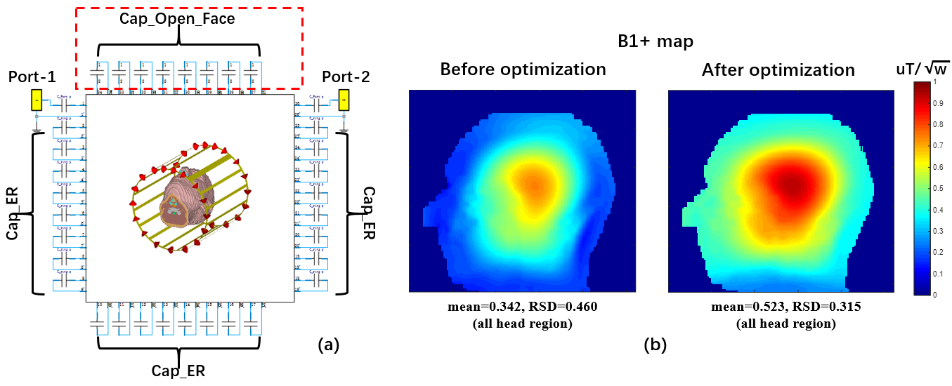
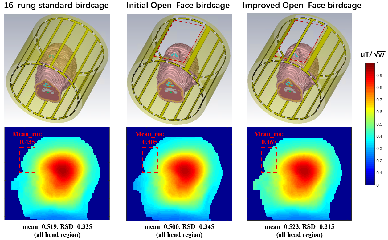
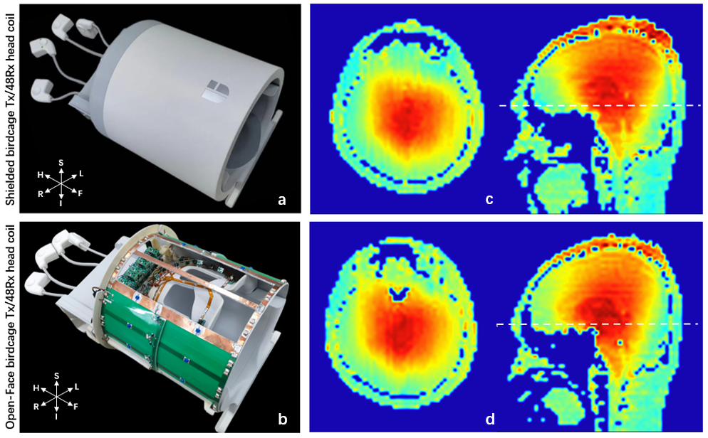
The EM simulations were performed using the finite-integration time-domain method and RF circuit co-simulation for optimizing capacitance values of coil (Microwave studio, CST, Darmstadt, Germany)3, as shown in Figure 1a. The realistic human head voxel model (Gustav) was used for the EM simulation study. The dielectric parameters are based on the Gabriel dispersion relationships4. Three configurations of transmit coil were evaluated: (1)a conventional shielded high-pass birdcage with 16 rungs; (2)an initial Open-Face birdcage, of which the top two legs was removed from the conventional birdcage structure to achieve a view window on the top face; (3)an improved Open-Face birdcage, which is designed based on the previous configuration with the addition of the leg at the top to improve transmit efficiency at the Open-Face window. All the configurations were driven in circular polarized mode. Both the simulated B1+ field maps and SAR10g were constructed by normalizing the accepted input power. The Open-Face coils were tuned at 210.8MHz by adjusting the values of capacitor (except at the open-window) on the end ring of coil. The capacitance value on the open ring was optimized for both the Open-Face birdcage coils.Figure 4c shows the photograph of the assembled Open-Face birdcage head coil for 5T. By the method of removing and adjusting the leg of the 16-rung high pass birdcage coil on the anterior, the Open-Face birdcage can be realized. Meanwhile, an open window (14cmx28cm) was removed on the RF shielding to achieve an open view of the eye. PIN diodes (Macom, MA4P7435NM-1091T) were added to each leg (except for the leg at the facial open-window) and port of the transmit coil to enable active detuning in the receive mode. The receive array was arranged on a close-fitting helmet former(A-P:230mm, L-R:210mm, I-S:266mm), of which 16 elements on the anterior and 32 elements on the posterior. More detailed information about receiver coil is provided in our previous work5. The measured B1+ field was characterized using a B1+ mapping DREAM sequence. All the experiments were performed on a whole-body 5T scanner (Shanghai United Imaging Healthcare, Shanghai, China).
Results
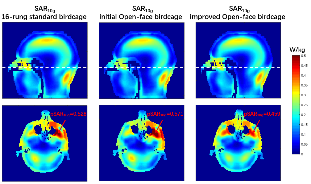
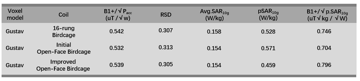
Figure 1b shows the simulated B1+ maps in the sagittal plane before and after the optimization of the capacitance value on the open ring. Figure 2 shows the simulated B1+ maps in the sagittal plane acquired using a standard birdcage and Open-Face birdcage coils. The mean values of transmit efficiency in the ROI are depicted in the maps. The results show that the improved Open-Face birdcage provides 15.3% better transmit efficiency compared to initial Open-Face configuration, and 7.3% better transmit efficiency compared to the standard shielded birdcage. As shown in Table 1, the transmit efficiency and homogeneity over the transversal, sagittal and coronal planes were comparable (within 3%) for all coils, represented by the average and RSD value over the 3 orthogonal slices, respectively. The quantitative analysis of the SAR is summarized in Figure 3 and Table 1. The improved Open-Face birdcage shows the best SAR efficiency. The improved Open-Face birdcage demonstrates 13.1% better SAR efficiency compared to initial Open-Face birdcage coil, and 6.7% better SAR efficiency compared to 16-rung standard birdcage coil.Figure 4 shows the photographs and measured B1+ maps of the conventional shielded birdcage and the improved Open-Face birdcage. The improved Open-Face birdcage transmit coil demonstrates better transmit efficiency in the prefrontal region of brain compared to conventional birdcage coil while the B1+ field distribution in the other region was very similar.
Discussions and Conclusions
The proposed Open-Face birdcage assembled with 48-channel receiver head coil provides better clinical feasibility without reducing the transmit efficiency and improving the local SAR safety efficiency. In the future work, imaging performance evaluation and functional imaging of the proposed coil will be performed at 5T MRI.Acknowledgements
This work was supported in part by National Key R&D Program of China, 2021YFE0204400, 2021YFC2400203; the Strategic Priority Research Program of Chinese Academy of Sciences (Grant No. XDB25000000); National Natural Science Foundation of China, U22A20344; Youth Innovation Promotion Association of CAS No. Y2021098; Guangdong Province grants 2018B030333001; Key Laboratory Project of Guangdong Province, 2020B1212060051; Shenzhen city grant, RCYX20200714114735123.References
1. Hayes, C. E., Edelstein, W. A., Schenck, J. F., Mueller, O. M., & Eash, M. (1985). An efficient, highly homogeneous radiofrequency coil for whole-body NMR imaging at 1.5 T. Journal of Magnetic Resonance (1969), 63(3), 622–628.
2. Kraff, O., & Quick, H. H. (2019). Radiofrequency Coils for 7 Tesla MRI. Topics in Magnetic Resonance Imaging, 28(3), 145–158.
3. Kozlov, M., & Turner, R. (2009). Fast MRI coil analysis based on 3-D electromagnetic and RF circuit co-simulation. Journal of Magnetic Resonance, 200(1), 147–152.
4. Gabriel S, Lau RW, Gabriel C. The dielectric properties of biological tissues: III. Parametric models for the dielectric spectrum of tissues. Phys Med Biol. 1996, 41(11):2271-93.
5. Wei Z, Chen Q, He Q, Zhang X, Liu X, Zheng H, Li Y. A quadrature birdcage/48-channel receiver coil assembly for human brain imaging at 5T. Proc. 30th Annual Meeting of ISMRM, London, 2022, p.3223.
Figures