4220
Dynamic T2 mapping following muscle exercise measured by fast spin-echo and ultrafast spin-echo EPI1Medical Imaging AI Research Center, Canon Medical Systems Korea, Seoul, Korea, Republic of, 2College of Medicine, Seoul National University, Seoul, Korea, Republic of, 3Department of Intelligent Precision Healthcare, Sungkyunkwan University, Suwon, Korea, Republic of, 4Magnetic Resonance Business Unit, Canon Medical Systems Korea, Seoul, Korea, Republic of
Synopsis
Keywords: Muscle, Muscle
The changes in T2 driven by water content, oxygenation, pH, and blood volume in tissue altered during intense physical activity provides chance to measure the muscle performance. In this study, we investigated feasibility of dynamic T2 measurement for exercise-induced thigh muscle activity using conventional fast spin-echo (FASE, Fast Advanced Spin Echo) and ultrafast spin-echo EPI (SE-EPI) with 3T MRI.Purpose
The muscle functional magnetic resonance imaging (mfMRI) is promising imaging technique to measure the muscle performance based on the changes in the transverse relaxation time (T2) driven by water content, oxygenation, pH, and blood volume in tissue altered during intense physical activity [1-3]. This can be useful to indirectly evaluate the spatial pattern and/or strength of exercise-induced muscle activity in sport medicine and rehabilitation medicine. However, conventional mfMRI using fast spin-echo sequence has a relatively long scan-time, so its use is often limited due to half-life of exercise-induced changes in muscle T2. Moreover, the detectability of mfMRI to T2 changes can be reduced if exercise is not performed until the onset of muscle fatigue. Therefore, it is difficult to use this approach for efficient evaluation during rehabilitation. Recently, mfMRI using ultrafast imaging demonstrated high detectability to focal muscle activity with short acquisition time [4, 5]. With the aim of evaluating the reliability of T2 measurement for exercise-induced thigh muscle activity, we demonstrated the feasibility of dynamic T2 mapping for both the sensitivity of temporal resolution and half-life effects using conventional fast spin-echo (FASE, Fast Advanced Spin Echo) and ultrafast spin-echo EPI (SE-EPI) with 3-Tesla MRI.Materials & Methods
Four healthy volunteers (male; 27~37 years old) were scanned on a 3T MRI system (Canon Medical Systems, Vintage Galan) with a 16-channel body coil. Functional imaging consistent of FASE and SE-EPI sequence with the same spatial resolution. The imaging parameters are summarized in Table 1. All participants performed right leg jump lunges with 5 sets of 10 times for each sequence, and imaging began immediately after the exercise. subjects then rested for approximately 40 minutes between each exercise set for full recovery of muscle T2 shift. The total acquisition time of FASE and SE-EPI in T2 mapping was performed for 6 min 16 sec and 2 min 16 sec, with four different echo times (30, 40, 50 and 60 ms) to distinguish the sensitivity of the time resolution effect. This T2 mapping protocol sets for FASE and SE-EPI were repeated 3 and 6 times, respectively, to evaluate the dynamic changes in muscle function. For the T2 mapping, multi-SE data were fitted by mono-exponential function, and then quantitative T2 values were measured from 4 different ROIs in vastus medialis and vastus lateralis of both thighs.Results
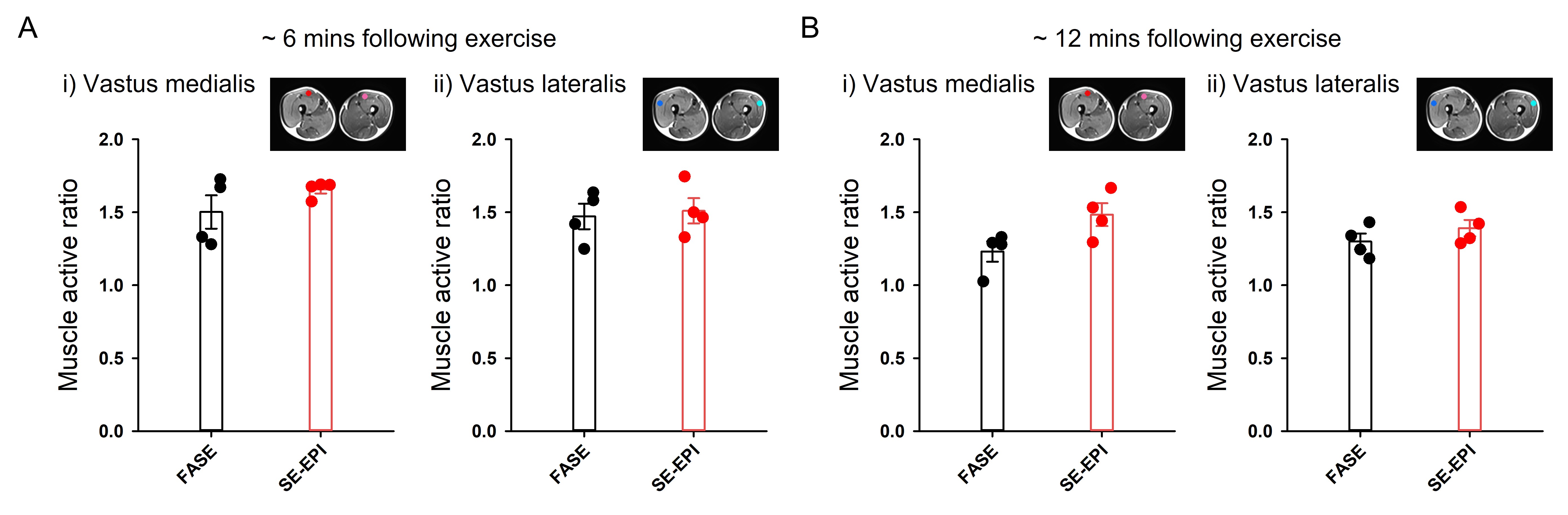
Figure1.A shows the representative T2 maps of both thigh muscles for 3 time-points of FASE and 6 time-points of SE-EPI, respectively; right muscle represents the exercise-induced activity and left muscle is inactivity. In initial T2 maps, SE-EPI can shorten the acquisition time by ~ 3 times compared to FASE, and showed higher T2 sensitivity than FASE, about 18.7 % and 9.77 % in the vastus medialis and lateralis, respectively. Figure1.B represents dynamic T2 values within two ROIs both thigh muscles from FASE and SE-EPI (red and purple for vastus medialis; blue and cyan for vastus lateralis). Overall, SE-EPI showed higher temporal T2 values than those of FASE. For the better interpretation, the muscle active ratio was calculated from each ROI pair (Fig 2). At ~ 6 mins following exercise, SE-EPI showed slightly higher muscle active ratio compared to FASE (Fig 2.A; 1.66 vs. 1.50 in vastus medialis; 1.51 vs. 1.47 in vastus lateralis), and this trend was continued at 12 mins following exercise (Fig 2.B; 1.48 vs. 1.23 in vastus medialis; 1.39 vs. 1.30 in vastus lateralis).Discussion & Conclusion
In this study, we demonstrated that SE-EPI provided higher T2 value than FASE both of initial acquisition data and during dynamic changes. We found that exercise-induced large muscle activity in T2 changes can be typically measured using conventional fast spin-echo, whereas the very small and/or focal muscle activity can be probably detected in the SE-EPI with high temporal resolving effects. That is, T2 measured from SE-EPI would be useful to detect the deep-layer muscles induced by acute exercise. The physician or trainer requires to know the normalized and calibrated levels of muscle activation in different tasks when choosing the specific training exercises and making rehabilitation decisions [3]. Using ultrafast mfMRI, they can obtain highly sensitivity and specificity in diagnosis. This study requires further technical optimizations such as B1 inhomogeneity correction or acquisition of pure T2 contrast as well as investigation of the underlying mechanisms on sensitivity of time resolution and half-life effects linked to muscular function in individuals with musculoskeletal disorders.Acknowledgements
No acknowledgement found.References
[1] Cagnie B, Elliott JM, O'Leary S, D'hooge R, Dickx N, Danneels LA. Muscle functional MRI as an imaging tool to evaluate muscle activity. J Orthop Sports Phys Ther. 2011;41(11):896-903. doi:10.2519/jospt.2011.3586
[2] Akima H, Takahashi H, Kuno SY, Katsuta S. Coactivation pattern in human quadriceps during isokinetic knee-extension by muscle functional MRI. Eur J Appl Physiol. 2004;91(1):7-14. doi:10.1007/s00421-003-0942-z
[3] Tawara N, Nitta O, Kuruma H, et al. Functional T(2) mapping of the trunkal muscle. Magn Reson Med Sci. 2009;8(2):81-83. doi:10.2463/mrms.8.81
[4] Tawara N. The ablility of muscle functional MRI to detect the slight effect of exercise on trunk muscle activity. iMRI 2022;26:117-124
[5] Tawara N, Nishiyama A. Muscle functional MRI of exercise-induced rotator cuff muscles. iMRI 2022;26:117-124
Figures
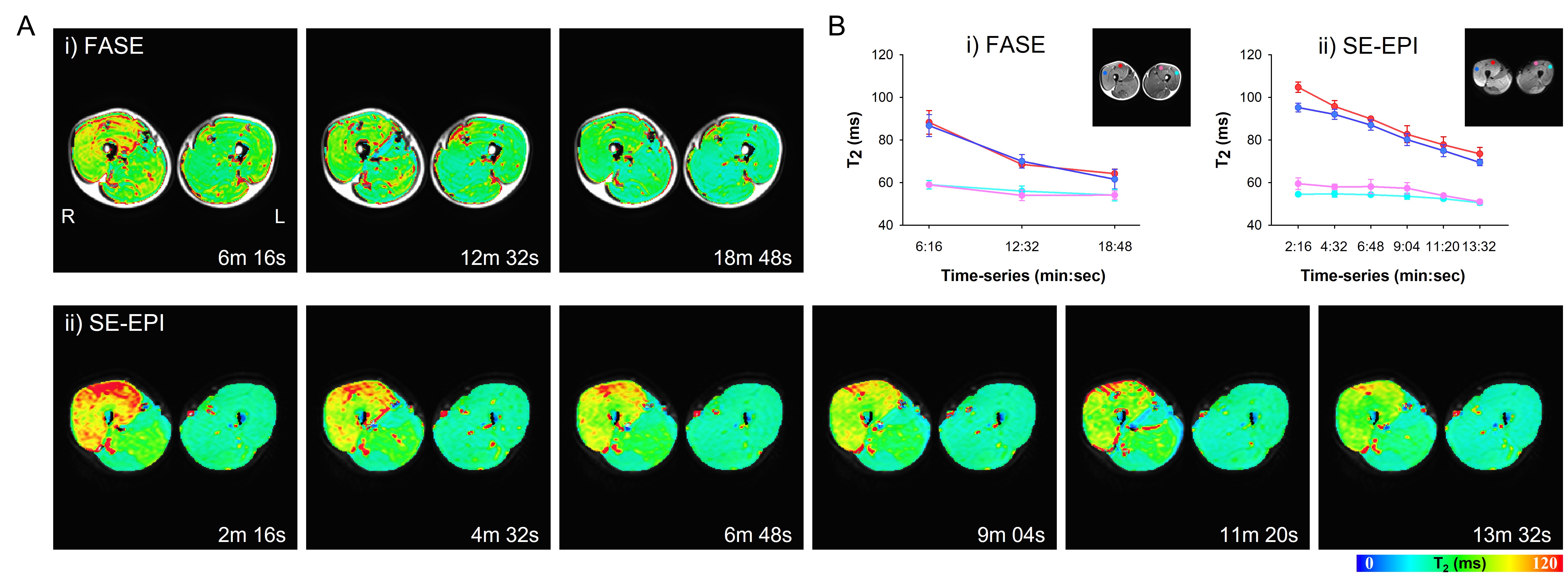
Figure 1. Dynamic T2 mapping following single leg jumping lunge with FASE and SE-EPI in the thigh muscles
A. Qualitative comparison between T2 maps with (i) FASE and (ii) SE-EPI
B. Dynamic T2 changes in vastus medialis and lateralis of active and inactive state with (i) FASE and (ii) SE-EPI