4204
Fast MRI of the cervical spinal cord by combining the deep learning-based denoising and Super-Resolution technique1Department of Radiological Technology, Hokkaido University Hospital, Sapporo, Japan, 2Department of Diagnostic and Interventional Radiology, Hokkaido University Hospital, Sapporo, Japan, 3Philips Japan, Ltd., Tokyo, Japan, 4Department of Diagnostic Imaging, Hokkaido University Graduate School of Medicine, Sapporo, Japan; Global Center for Biomedical Science and Engineering, Faculty of Medicine, Hokkaido University, Sapporo, Japan
Synopsis
Keywords: Spinal Cord, Spinal Cord
SmartSpeed Precise Image (SSPI) is one of novel image reconstruction technique which combined the physics driven type deep learning-based denoising process and Super-Resolution techniques. We investigated the utility of SSPI-based single-shot turbo spin-echo sequence of the cervical spinal cord T2-weighted image with the short acquisition time. High quantitative signal-to-noise ratio and qualitative image sharpness were successfully demonstrated by using SSPI. This methodology can be useful for the better image quality and the short acquisition time in the assessment of patients with cervical spinal cord diseases.
Introduction
Sagittal T2-weighted image (T2WI) is one of the most frequently used sequences for the evaluation of lesions in spine and spinal cord. Single-shot turbo spin echo (ssh-TSE) can acquire images with relatively few artifacts by rapidly collecting all echoes in the k-space with a single excitation pulse. However, because this scheme requires a large number of echo trains (ET), the image quality tends to be degraded particularly by blurring when the k-space is filled with echoes of various signal intensities. Although setting the increased number of reduction factors in parallel imaging might be effective to decrease the number of ET for the reduction of image blurring, this methodology suffers from the reduction of signal-to-noise ratio (SNR).Recently, SmartSpeed Precise Image (SSPI) is introduced as hybrid type of the deep-learning (DL)-based reconstruction; SSPI combined the physics driven type DL-based denoising and DL-based Super-Resolution technique. SSPI may achieve both higher image quality and image contrast simultaneously even with short scanning time.
The aim of this study was to assess the rapidly acquired T2WI of the cervical spinal cord by ssh-TSE with the SSPI reconstruction, by comparing the other major reconstruction techniques.
Materials and Methods
Five healthy volunteers were included in this prospective study. All scanning was performed using a 3.0T unit (Ingenia Elition, Philips Healthcare, Best, Netherland) with a 16-channel Head and Neck coil. The ssh-TSE-based sagittal T2WI of the cervical spinal cord was conducted with the following parameters: TR, 20000ms; equivalent TE, 100ms; FOV, 240×240mm; acquisition matrix, 1.00×1.20mm; reconstruction matrix, 0.5×0.5mm; Slice thickness, 3 mm×13 slices; reduction factor, 4.We used the SSPI technique for the image reconstruction as follows; images have been reconstructed using vendor provided prototype (Philips SmartSpeed Precise Image). This AI-based reconstruction technique consists of a series of convolutional neural networks (CNNs): Adaptive-CS-Net1 allows to reconstruct images acquired with compressed sensitivity-encoding (CS)-based variable intensity undersampling patterns. This CNN is applied prior to coil combination, removing the noise from the images in order to obtain good image quality from accelerated acquisitions2. Subsequently, Precise Image Net is an AI-model applied to remove ringing artifacts and to replace the traditional zero-filling strategy to increase the matrix size and therewith the sharpness of the images; these types of networks are known as Super-Resolution networks3,4. This network is trained on pairs of low- and high-resolution data with k-space crops to induce ringing. Data consistency checks are implemented to match the resulting k-space with the measured k-space data. The full reconstruction pipeline generates images with improved SNR and sharpness, higher matrix size and reduced ringing artifacts, and can be applied to all 2D cartesian acquisitions. The noise reduction can be controlled using a parameter to the users’ preference.
For the comparison to SSPI, we also performed the other three reconstruction techniques: 1) conventional sensitivity-encoding (SENSE), 2) CS, and 3) SmartSpeed (SS); a DL reconstruction with CNN-based method with Adaptive-CS-Net as mentioned above without Super-Resolution networks. In addition, as reference images for the qualitative image sharpness assessment (see below), a multi-shot (msh) TSE sequence with conventional SENSE reconstruction which was used for routine clinical imaging was acquired.
The image quality of these reconstruction techniques were evaluated by 1) quantitative SNR and 2) qualitative visual image sharpness. For the SNR measurement, the ROI was placed on the spinal cord tissue, thereafter the SNR was calculated using the mean signal intensity and the standard deviation in the ROI.
The qualitative image sharpness was visually assessed using three-scoring system (1: poor, 2: moderate, 3: good) by two experts (experienced radiological technologist and radiologist).
The value of SNR in SSPI was compared to the other three reconstruction techniques using Wilcoxon signed-rank test. The visual score of qualitative image sharpness in SSPI was compared to other three reconstruction techniques and also the reference image (i.e., the msh-TSE with SENSE reconstruction) using Mann–Whitney’s U-test. In both assessment of the SNR and the qualitative image sharpness, statistical significance was set to p<0.01 (*the value with the Bonferroni’s correction).
Results
Representative images obtained by various reconstruction techniques were shown in Fig.1.In the SNR assessment, the value of SNR in SSPI was significantly higher than that in SENSE and SS, although CS showed non-significant difference. The detail of measured SNR was presented as median and interquartile range (IQR) in Fig.2.
In the assessment of qualitative image sharpness, the score in SSPI was significantly higher than the other three reconstruction techniques (SENSE, CS, and SS), additionally, qualitative image sharpness was indicated to maintain its quality compared to msh-TSE with SENSE reconstruction (i.e., a long-time scanning which was clinically used). The detail of qualitative image sharpness was presented in Fig.3.
Disucussion
The use of novel DL-based SSPI reconstruction technique with the ssh-TSE sequence demonstrated the sufficient SNR and the superb qualitative image sharpness even in a short time acquisition protocol. We speculated the sufficient SNR was achieved by effective denoising in this DL-based SSPI reconstruction technique. Image sharpness was also achieved by this technique particularly due to Super-Resolution networks, as one of important structure of the SSPI.Conclusion
The SSPI can be useful for the better image quality and a short acquisition time in acquiring images of cervical spinal cord.Acknowledgements
No acknowledgements.References
- Pezzotti N, de Weerdt E, Yousefi S, et al. Adaptive-CS-Net: FastMRI with Adaptive Intelligence. arxiv. 2019;(NeurIPS).
- Philips SmartSpeed. No compromise Image quality and speed at your fingertips. Hans Peeters PhD, Hayley Chung PhD, Giuseppe Valvano PhD, Deniz Yakisikli MSc., Jeroen van Gemert PhD, Elwin de Weerdt PhD and Kim van de Ven PhD. https://www.philips.com/c-dam/b2bhc/master/landing-pages/mri-innovations/philips-smart-speed-brochure-lr.pdf
- Super-Resolution Using Deep Convolutional Networks. Chao Dong, Chen Change Loy, Kaiming He, Xiaoou Tang. 2014 arXiv:1501.00092.
- Review of the deep learning methods for medical images super resolution problems. Y. Li, B. Sixou, F.Peyrin. IRBM, Volume 42, Issue 2, April 2021, Pages 120-133.
Figures
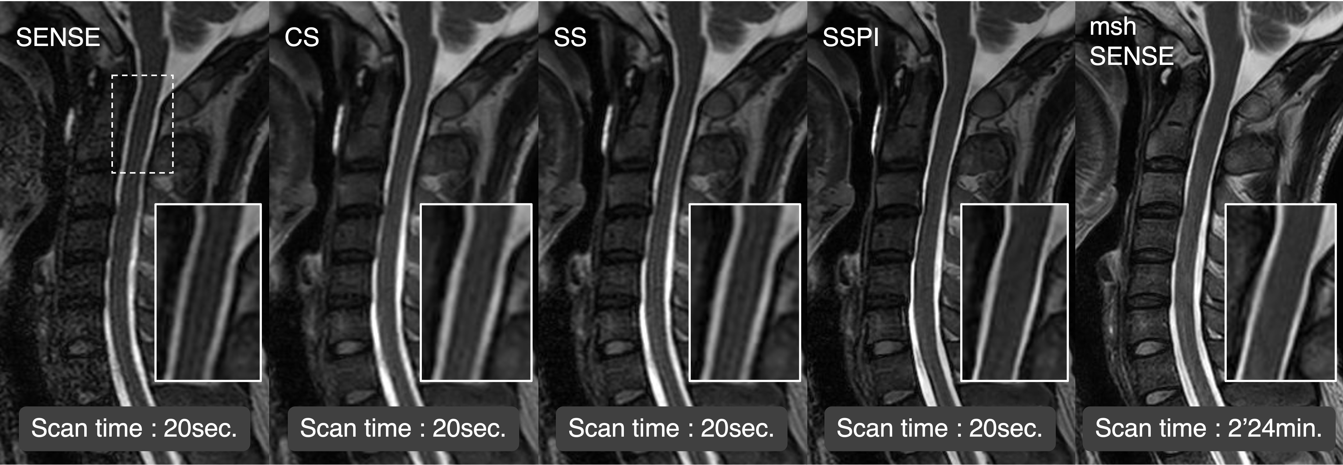
Fig.1 Representative images with various reconstruction techniques
Representative images obtained with SENSE, CS, SS, SSPI, and msh-SENSE were presented. The cervical cord was clearly depicted with SSPI as well as msh-SENSE, whereas a certain degree of blurring was presented in SENSE, CS, and SS. The scanning times of the respective reconstruction techniques were as follows: SENSE (0’ 20), CS (0’20), SS (0’20), SSPI (0'20) and msh-SENSE (2’24).
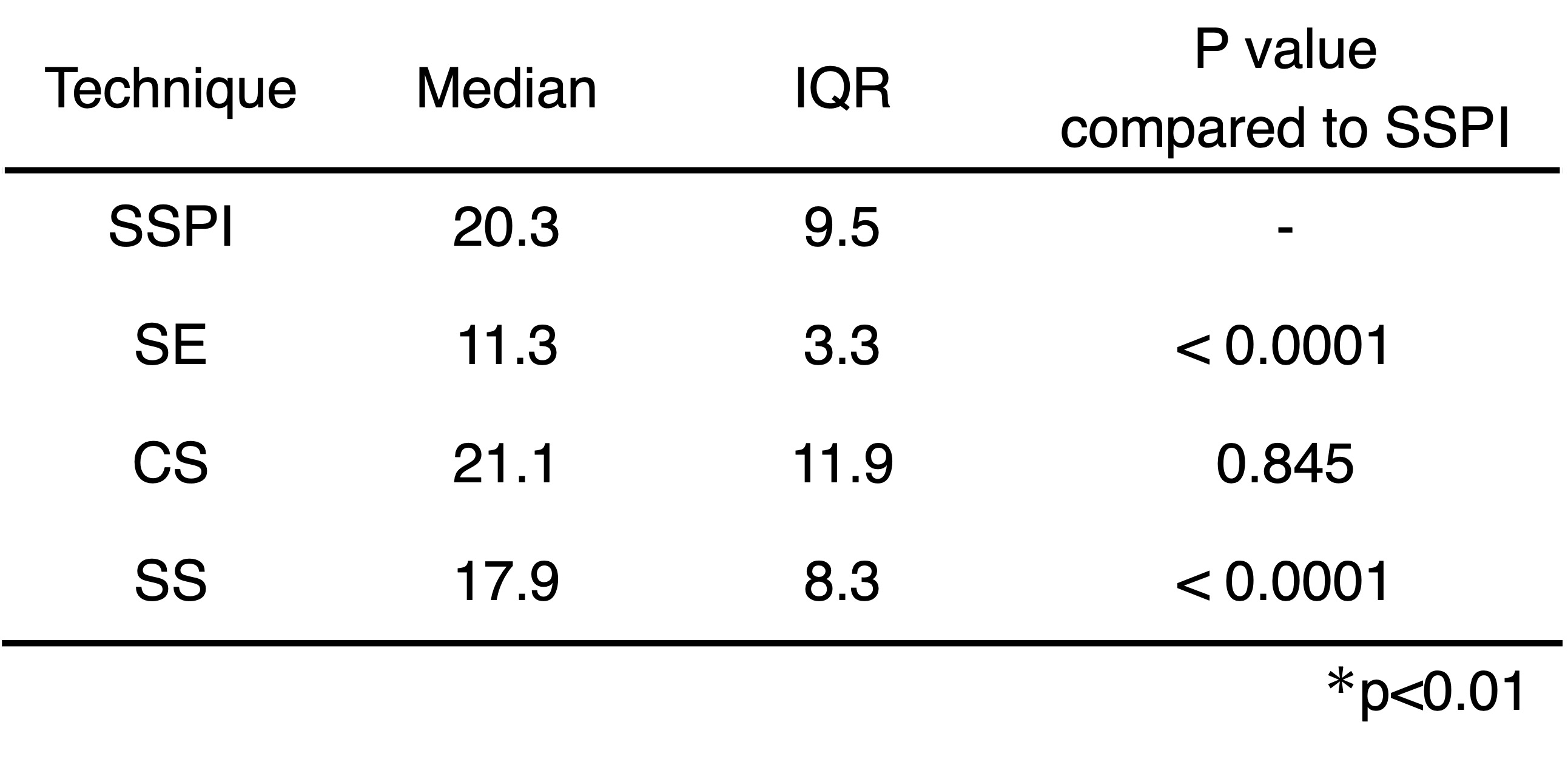
Fig.2 SNR in various reconstruction techniques.
The value of SNR with SSPI reconstruction was significantly higher than that with SENSE and SS. In contrast, non-significant difference was observed between the SSPI and CS.
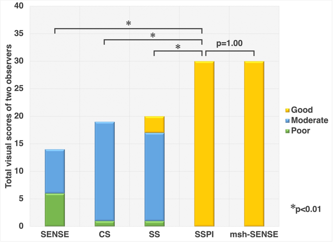
Qualitative image sharpness in SSPI reconstruction was significantly higher than SENSE, CS, and SS (*p<0.01). In addition, non-significant difference was observed between the SSPI and msh-SENSE (i.e., clinically used long scanning time sequence) (p=1.0).