4203
Assessment of Multi-modal MR Imaging for Glioma Based on a Deep Learning Reconstruction Approach with the Denoising Method1Capital Medical Universtiy, Beijing Tiantan Hospital, Beijing, China, 2MR Research, GE Healthcare, Beijing, China, 3BioMind Inc., Beijing, China, 4Department of Medical Imaging Product, Neusoft, Group Ltd., Shenyang, China, 5Department of Medical Imaging Product, Neusoft, Group Ltd., Beijing, China
Synopsis
Keywords: Tumors, Brain
Deep learning reconstruction (DLR) approach with denoising can improve image quality of magnetic resonance (MR) images. However, its applications on multi-modal glioma imaging have not been assessed.Multi-modal images of 107 glioma patients were evaluated by signal-to-noise ratio (SNR), contrast-to-noise ratio (CNR), edge sharpness, visual assessment, diagnosis accuracy and efficiency. Contrasted with conventionally reconstructed images, the DLR images showed higher tumor/residual tumor SNR, higher tumor to white/gray matter CNR, better results of the visual assessment, and a trend of improved diagnosis efficiency and comparable accuracy. DLR can improve image quality of multi-modal glioma images which should benefit the glioma diagnosis.Background or Purpose
The deep learning reconstruction (DLR) approach with denoising can improve the image quality of magnetic resonance (MR) images. However, its applications on multi-modal glioma imaging, including T1-weighted (T1w), contrast enhanced T1w (CE-T1), T2w, T2 Fluid Attenuated Inversion Recovery (T2-FLAIR), and Diffusion Weighted Imaging (DWI) have not been assessed.Methods
We assessed multi-modal images of 107 patients with glioma, including 49 preoperative and 58 postoperative patients with available T1w, CE-T1, T2w, T2-FLAIR, and DWI images. All these images were reconstructed with both DLR and conventional reconstruction methods. The image quality was evaluated by signal-to-noise ratio (SNR), contrast-to-noise ratio (CNR) for all cases, and edge sharpness regarding preoperative cases. Visual assessment of the glioma imaging quality was conducted blindly by three radiologists on all cases. Assessment of diagnosis accuracy and efficiency on preoperative cases was conducted by other six neuroradiologists who were blind to the final pathological diagnosis based on randomly rearranged DLR and conventionally reconstructed images. The differences between the DLR and conventionally reconstructed images were compared by paired t-test or Wilcoxon test.Results
All tumor/residual tumor SNR from multi-modal DLR images were higher than those from conventionally reconstructed images. The CNR of tumor to white matter and tumor to gray matter from DLR images, including T1w, T2w, and T2-FLAIR, were also higher. There is no difference in tumor edge sharpness for preoperative cases. The visual assessment of DLR images demonstrated the better presence of tumor on T2w, edema on T2-FLAIR, enhanced tumor part and necrosis on CE-T1, tumor boundary on DWI, and fewer artifacts in all modalities. A trend of improved diagnosis efficiency and comparable accuracy was observed for preoperative cases with DLR images.Conclusions
The DLR can improve image quality of multi-modal glioma images, including T1w, CE-T1, T2w, T2-FLAIR, and DWI, which should benefit the glioma diagnosis.Acknowledgements
We acknowledged all the colleagues who help the patient recruitment and MR imaging.References
1. GBD 2016 Neurology Collaborators. Global, regional, and national burden of neurological disorders, 1990-2016: a systematic analysis for the Global Burden of Disease Study 2016. Lancet Neurol 2019;18(5):459-480.
2. Gupta RK, Bharti S, Kunhare N, et al. Brain Tumor Detection and Classification Using Cycle Generative Adversarial Networks. Interdiscip Sci Comput Life Sci 2022;14, 485-502.
3. Molinaro AM, Taylor JW, Wiencke JK, et al. Genetic and molecular epidemiology of adult diffuse glioma. Nat Rev Neurol 2019; 15(7):405-417.
4. Tripathi PC, Bag S. A computer-aided grading of glioma tumor using deep residual networks fusion. Comput Methods Programs Biomed 2022;215:106597.
5. Guan X, Yang G, Ye J, et al. 3D AGSE-VNet: an automatic brain tumor MRI data segmentation framework. BMC Med Imaging 2022;22(1):6.
6. Koch KM, Sherafati M, Arpinar VE, et al. Analysis and Evaluation of a Deep Learning Reconstruction Approach with Denoising for Orthopedic MRI. Radiology: Artificial Intelligence 2021;3:e200278.
7. Wang X, Ma J, Bhosale P, et al. Novel deep learning-based noise reduction technique for prostate magnetic resonance imaging. Abdom Radiol (NY) 2021;46(7):3378-3386.
8. Johnson PM, Tong A, Donthireddy A, et al. Deep Learning Reconstruction Enables Highly Accelerated Biparametric MR Imaging of the Prostate. J Magn Reson Imaging 2021.
9. Park JC, Park KJ, Park MY, et al. Fast T2-Weighted Imaging With Deep Learning-Based Reconstruction: Evaluation of Image Quality and Diagnostic Performance in Patients Undergoing Radical Prostatectomy. J Magn Reson Imaging 2022;55(6):1735-1744.
10. Zormpas-Petridis K, Tunariu N, Curcean A, et al. Accelerating whole-body diffusion-weighted MRI with Deep Learning–based Denoising Image Filters. Radiology: Artificial Intelligence 2021; 3:e200279.
11 Gudbjartsson H, Patz S. The Rician distribution of noisy MRI data. Magn Reson Med 1995;34(6):910–914.
12. Yasaka K, Tanishima T, Ohtake Y, et al. Deep learning reconstruction for 1.5 T cervical spine MRI: effect on interobserver agreement in the evaluation of degenerative changes. Eur Radiol 2022;32(9):6118-6125.
13. Sun S, Tan ET, Mintz DN, et al. Evaluation of deep learning reconstructed high-resolution 3D lumbar spine MRI. Eur Radiol 2022;32(9):6167-6177.
14. Herrmann J, Keller G, Gassenmaier S, et al. Feasibility of an accelerated 2D-multi-contrast knee MRI protocol using deep-learning image reconstruction: a prospective intraindividual comparison with a standard MRI protocol. Eur Radiol 2022;32(9):6215-6229.
15. Kim M, Kim HS, Kim HJ, et al. Thin-Slice Pituitary MRI with Deep Learning–based Reconstruction: Diagnostic Performance in a Postoperative Setting. Radiology 2021; 298:114–122.
16. Zhou Z, Sanders JW, Johnson JM, et al. Computer-aided Detection of Brain Metastases in T1-weighted MRI for Stereotactic Radiosurgery Using Deep Learning Single-Shot Detectors. Radiology 2020;295(2):407-415.
17. Zhao R, Yaman B, Zhang Y, et al. fastMRI+, Clinical pathology annotations for knee and brain fully sampled magnetic resonance imaging data. Sci Data 2022;9(1):152.
18 Kumar P, VijayKumar B. Brain tumor MRI segmentation and classification using ensemble classifier. IJRTE 2019; 8(1S4).
19. Uetani H, Nakaura T, Kitajima M, et al. Hybrid deep-learning-based denoising method for compressed sensing in pituitary MRI: comparison with the conventional wavelet-based denoising method. Eur Radiol 2022;32(7):4527-4536.
20. Kim EH, Choi MH, Lee YJ, et al. Deep learning-accelerated T2-weighted imaging of the prostate: Impact of further acceleration with lower spatial resolution on image quality. Eur J Radiol 2022; 145:110012.
21. Vranic JE, Cross NM, Wang Y, et al. Compressed sensing-sensitivity encoding (CS-SENSE) accelerated brain imaging: reduced scan time without reduced image quality. AJNR Am J Neuroradiol 2019; 40(1):92-98.
22. Hu Y, Ren J, Yang J, et al. Noise reduction by adaptive-SIN filtering for retinal OCT images. Sci Rep 2021;11(1):19498.
Figures
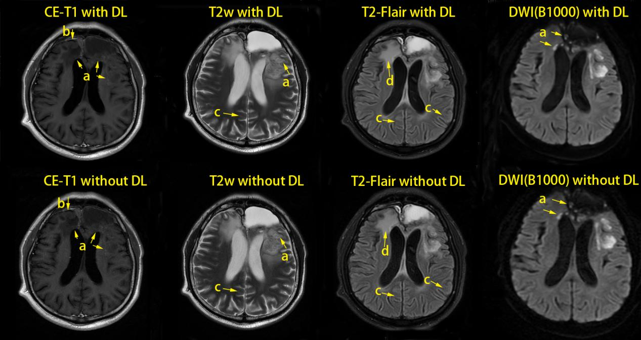
Figure 1 One case’s multi-modal images of DL and conventionally reconstructed (non-DL) methods
The patient was male with age of 39 years.
The mark a and b is lesion, c is artifact, and d is edema.
DL: deep learning
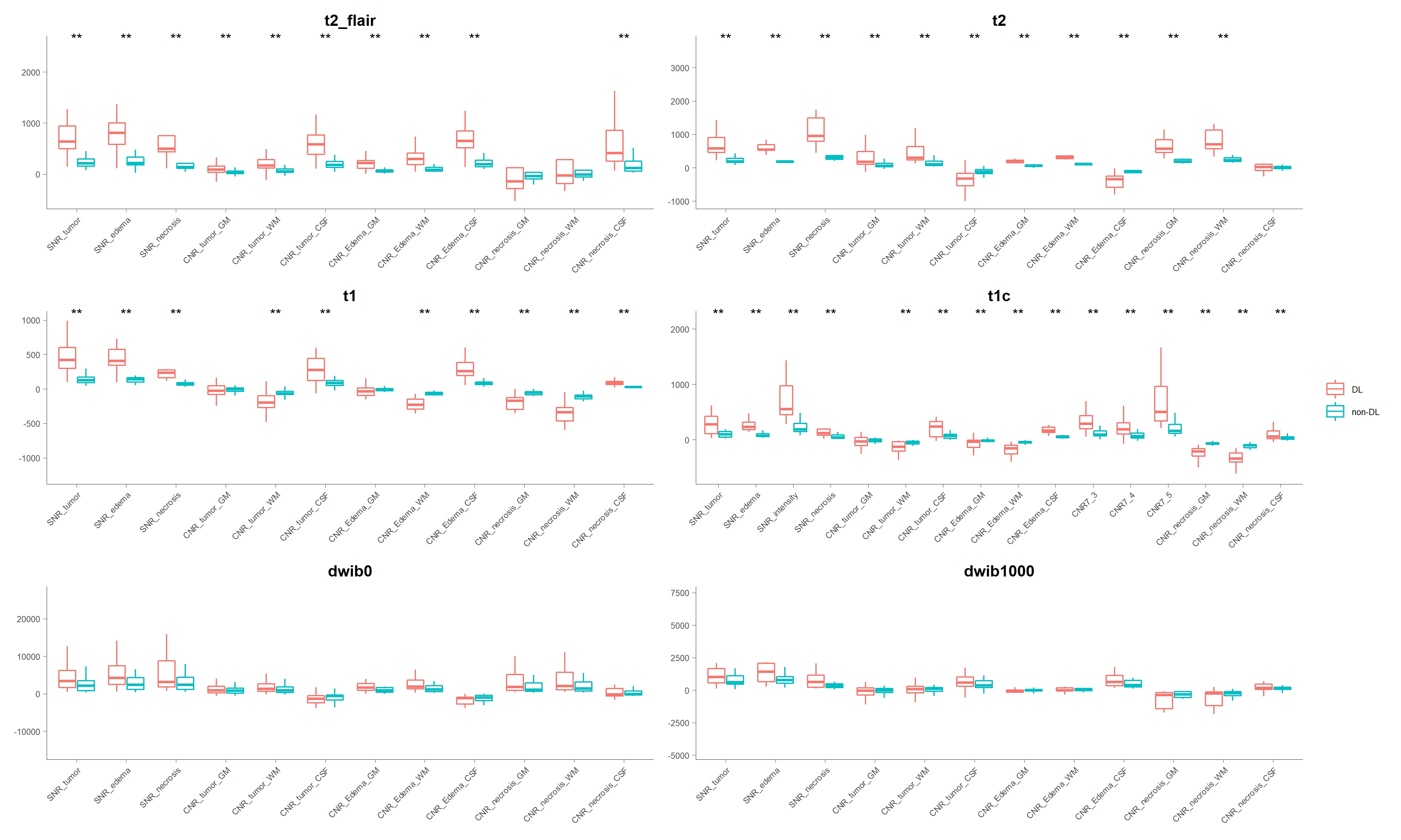
Figure 2 Mean SNR and CNR of multi-modal DL and conventionally reconstructed (non-DL) images in preoperative glioma cases
SNR:signal-to-noise ratioCNR:contrast-to-noise ratio
DL: deep learning
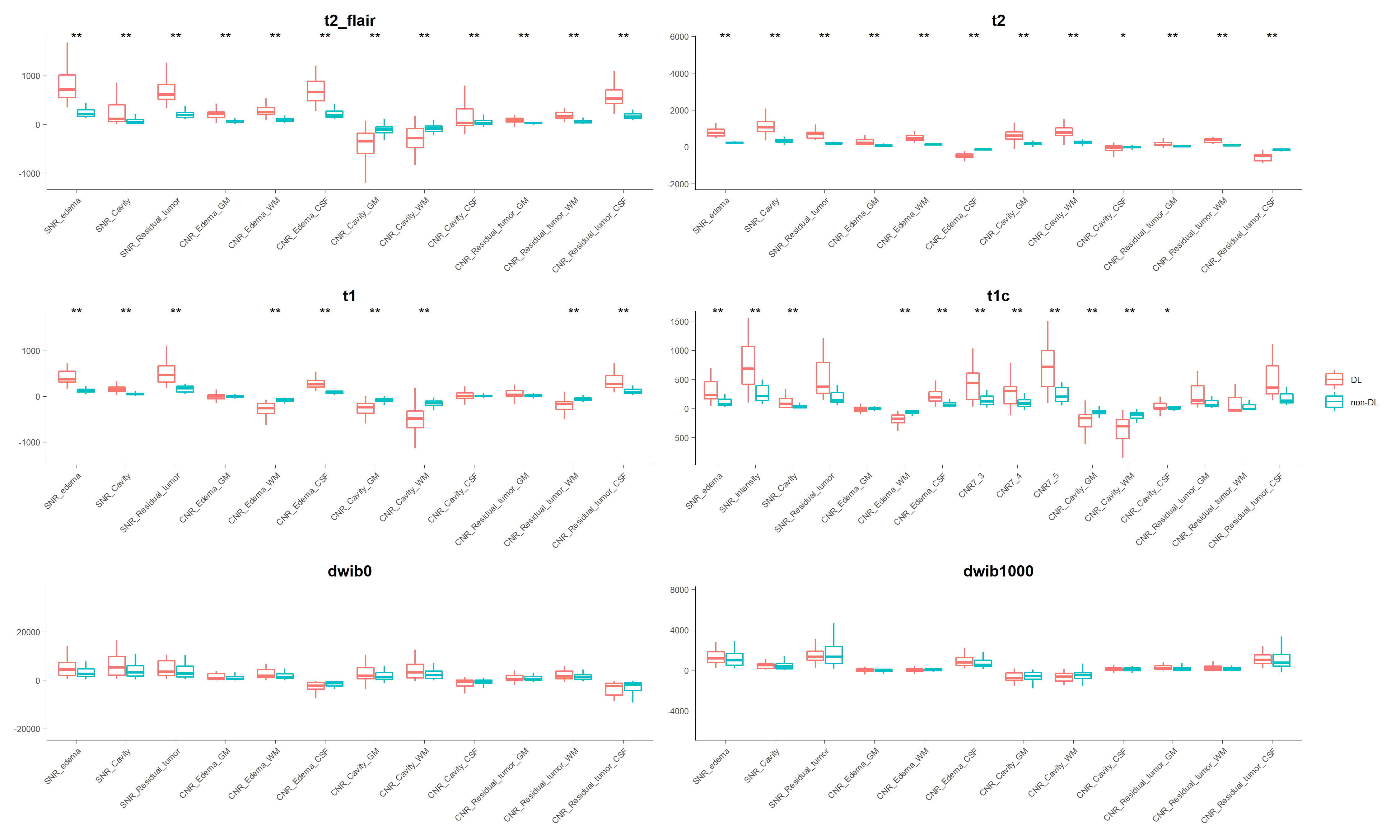
Figure 3 Mean SNR and CNR in multi-modal DL and conventionally reconstructed (non-DL) images in postoperative glioma cases
SNR:signal-to-noise ratio
CNR:contrast-to-noise ratio
DL: deep learning
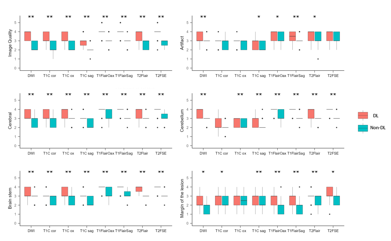
Figure 4 The visual assessment scores of DL and conventionally reconstructed (non-DL) images for preoperative cases
DL: deep learning
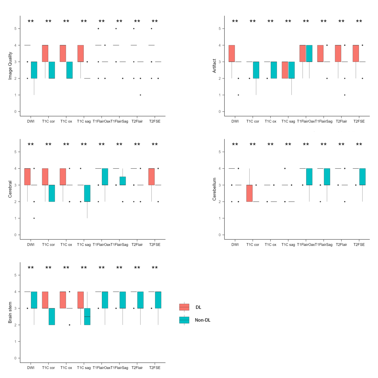
Figure 5 The visual assessment scores of DL and conventionally reconstructed (non-DL) images for postoperative cases
DL: deep learning