4193
Convolutional-neural-network-based denoising with estimated noise-based normalization to effectively reduce noise for various noise levels1Imaging Technology Center, FUJIFILM Corporation, Tokyo, Japan
Synopsis
Keywords: Machine Learning/Artificial Intelligence, Machine Learning/Artificial Intelligence, Noise reduction
To develop a convolutional neural network (CNN)-based denoiser for various noise levels, we propose the use of estimated noise-based normalization in denoising. When the CNN-based denoiser with estimated noise-based normalization was applied to brain FLAIR images with various noise levels, it resulted in values closer to the normalized root mean square error (NRMSE) between the denoised and the target images compared with a conventional CNN-based denoiser trained with the same noise level as that in the input image. In conclusion, our method effectively reduced the noise in an image with various noise levels in terms of minimization of the NRMSE.Introduction
Convolutional neural network (CNN)-based denoising with image post-processing has the potential to improve image quality in short-scan images [1-4]. In general, training of CNN-based denoisers uses image pairs of target and noise images with specific noise levels to minimize the normalized root mean square error (NRMSE) between the target and the denoised images. Thus, the trained CNN-based denoisers can most effectively reduce noise for images with the same noise level as the training data. However, when the noise levels of the images are different from those of the training data, effective noise reduction may not be achieved due to noise level mismatch. Since the noise level varies depending on the scan parameters, a denoising that is robust to different noise levels is required. To develop a denoising method that can effectively reduce noise for images with various noise levels, we propose a denoising method with estimated noise-based normalization.Methods
For estimated noise-based normalization, the input image was normalized using the following equation.Iin'= β×Iin=α/Nair×Iin (1)
Here, Iin, Iin', Nair, α, and β respectively represent input image, normalized input image, estimated noise level in the air, certain coefficient, and α/Nair. Figure 1 shows a histogram of a FLAIR brain image of a volunteer. The peak is in the low pixel region. This peak value is defined to be the estimated noise level (Nair) in the air. α was obtained as follows. A noisy image was generated by adding Gaussian noise to a target image, and N’air of the noisy image was obtained. Various coefficients β' (0.1 to 10.0) were multiplied to the noisy image, and then the noisy images were denoised using a CNN-based denoiser. For each β', the NRMSE between the denoised and the target images was calculated, and the optimal β' for the NRMSE to be minimized was obtained. Similarly, noisy images with various noise levels were generated, and then N’air and optimal β' were obtained. N’air and optimal β' for each noise level were plotted, and α was obtained by fitting the plot with a linear line.
A super resolution convolutional neural network (SRCNN) was used as the CNN-based denoiser [6]. FLAIR brain images of five volunteers were measured using a 3T MRI (FUJIFILM Healthcare Corporation). Images from four volunteers were used for training, and those from the remaining volunteer for evaluation. A target image was reconstructed from full-sampling data, and a noisy image was generated by adding Gaussian noise to the target image. Also, conventional maximum normalization was used for comparison. To generate the CNN-based denoiser with maximum normalization, we generated noisy images with different noise levels (2, 3, 4, and 5). A noise level of 2 means that the signal to noise ratio (SNR) of the noisy image was 1/2 that of the target image. To generate the CNN-based denoiser with the estimated noise-based normalization, we used noisy images with noise level of 3. To evaluate the performance of our method, we used noisy FLAIR brain images (noise level = 2, 3, 4, 5). This study was approved by the ethics committee of FUJIFILM Healthcare Corporation, following receipt of written informed consent from the volunteers.
Results
Figure 2 (a) shows β' versus NRMSE at each noise level (noise level: 2, 3, 4, 5) for a single volunteer. The higher the noise level, the smaller the optimal β'. Figure 2 (b) shows 1/N’air versus optimal β'. α was obtained to be 1.04 by fitting the plot with a linear line with an intercept of zero.Figure 3 shows the denoised images for the noise level of 2. The image denoised by using the estimated noise-based normalization (Figure 3 (g)) has similar image quality to that by using the maximum normalization with noise level of 2 (Figure 3 (c)). Likewise, Figure 4 shows the denoised images for the noise level of 3. The image denoised by using the estimated noise-based normalization (Figure 4 (g)) has similar image quality to that by using the maximum normalization with noise level of 3 (Figure 4 (c)).
Table 1 shows the NRMSE between the denoised and the target FLAIR brain images. For each noise level, the maximum normalization with the same noise level resulted in the lowest NRMSE value, while the estimated noise-based normalization resulted in values similar to those with the maximum normalization with the same noise level.
Discussion
The CNN-based denoiser provided different smooth strengths depending on β', as shown Figure 2 (a). Since the use of the smaller β' reduced the higher noise, the input image was divided by the noise level in the air. Although the maximum normalization with the same noise level resulted in the lowest NRMSE values, the estimated noise-based normalization resulted in values close to those obtained by the maximum normalization with the same noise level. Therefore, the estimated noise-based normalization enabled successful denoising at various noise levels.Conclusion
The CNN-based denoiser with estimated noise-based normalization was able to effectively reduce the noise in the image with various noise levels in terms of minimization of the NRMSE.Acknowledgements
No acknowledgement found.References
1. Jiang D., Dou W., Vosters L., et al. Denoising of 3D magnetic resonance images with multi-channel residual learning of convolutional neural network. Jpn J Radiol. 2018; 36: 566–574.
2. Kawamura M., Tamada D., Funayama S., et al. Accelerated Acquisition of High-resolution Diffusion-weighted Imaging of the Brain with a Multi-shot Echo-planar Sequence: Deep-Learning-based Denoising. Magn Reson Med Sci. 2020; 10.2463/mrms.tn.2019-0081.
3. Rao G.S., Srinivas B., et al. De-Noising of MRI Brain Tumor Image using Deep Convolutional Neural Network. Proceedings of International Conference on Sustainable Computing in Science, Technology and Management (SUSCOM). 2019.
4. Kidoh M., Shinoda K., Kitajima M., et al. Deep Learning Based Noise Reduction for Brain MR Imaging: Tests on Phantoms and Healthy Volunteers. Magn Reson Med Sci. 2020; 19 (3):195–206.
5. Dong C., Loy C., He K., et al. Image Super-Resolution Using Deep Convolutional Networks. IEEE Trans Pattern Anal Mach Intell. 2016; 38 (2): 295–307.
Figures
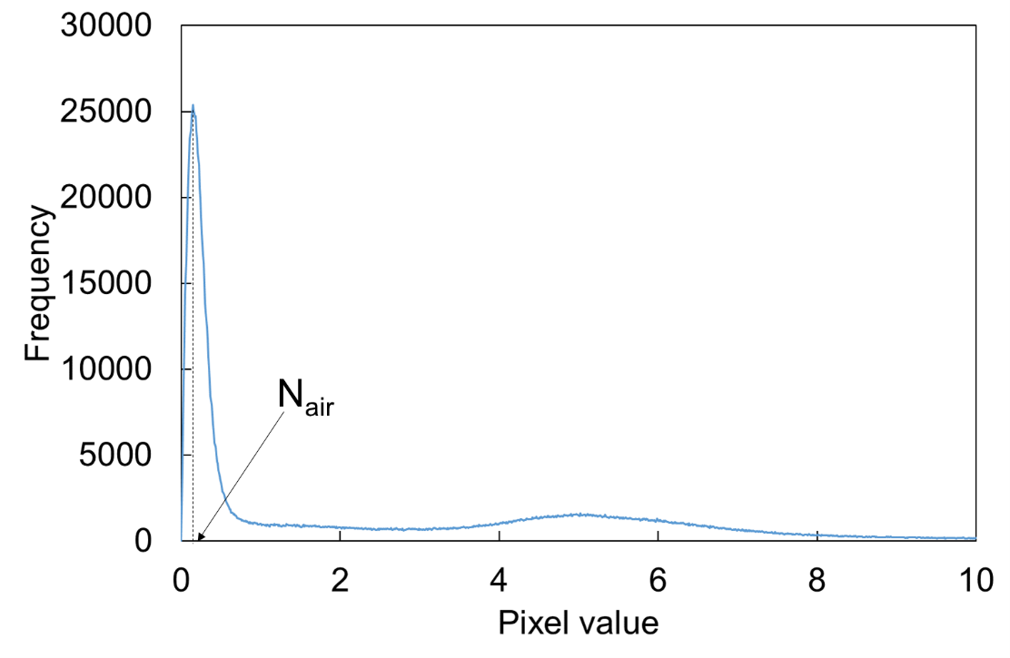
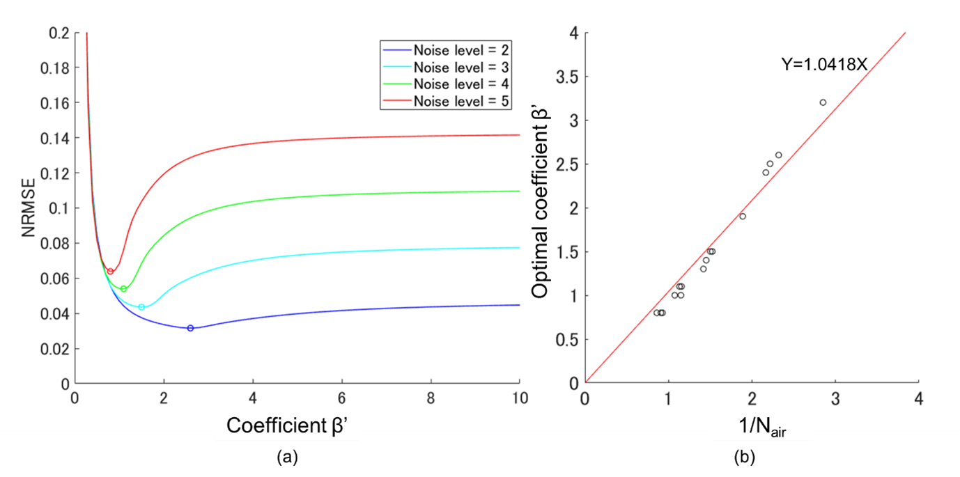

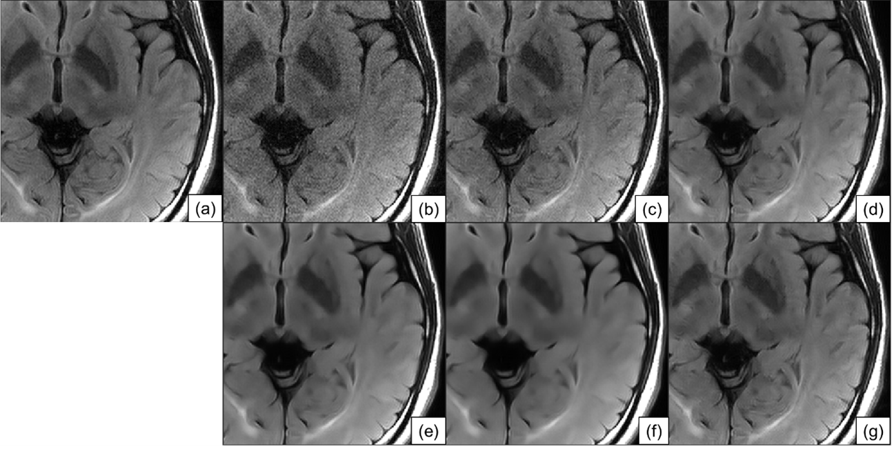
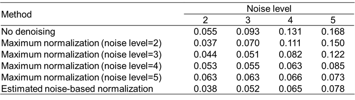
Table 1. NRMSE between the denoised and the target FLAIR brain images for each noise level.