4191
Artificial intelligence-based denoising for clinical magnetic resonance imaging: from head to toe
Dallas Turley1,2, Pattana Wangaryattawanich2, Jalal Andre2, Majid Chailan2, Johannes M. Peeters3, Kim Van de Ven3, and Orpheus Kolokythas2
1Philips Healthcare, Seattle, WA, United States, 2Department of Radiology, University of Washington, Seattle, WA, United States, 3Philips Healthcare, Best, Netherlands
1Philips Healthcare, Seattle, WA, United States, 2Department of Radiology, University of Washington, Seattle, WA, United States, 3Philips Healthcare, Best, Netherlands
Synopsis
Keywords: Machine Learning/Artificial Intelligence, Machine Learning/Artificial Intelligence
Artificial intelligence in MR is a diverse and growing field. In this work, we apply convolutional neural networks (CNN) to accelerated routine clinical protocols to investigate potential image quality improvements. For moderate increase in reconstruction time, CNNs were judged by experienced radiologists to significantly improve image quality by reducing noise and artifact.Purpose
To investigate utility and performance of artificial intelligence convolutional neural networks for image reconstruction in routine clinical imaging for the purpose of increasing signal to noise ratio.Methods
Images were reconstructed with Compressed SENSE in the clinical workflow, and at the end of the day with a vendor-provided prototype for retrospective reconstruction to avoid disruption of clinical workflow. The artificial intelligence (AI)-based reconstruction technique consists of a series of convolutional neural networks (CNN): Adaptive-CS-Net1 (ACN) allows for reconstruction of images acquired with CompressedSENSE-based variable-density undersampling techniques. This CNN is applied during to coil combination, removing noise and undersampling artifacts from accelerated images to improve image quality. Subsequently, Precise-Imaging-Net (PIN) is applied to remove ringing artifacts and to replace traditional zero-filling strategy to increase image matrix size and sharpness2,3. This network was trained on pairs of low- and high- resolution data with k-space crops to reduce ringing. Images were reconstructed on the scanner host using a NVIDIA GeFORCE RTX5000 GPU on a HP Z4G4 computer with an 8-core Intel Xeon 3.9GHz processor. 3D sequences were reconstructed with ACN at three denoising levels: low, medium and high. 2D sequences were also reconstructed with the three different denoising levels, as well as with and without a two-fold increased matrix size using the PIN CNN. AI-reconstructed images were independently scored by four radiologists, rating various AI denoising levels against standard clinical images. Images were rated on a scale of 1-5 (1-much worse, 2-worse, 3-equal, 4-better, 5-much better). A total of 120 image volumes were reviewed by radiologists in various anatomies, including brain, liver, knee, spine, and prostate.Results
Representative images from a routine brain exam and knee exam are shown in Figure 1 and Figure 2 respectively. Reconstruction time for ACN took 32 seconds (0.5 seconds/slice) on average regardless of denoising level, compared to 24 seconds (0.3 seconds/slice) for standard clinical image reconstruction. Combined ACN-PIN reconstruction took 28 seconds (1 second/slice) on average. Note that ACN-PIN was only applied to 2D multi-slice acquisitions, which had fewer slices to reconstruct than 3D acquisitions (ACN only). Table 1 shows SNR averaged over the image volume for each of the 6 image reconstruction combinations. Results from image assessment by independent radiologists are shown in Figure 3.Discussion/Conclusions
In clinical examinations, radiologists ranked AI-based denoising to have equivalent or superior image quality in every case, especially in highly accelerated sequences (e.g. 3D FLAIR acquired with undersampling factor R=12). ACN reconstructions improved signal to noise compared to standard clinical images, especially when the clinical images were of poor quality. Combined ACN-PIN reconstructed images with high denoising and 2x matrix enhancement showed highest SNR and lesion conspicuity, leading to improved diagnostic confidence compared to standard clinical imaging, although in some cases, excessive noise suppression led to an “artificial” or “cartoonish” image quality in both ACN and ACN-PIN reconstructions. Additionally, AI reconstruction sharpened some artifacts present in the standard clinical images, such as bulk motion and physiological motion around blood vessels, but in every case, the artifacts were present in the standard clinical image and did not reduce the overall diagnostic quality of the image. Artificial intelligence was shown to reduce noise and improve image quality in a variety of anatomies for routine clinical imaging.Acknowledgements
No acknowledgement found.References
1. Pezzotti N et al. Adaptive-CS-Net: FastMRI with Adaptive Intelligence. NeuIPS 2019. 2. Chao D et al. arXiv:1501.00092. 3. Y Li et al. doi:10.1016/j.irbm.2020.08.004.Figures
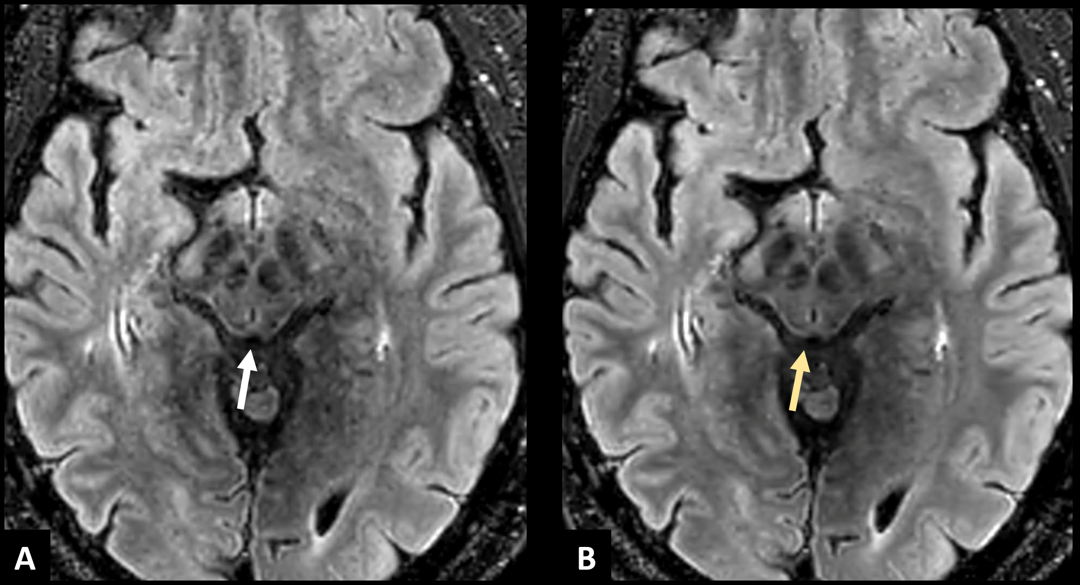
Figure 1: Routine axial 3D FLAIR
(A) shows suboptimal signal to noise ratio with noise and blurriness in brain
parenchyma, especially in the midbrain (white arrow). High denoising with ACN (B)
reconstructed from the same raw data in A shows increased sharpness of the
image, with improved visibility and better delineation of fine structures in
the midbrain (yellow arrow) as well as grey matter contour. Resolution: 0.9 x
0.9 x 1.8 mm. FOV: 240 x 240 x 170 mm. Undersampling R=12. Scan time 1:45.
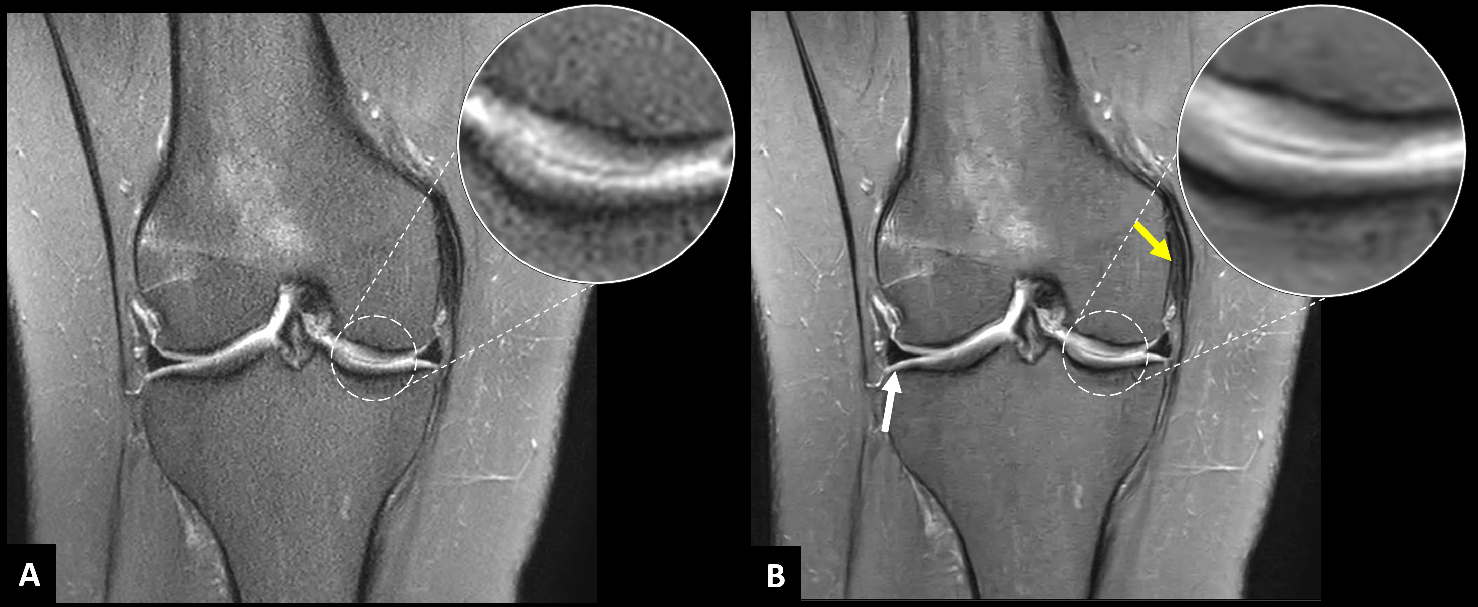
Figure 2: Original coronal 2D
fat saturated proton density image (A) demonstrates good signal to noise ratio,
however with blurring in bone marrow, cartilage, and meniscus. Combined ACN-PIN
reconstruction with high denoising and 2x image matrix enhancement (B) shows
increased conspicuity of the image with improved delineation of the menisci
(white arrow) and sharper demarcation of the cartilage (inset), as well as fine
structures such as deep meniscofemoral ligament (yellow arrow). Resolution: 0.4
x 0.4 x 3 mm. FOV: 140 x 140 x 110 mm. Undersampling R=3.8. Scan time 1m:28s.
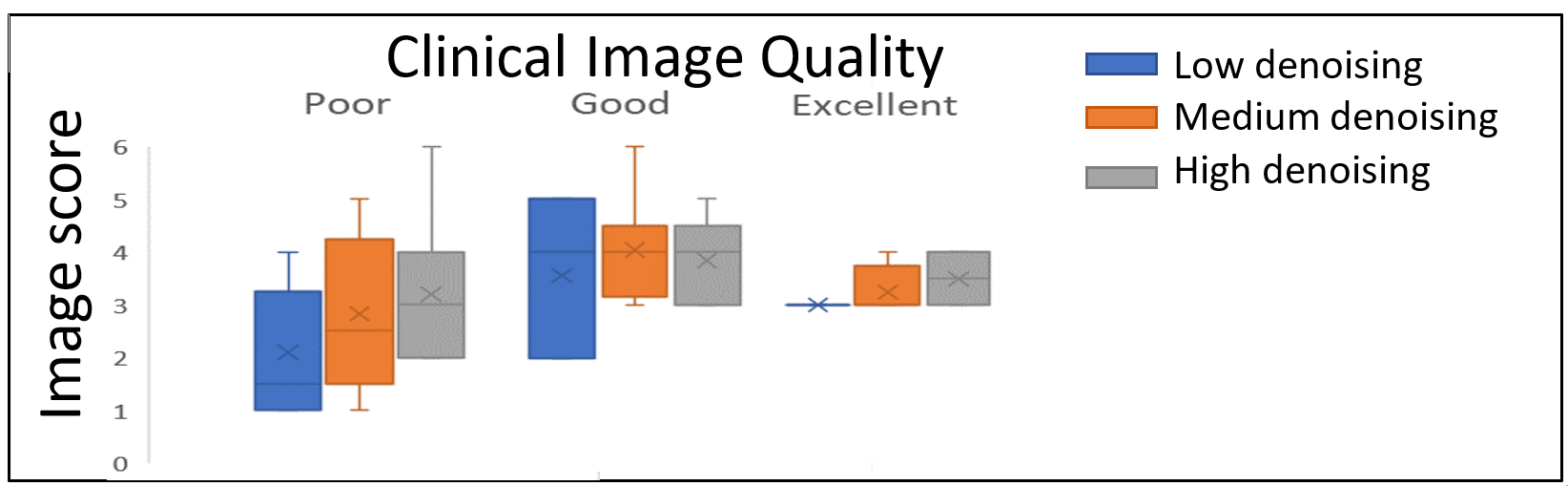
Figure 3. Radiologists scored
image quality compared to standard clinical protocols. Clinical images with a
“poor” score showed the most improvement from AI-based denoising. The highest
denoising level had the highest average score and was rated as equivalent or
superior image quality in every reviewed case.
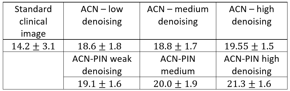
Average SNR across
reconstructed volumes showed improved SNR across all AI-based reconstruction
methods. ACN-PIN with strong denoising and 2x matrix enhancement had the
highest average SNR, showing 50% increase in SNR over standard clinical images.
DOI: https://doi.org/10.58530/2023/4191