4146
Applications of artificial intelligence-assisted compressed sensing cardiac cine in left heart function imaging: Quality and Efficiency1Peking University Shenzhen Hospital, ShenZhen, China
Synopsis
Keywords: Heart, Cardiomyopathy
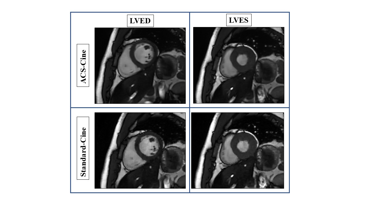
Cardiac magnetic resonance (CMR) imaging is the gold standard for non-invasive evaluation of cardiac structure and function. Conventional 2D segmented steady-state free precession cine imaging(Standrad-Cine)is widely used but the scanning time is long. Artificial intelligence-assisted compressed sensing(ACS)is a new technology that can reduce the scanning time and improve image quality. By comparing the image quality and left ventricular function (LVF) of CMR sequence based on Standrad-Cine and ACS-Cine, we found that the use of ACS-Cine can significantly reduce the scanning time while obtaining high-quality and accurate parameters images.Cardiac magnetic resonance (CMR) is the favored modality for cardiac structure and function because of its multiplanar capabilities, repeatable and excellent soft-tissue contrast. The use of 2D segmented steady-state free precession cine sequence(Standard-Cine) for imaging can obtain high-quality left heart images, but the conventional Standard-Cine sequence requires the patient to hold breath for many times, which need scan long time and may cause spontaneous or involuntary movement due to the patient's discomfort resulting in artifacts in the image. ACS-Cine imaging has the characteristics of real-time, real imaging, sparse data sampling and iterative reconstruction, and can obtain the whole left ventricle image with a single breath hold. This study is based on artificial intelligence-assisted compressed sensing (ACS) MR imaging sequence (artificial intelligence-assisted compressed sensing 2D steady-state free precession cine) to explore the feasibility of this technique in evaluating left ventricular function.
Materials and Methods
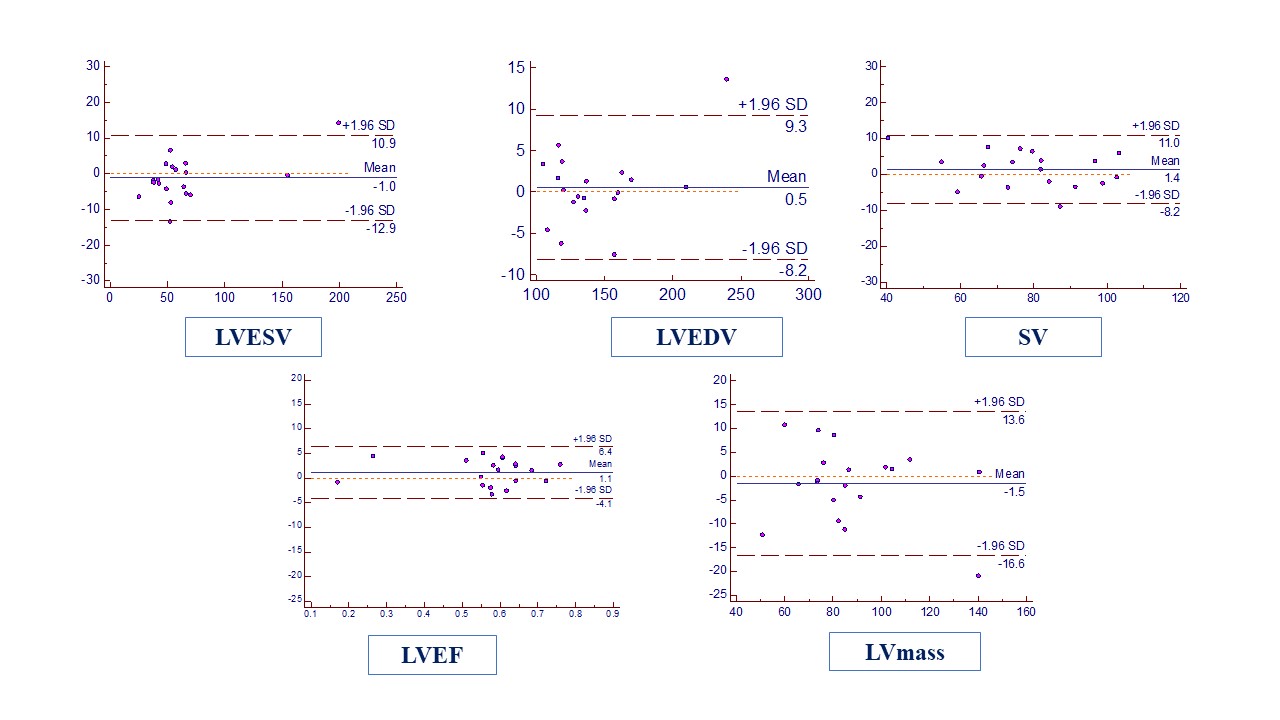
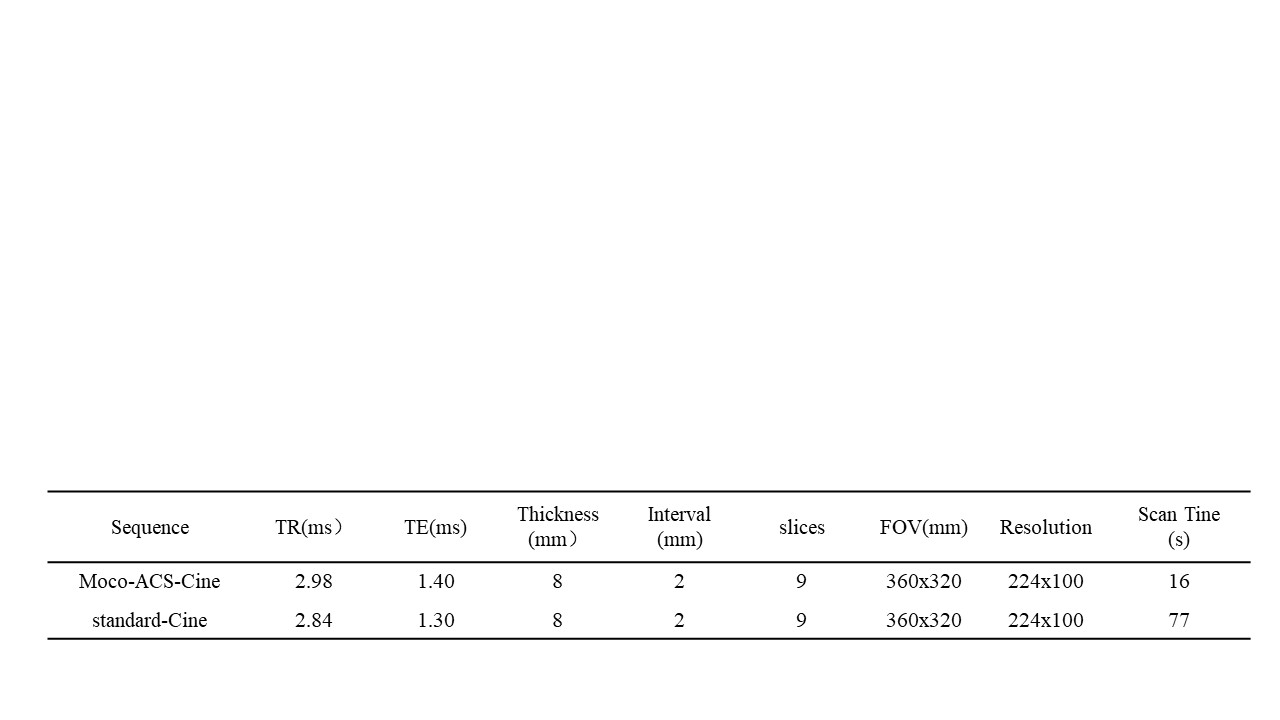
This study has been approved by the local IRB. Recruit 20 healthy volunteers (male=7, female=9, age 21-61, 29.85±10.67), using uMR 780 3.0 T from United Imaging, China, using ACS-Cine and Standard-Cine sequence for left heart function imaging, scanning parameters were shown in Table 1. Each volunteer obtains 2 sets of data and records the scanning time of each set. Use uWS-780 workstation (United Imaging, China) to obtain cardiac function parameters including left ventricular end systolic volume (LVESV), left ventricular end diastolic volume (LVEDV), stroke volume (SV), left ventricular ejection fraction (LVEF) and left ventricular mass, (LVmass) of two sequences. The ICC and Bland-Altman were applied to evaluate the consistency of the functional imaging parameters of the two imaging methods. A five-point scoring criterion was used for subjective scoring, and the kappa test and Wilcoxon paired rank sum test were applied for comparative analysis of image quality.
Result
The subjective scores of image quality of the two groups were highly consistent (P<0.05), and there was no significant difference between the two groups (P>0.05). The results of cardiac function measured by two observers were in good agreement (P<0.05). The Bland Altman analysis results show that more than 95% of the cardiac function parameters measured are within the consistency range (fig.1-2).
Discussion and Conclusion
The use of ACS-Cine sequence for left heart function scan can significantly reduce the scanning time (16s vs, 77s, 79.22%) and is comparable to the conventional plan image.
Acknowledgements
N/AReferences
Reference [1] Klinke V, Muzzarelli S, Lauriers N, et al. Quality assessment of cardiovascular magnetic resonance in the setting of the European CMR registry: description and validation of standardized criteria[J/OL]. J Cardiovasc Magn Reson, 2013, 15(1) [2022-07-07]. DOI:10.1186/1532-429x-15-55.
[2] Wang J, Li X, Lin L, et al. Diagnostic efficacy of 2-shot compressed sensing cine sequence cardiovascular magnetic resonance imaging for left ventricular function[J]. Cardiovasc Diagn Ther, 2020, 10(3): 431-441. DOI:10.21037/cdt-20-135.
[3] Petersen SE, Aung N, Sanghvi MM, et al. Reference ranges for cardiac structure and function using cardiovascular magnetic resonance (CMR)in Caucasians from the UK Biobank population cohort[J/OL]. J Cardiovasc Magn Reson, 2017, 19(1) [2022-07-07]. DOI:10.1186/s12968-017-0327-9.
[4] Nguyen KL, Hu P, Finn JP. Cardiac Magnetic Resonance Quantification of Structure-Function Relationships in Heart Failure[J]. Heart Fail Clin, 2021, 17(1): 9-24. DOI:10.1016/ j. hfc. 2020.08.001.
Figures