4140
Evaluation of right ventricular myocardial microcirculation in diabetes patients by using optimized intravoxel incoherent motion imaging1Department of Magnetic Resonance, Lanzhou University Second Hospital, Lanzhou, China, 2Philips Healthcare, Xi’an, China
Synopsis
Keywords: Myocardium, Diffusion/other diffusion imaging techniques, IVIM, diabetes, myocardial microcirculation, right ventricular myocardial
Intra-voxel incoherent motion imaging (IVIM) can noninvasively and quantitatively evaluate myocardial microcirculation. At present, some scholars have demonstrated that IVIM can observe left ventricular myocardial microcirculation. However, due to the thickness of the right ventricular wall, IVIM imaging is difficult to assess it. This study proposed an optimized navigator-based and free-breathing acquisition scheme for cardiac IVIM. The results found that right ventricular myocardial microcirculation was impaired, pseudo-diffusion coefficient (Dfast) were significantly lower than healthy volunteers. It is helpful to monitor cardiovascular risk and evaluate prognosis.
Introduction
Diabetes is a very important risk factor for cardiovascular diseases, it has been reported that 70-80% people died from cardiovascular complications1, mainly due to the impaired self-regulation mechanism of myocardial microcirculation caused by hyperglycemia, which can further develop into cardiac hypertrophy and heart failure. Mou A et al2 found that Dfast values of left ventricle in diabetic patients or hyperpietic patients were lower than healthy volunteers. However, due to the thickness of the right ventricular wall, the microcirculation of right ventricle remains unclear. In this study, we proposed an optimized navigator-based and free-breathing acquisition scheme for cardiac IVIM. The proposed method was used to detect right ventricular myocardial microcirculation in diabetic patients. In addition, the consistency of IVIM derived parameters was observed under the condition of diaphragmatic navigation.Materials and Methods
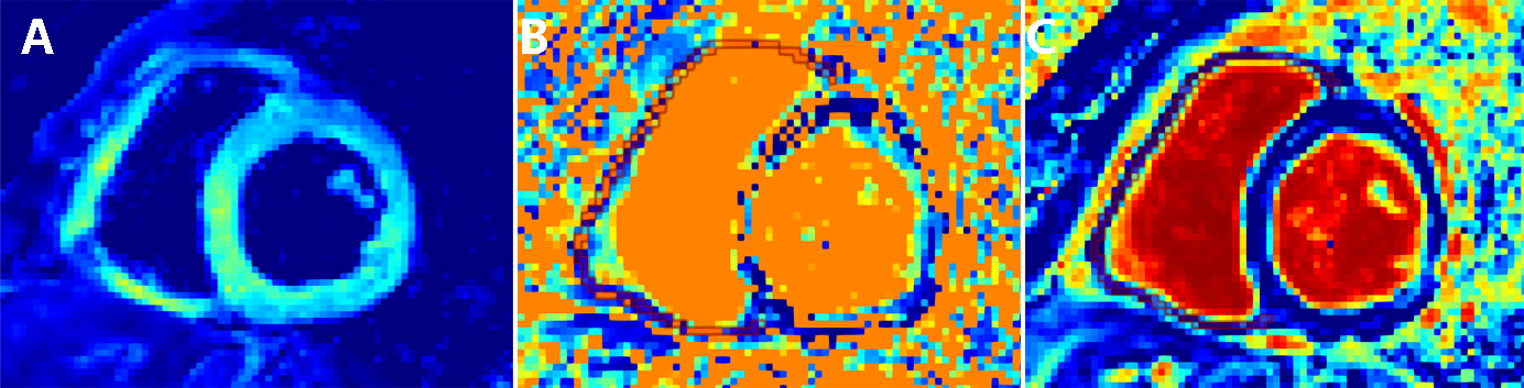
Ten healthy volunteers and eight diabetics patients were enrolled from the Second Hospital of Lanzhou University. The diagnostic criteria for type II diabetes mellitus (fasting blood glucose ≥7.0 mmol/l, oral glucose tolerance test (OGTT≥11.0 mmol/l). All data were acquired by using a 3.0T MR scanner (Ingenia CX, Philips Healthcare, the Netherlands) with a 16-channel phase array body coil. The free-breathing IVIM imaging with 9 values (0, 20, 50, 80, 100, 120, 200, 300, 500 s/mm2) were performed from Right ventricle apex to basal slices. by diaphragmatic navigationThe scan time was 2min. The imaging parameters were list as below: FOV was set to 310×310mm, number of slices was 3, slice thickness was 8mm. The raw IVIM images were transmitted to the post-processing software MIKT (https://www.mitk.org), and IVIM related parameter values (Dslow, Dfast, f) were obtained by using the double exponential model. The ROI of myocardium which placed at the mid-wall area were manually drawn three times to obtain the average values. The difference of IVIM derived parameters between healthy volunteers and diabetics patients was analyzed by independent-samples t-test. The intraclass correlation coefficient (ICC) test was used to assess the consistency of IVIM derived parameters.Results
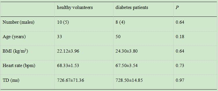
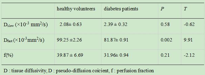
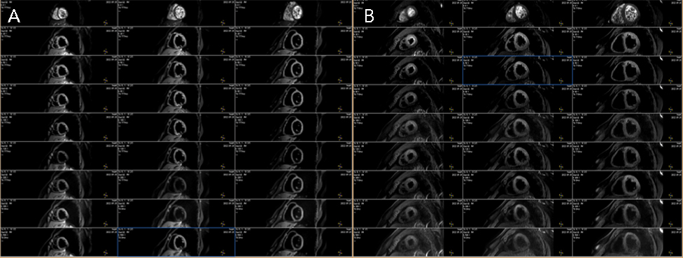
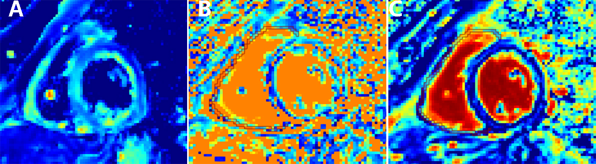
Basic characteristics of study population, were shown in Table 1. The sex ratio, age, BMI, heart rate and TD(Trigger Delay) of the two groups had no significant difference. IVIM derived parameters of right ventricle had good consistency (ICC≥ 0.85). The typical IVIM examples of healthy volunteer and diabetes patient were shown in Fig 1. The related IVIM derived parameters maps were shown in Fig 2 and Fig 3. Statistical values of Dslow, Dfast and f were shown in table 2. As can be seen from table 2, Dslow and f value showed no significant different between healthy volunteers and diabetic patients. And Dfast value of right ventricle in diabetic patients (81.87± 0.91) were significantly lower (P=0.002) than healthy volunteers (99.25 ±2.26).Discussion
Myocardial microcirculation can reflect the coronary microvascular networks and blood flow distribution in heart. The ability to detect more precise abnormalities in myocardial microcirculation requires more stable imaging method.. Conventional imaging technologies (CT, MRI, and ultrasound) cannot accurately reflect damage of myocardial microcirculation in diabetic patients. At present, Mou A2 et al observed the left ventricular myocardial microcirculation in hypertensive and diabetic patients. Li S3 et al observed the left ventricular myocardial microcirculation in normal subjects. However, due to the thin right ventricular wall and the influence of physiological movement, right ventricle is difficult to image. Therefore, it is important to seek a suitable method for quantitative monitoring myocardial microcirculation in diabetic patients. IVIM can non-invasively and quantitatively assess micro-perfusion and water diffusion without contrast agents2. This study found that the Dfast of right ventricular in diabetic patients (81.87±0.91) was significantly lower than that of normal subjects (99.25±2.26 P=0.002). The probable reason is that hyperglycemia causes cardiomyocytes to release advanced glycation end products (AGEs), which lead to the expansion of extracellular matrix, thickening of vascular basement membrane, increased microvascular permeability and other typical features of diabetic microangiopathy. Besides, the common feature of glucose metabolism defects is that oxidative stress directly or indirectly induces endothelial cell dysfunction4. In a word, the increase of blood glucose affects the function of vascular endothelium, which leads to the damage of the autoregulation mechanism of microcirculation5. The proposed 9 b values’ acquisition scheme for cardiac IVIM is a navigator-based, free-breathing method. It offered an efficient way to evaluate the right ventricular myocardial microcirculation. Moreover, the results had good consistency.Conclusions
This study proposed an optimized, navigator-based free-breathing IVIM acquisition scheme for cardiac IVIM. And it’s helpful to evaluate myocardial microcirculation of right ventricle in diabetic patients.Acknowledgements
No acknowledgement foundReferences
1. Guariguata L, Whiting DR, Hambleton I, et al. Global estimates of diabetes prevalence for 2013 and projections for 2035. Diabetes Res Clin Pract, 2014, 103(2): 137-149.
2. Mou A, Zhang C, Li M, et al. Evaluation of myocardial microcirculation using intravoxel incoherent motion imaging[J]. J Magn Reson Imaging, 2017, 46(6): 1818-1828.
3. Li S, Mou A, Li X, Guo Y, et al. Myocardium microcirculation study in a healthy Chinese population using 30-T cardiac magnetic resonance intravoxel incoherent motion imaging[J]. Acta Radiol, 2022, 63(5):596-605.
4. Labazi H, Trask AJ. Coronary microvascular disease as an early culprit in the pathophysiology of diabetes and metabolic syndrome. Pharmacol Res. 2017, 123: 114-121.
5. Brodsky SV, Morrishow AM, Dharia N, et al. Glucose scavenging of nitric oxide. Am J Physiol Renal Physiol, 2001, 280(3): F480-486.
Figures