4126
Diffusion Kurtosis Imaging Combined with Intravoxel Incoherent Motion Imaging in Prediction of P53 Status in Rectal Cancer
Anliang Chen1, Deshuo Dong1, Changjun Ma1, Ailian Liu1, and Qingwei Song1
1radiology, The First Affiliated Hospital of Dalian Medical University, Dalian, China
1radiology, The First Affiliated Hospital of Dalian Medical University, Dalian, China
Synopsis
Keywords: Digestive, Pelvis
Mutant P53 promotes tumor cell proliferation, invasion, and resistance to chemotherapy. The purpose of this study was to evaluate the value of combination of DKI and IVIM for prediction of P53 expression status in rectal cancer. The results indicate that the combination of MK and D* improved the diagnostic efficiency than single parameters. DKI and IVIM imaging allows non-invasive visualization and quantification of tissue composition for prediction of P53 status in rectal cancer.Introduction
Colorectal cancer (CRC) ranks the third estimated new cancer cases and deaths in men and wemen, and rectal cancer (RC) accounts for approximately three-tenth of all CRC cases1. P53 has significant prognostic value in patients with rectal cancer. And positive P53 status tumors exhibit more aggressive behavior2,3. DKI and IVIM imaging can noninvasively and quantitatively evaluate the diffusion and microcirculation perfusion of the water molecules in the voxel without the need for an exogenous contrast agent, and were used in all parts of human body. This study is aimed to evaluate the value of DKI combined IVIM imaging for prediction of P53 status in rectal cancer.Methods and Materials
A retrospective analysis was performed on 82 rectal cancer patients confirmed with P53 expression according to their postoperative pathology results. They were divided into group 1 (positive P53 status; 60 patients; 45 male, 15 female, mean age: 66.57±9.91 years, range: 45-90 years) and group 2 (negative P53 status; 22 patients, 17 male, 5 female, mean age: 62.55±10.13 years, range: 43-89 years) according to their pathological P53 status. All participants underwent DKI, IVIM and conventional scans on a 3.0T MRI scanne(GE Signa HDXT) before surgery. The DKI and IVIM images were post-processed using FuncTool software on AW4.6 workstation. Three region of interests (ROIs) were put on the largest slice of tumors, avoiding bleeding, necrosis, and cystic degeneration. Statistical analyses were carried out with SPSS 26.0 (IBM) and MedCalc 12.5.5. Mann-Whitney U test was used to compare the differences between two groups. Logistic regression and receiver operating characteristic (ROC) curve analyses were performed to evaluate the diagnostic efficiency of the parameters. Differences in the area under the curves (AUCs) of different parameters were compared using the DeLong test.Results
Group 1 had significantly higher MK and D* values, while lower MD, D and f values than group 2 (P < 0.05)(Table 1). There was no difference of FA and ADC values between two groups. The AUC values for the ROC analyses of MD, MK,D, D* and f values for differentiation two groups was 0.650, 0.676, 0.702, 0.712 and 0.649, respectively (Figure 2). The AUC value of MK combined with D* parameter was 0.812, with a sensitivity of 85.0% and specificity of 68.18% (Table 2). The combination of MK and D* improved the diagnostic efficiency than single parameters(P < 0.05).Discussion and Conclusions
Group 1 had significantly higher MK value than group 2, which might be related to the complexity of the microstructure in positive P53 status group (such as cell density, cell atypia and nuclear pleomorphism, etc.). Group 1 had significantly higher D* value than group 2, which might because the tumor with positive P53 status had more blood microcirculation in rectal cancer. MK combined D* values explained the characteristics of positive P53 status tumors from different angles which showed the greatest diagnostic efficiency. DKI combined IVIM might be a noninvasive way to evalute the P53 status in rectal cancer, which is of guiding value for clinical diagnosis and treatment.Acknowledgements
No acknowledgement found.References
[1] Siegel RL, Miller KD, Fuchs HE, Jemal A. Cancer statistics, 2022. CA Cancer J Clin. 2022 Jan;72(1):7-33.
[2] Nakayama M, Oshima M. Mutant p53 in colon cancer. J Mol Cell Biol 2019;11(4):267-276.
[3] Meyer HJ, Höhn A, Surov A. Histogram analysis of ADC in rectal cancer: associations with different histopathological findings including expression of EGFR, Hif1-alpha, VEGF, p53, PD1, and KI 67. A preliminary study. Oncotarget. 2018 Apr 6;9(26):18510-18517.
Figures
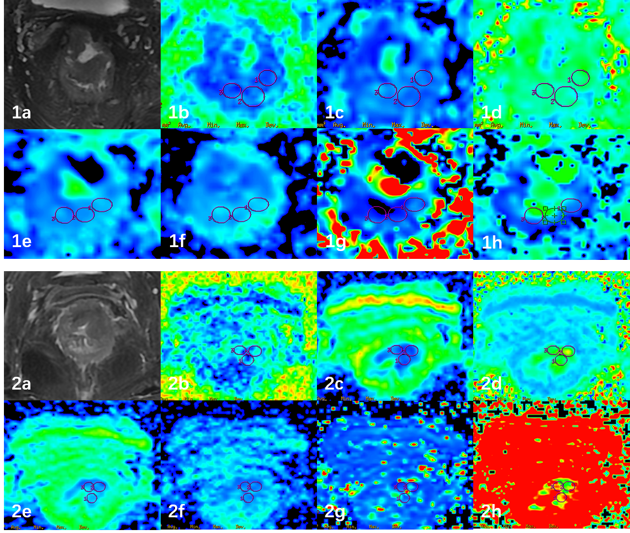
Figure 1. A 66-year-old male rectal cancer patient with negative P53 status. T2WI(1a), FA(1b), MD(1c), MK(1d), ADC(1e), D(1f), D*(1g), f(1h) images were shown, and the average values were 0.199, 1.570 μm2 /ms, 0.706, 0.923×10⁻³ mm2/s, 0.704×10⁻³ mm2/s, 2.667×10⁻³ mm2/s and 0.586.A 78-year-old female rectal cancer patient with positive P53 status. T2WI(2a), FA(2b), MD(2c), MK(2d), ADC(2e), D(2f), D*(2g), f(2h) images were shown, and the average values were 0.300, 1.019 μm2 /ms, 0.983, 0.570×10⁻³ mm2/s, 0.452×10⁻³ mm2/s, 6.320×10⁻³ mm2/s and 0.286.
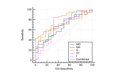
Figure 2. ROC curves of diagnostic efficacy of MD, MK, D, D*, f value and MK combined D* in prediction of P53 status in rectal cancer.
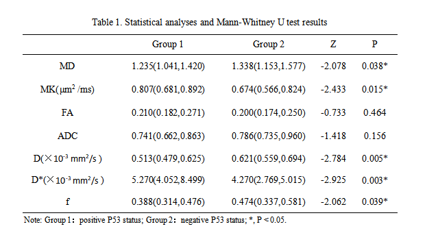
Table 1. Statistical analyses and Mann-Whitney U test results
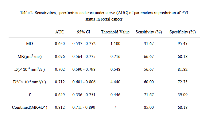
Table 2. Sensitivities, specificities and area under curve (AUC) of parameters in prediction of P53 status in rectal cancer
DOI: https://doi.org/10.58530/2023/4126