4124
Comparation of DCE-MRI and dual-energy CT imaging in discriminating tumor deposits from lymph node metastasis in rectal cancer.
WEN JUN HU1, Anliang Chen1, and AIlian Liu1
1The First Affiliated Hospital of Dalian Medical University, Dalian, China
1The First Affiliated Hospital of Dalian Medical University, Dalian, China
Synopsis
Keywords: Digestive, Cancer
Tumor deposits (TDs) in rectal cancer have been shown to be an important marker of poor prognosis. Although very similar to lymph node metastasis (LNMs), TDs have unique features in terms of biology and outcome, suggesting that distinguishing between these two entities may be of great importance. Results of this study indicate iodine-based material-decomposition (MD) images and DCE-MRI can effectively differentiate TDs and LNMs in rectal cancer, and iodine-based MD images showed better diagnostic efficiency.Introduction
Rectal cancer (RC) is a common gastrointestinal malignancy that cause signifcant morbidity and mortality1. Tumor deposits (TDs), a powerful prognostic factor of RC patients2, has similar performance to lymph node metastasis (LNMs) on conventional CT/MRI images, which makes it difficult to distinguish them. Currently, the diagnosis of TDs still depends on the pathology after surgery, which is not conducive to the early evaluation of tumor characteristics. Dynamic contrast-enhanced MRI (DCE-MRI) is a functional imaging that can provide valuable information about tumour aggressiveness and the degree of angiogenesis. Dual-energy CT imaging can provide multiple parameters to be used as analytical tools and quantitative indicator. The iodine concentration (IC) measured in the material-decomposition (MD) images can obviously reflect the vascularization in various tissues. Thus, the purpose of this study is to explore the value of DCE-MRI and iodine-based MD images with dual-energy CT imaging in discriminating TDs from LNMs.Methods
This study has been approved by the local IRB. 23 patients with pathologically confirmed with rectal cancer were recruited in this study. The patients were divided into two groups: TDs group (8 patients) and LNMs group (15 patients). They underwent examinations on 3.0T MR and contrast-enhanced dual-energy CT scans. The MR sequences included T2WI, DWI, LAVA, etc. Detailed parameters were listed in Table 1. The dual-energy CT scan parameters were as follows: rapid switching between tube voltages of 80 kVp and 140 kVp; tube current, 375 mA; helical pitch, 1.375:1; rotation time, 0.8 s. The original axial digital images were transmitted to the ADW 4.6 workstation. GenIQ software and GSI viewer software were used to perform post-processing to obtain DCE maps (Ktrans, Kep, Ve) and iodine-based MD images(IC in arterial, venous, and delayed phase), respectively. Two radiologists used a double-blind method to place three ROIs on the largest layer of the lesion to measure DCE parameters and IC values. The average values were calculated to minimize measurement bias. An unpaired t-test was used to analyze the differences of DCE parameters and IC values between the two groups. The ROC curves were used to evaluate the diagnostic efficacy of these parameters in the differential diagnosis of the two groups.Results
The consistency of the data obtained by the two observers was good (ICC value > 0. 75). The Kep values (1.64±0.62 vs. 2.27±0.67/min) and IC values in arterial phase (16.89±4.56 vs. 26.17±2.74) of TDs group were significantly lower than that of LNMs group (P<0.05). The area under the ROC curve (AUC) for the ROC analyses of Kep and IC values in arterial phase for differentiation between TDs and LNMs groups was 0.775 and 0.950, respectively.Discussion
The significant difference in DCE parameters and IC values between the two groups may be due to the more angiogenesis in rectal cancer patients with LNMs than in those with TDs. Moreover, IC values showed better diagnostic efficiency than DCE parameters.Conclusion
DCE-MRI and iodine-based MD images with dual-energy CT imaging have the potential to be used in routine clinical practice as new indicators to differentiate TDs from LNMs in RC, and to help personalized treatment of RC.Acknowledgements
No acknowledgement found.References
1. Mattiuzzi C, Sanchis-Gomar F, Lippi G. Concise Update on Colorectal Cancer Epidemiology. Ann Transl Med. 2019;7(21):609.
2. Lord AC, D'Souza N, Pucher PH, et al. Significance of Extranodal Tumour Deposits in Colorectal Cancer: A Systematic Review and Meta-Analysis. Eur J Cancer. 2017;82:92–102.
Figures
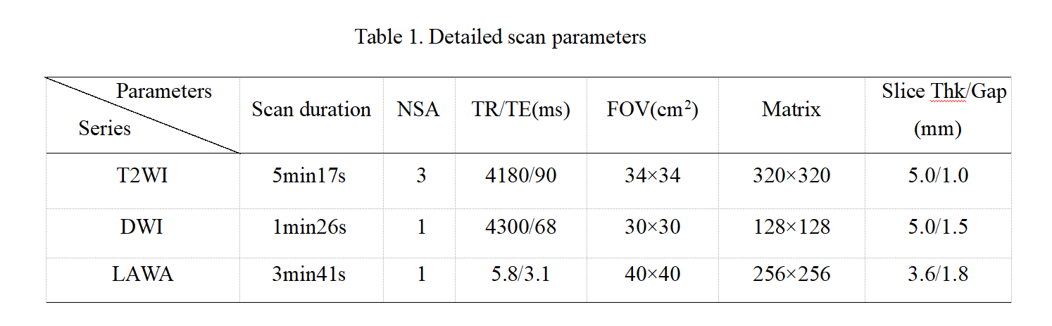
Table 1. Detailed scan parameters

Figure 1. A 66-year-old male, rectal cancer with TDs. The LAVA (A) image, Ktrans map(B), Kep map(C), Ve(D) map, 70 KeV monochromatic CT image (E), iodine-based MD image (F), scatter diagram of iodine concentration (G) and spectral Hounsfield unit curve (H) in arterial phase were shown, Ktrans, Kep, Ve, and IC values are 0.91/min, 1.6/min, 0.58, and 12.91, respectively.
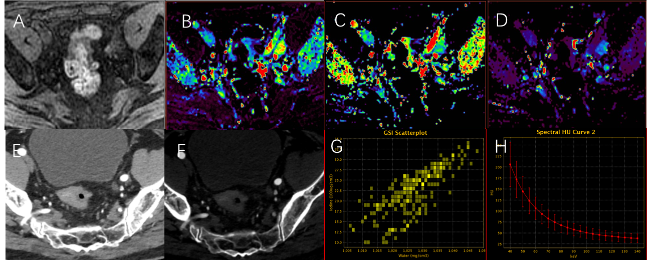
Figure 2. A 62-year-old male, rectal cancer with LNMs. The LAVA (A) image, Ktrans map(B), Kep map(C), Ve(D) map, 70 KeV monochromatic CT image (E), iodine-based MD image (F), scatter diagram of iodine concentration (G) and spectral Hounsfield unit curve (H) in arterial phase were shown, Ktrans, Kep, Ve, and IC values are 0.22/min, 1.73/min, 0.15, and 21.21, respectively.
DOI: https://doi.org/10.58530/2023/4124