4106
Three-dimensional radial echo-planar spectroscopic imaging for in vivo hyperpolarized 13C MRSI at 3 T1Division of Medical Physics in Radiology, German Cancer Research Center (DKFZ), Heidelberg, Germany, 2Faculty of Physics and Astronomy, University of Heidelberg, Heidelberg, Germany, 3Max-Planck-Institute for Nuclear Physics, Heidelberg, Germany, 4Division of Medical Physics in Radiation Oncology, German Cancer Research Center (DKFZ), Heidelberg, Germany, 5Faculty of Chemistry and Earth Sciences, University of Heidelberg, Heidelberg, Germany, 6Faculty of Medicine, University of Heidelberg, Heidelberg, Germany
Synopsis
Keywords: Pulse Sequence Design, Hyperpolarized MR (Non-Gas), Carbon-13
In this study, a novel radially-sampled echo-planar spectroscopic imaging (rEPSI) sequence was implemented at a clinical 3T MR scanner to enable the non-invasive investigation of metabolic processes via hyperpolarized 13C substrates in real-time.
Customized data analysis pipelines yielded high quality spectra and volumetric intensity maps for in vivo experiments using hyperpolarized [1-13C]pyruvate. Data extracted from k-space center points enabled a non-localized quantification of T1 values.
Introduction
MRI of hyperpolarized (HP) 13C is an emerging molecular imaging technique with enormous clinical potential. Despite spectrally-separated fast imaging approaches being typically used for dynamic imaging nowadays, spectroscopic imaging techniques might still be useful, particularly when good spectral resolution is required, as is the case for imaging chemical shift based pH sensors like zymonic acid [1]. For this purpose, echo-planar spectroscopic imaging (EPSI) with radial k-space coverage might be beneficial as it is (I) suitable for spatial undersampling to enable further acceleration, and (II) as the continuous passage through k-space center allows tracking the global T1 decay.The purpose of this study was to demonstrate feasibility of an EPSI technique with 3D radial k-space coverage for use in HP 13C MRSI.
Methods
Figure 1 illustrates the scheme of the radial echo-planar spectroscopic imaging (rEPSI) sequence, implemented on a 3T human PET/MR system (Biograph mMR, Siemens Healthineers). After a single rectangular RF pulse was used for whole-body excitation, a sinusoidal-shaped readout gradient was used for simultaneous sampling of the spatial and temporal dimension. To cover the 3D k-space in a spherical manner, the scheme described in Figure 1 is executed multiple times while rotating the readout direction. The k-space center is hereby sampled in every spoke.For proof-of-concept, a 50-ml phantom filled with thermally polarized ethylene glycol (EG) was measured with the following parameters: FOV: 200 mm, image matrix: 163, spectral BW: 1000 Hz, echoes: 512, TR: 520 ms, Ttot: 3:42 min, spokes: 427. For HP 13C MRSI in vivo, the following sequence parameters were used: FOV: 120mm, image matrix: 83, spectral BW: 1000 Hz, echoes: 256, TR: 300 ms, Ttot: 19.5 s, spokes: 65, undersampling factor: 1.55. To this end, a solution of about 80 mM of HP [1-13C]pyruvate was prepared in a SPINlab polarizer (GE Healthcare), and administered to six rats in total (animal approval: G197/17). The rEPSI sequence was started directly after injection of the HP [1-13C]pyruvate into a rat with four repeated acquisition blocks (4 x 19.5 s = 1:18 min). All measurements were performed using a double-resonant 13C/1H volume resonator with 72 mm inner diameter (Rapid Biomedical).
Data reconstruction and processing were performed with custom-written MATLAB scripts. The acquired data was divided first into odd and even datasets that were reconstructed separately.
A gridding reconstruction [2] was applied that distributes the radially sampled data points onto a Cartesian grid in a distance-weighted manner. Here, one-fold zero-filling in spatial as well as temporal domain was used.
The peak amplitudes of the resulting 13C MRSI data were quantified using a customized implementation [3] of the AMARES fitting algorithm [4] with a Gaussian lineshape model, and parameters optimized to ensure stable fitting.
Finally, dynamic intra-acquisition data was extracted through the use of the k-space center points, which are traversed with each gradient, resulting in a non-localized spectrum for each spoke. To quantify the corresponding T1eff values of each metabolite, an exponential fitting model was applied to the quantified amplitudes.
Results
Figure 2 illustrates representative 13C spectra and metabolite maps of EG acquired with 3D rEPSI. The 13C resonances are well-resolved and a well-defined distinction of the sample vial with respect to the background noise is possible, demonstrating the robustness of the acquisition and reconstruction.Figure 3 shows an example in vivo measurement of one rat. Metabolite maps of the administered HP [1-13C]pyruvate, as well as [1-13C]lactate and [1-13C]alanine are illustrated from a single measurement block with duration of 19.5 s. Also, good in vivo spectral quality is achieved. The intensity maps resemble the coarse shape of the rat quite well, but suffer from blurring due to T1 decay.
Figure 4 demonstrates the possibility to extract the effective signal decay within a single acquisition block, yielding T1eff values of (6.05±0.23) s for [1-13C]pyruvate, (12.90±1.44) s for [1-13C]lactate and (17.49±2.43) s for [1-13C]alanine.
Discussion
2D versions of rEPSI have been described before [5], and also applied to HP 13C [6]. But a 3D version as described in [7] has not been applied to HP 13C imaging yet. The obtained data proved the functionality of the implemented rEPSI sequence, though blurring of the in vivo data was not tackled in reconstruction, yet. In the future, the determined T1eff times can be utilized to account for the blurring artifact arising from T1-related decay of the HP state.This study demonstrated only the general applicability of the sequence, and further optimization of sequence parameters is required to be competitive with other fast imaging approaches. A higher undersampling factor in combination with compressed sensing algorithms could reduce the acquisition time for dynamic 13C MRSI datasets drastically. Additionally, the odd and even datasets could be combined by either summation or interleaving. The first approach would result in an increased SNR by a factor of $$$\sqrt2$$$ , while the second one would yield a doubled spectral bandwidth.
Conclusion
In this study, we demonstrated the feasibility of a three-dimensional radial echo-planar spectroscopic imaging sequence for its use in HP 13C MRSI. Customized data analysis pipelines yielded high quality spectra and volumetric intensity maps for in vivo experiments using hyperpolarized [1-13C]pyruvate. Data extracted from k-space center points enabled a non-localized quantification of T1 values.Acknowledgements
No acknowledgement found.References
[1] Christian Hundshammer, Stephan Düwel, and Franz Schilling. „Imaging of Extracellular pH Using Hyperpolarized Molecules“, volume 57. Wiley Online Library, 2017.
[2] John D O'Sullivan. „A Fast Sinc Function Gridding Algorithm for Fourier Inversion
in Computer Tomography“, volume 4. IEEE, 1985.
[3] Andreas Korzowski, et al. „Volumetric Mapping of Intra- and Extracellular pH in the Human Brain Using 31P MRSI at 7T“, volume 84. Wiley Online Library, 2020.
[4] Leentje Vanhamme, Aad van den Boogaart, and Sabine Van Huffel. „Improved Method for Accurate and Efficient Quantification of MRS Data with Use of Prior Knowledge“, volume 129. Academic Press, 1997.
[5] Saucedo, Andres, Paul M. Macey, and M. Albert Thomas. "Accelerated radial echo‐planar spectroscopic imaging using golden angle view‐ordering and compressed‐sensing reconstruction with total variation regularization", Magnetic Resonance in Medicine 86.1, 2021.
[6] Ramirez, Marc S., et al. "Radial spectroscopic MRI of hyperpolarized [1‐13C] pyruvate at 7 tesla", Magnetic Resonance in Medicine 72.4, 2014.
[7] Ludwig, Dominik, et al. "Three‐dimensional 31 P radial echo‐planar spectroscopic imaging in vivo at 7T", ISMRM 25th Annual Meeting and Exhibition, 2017.
Figures
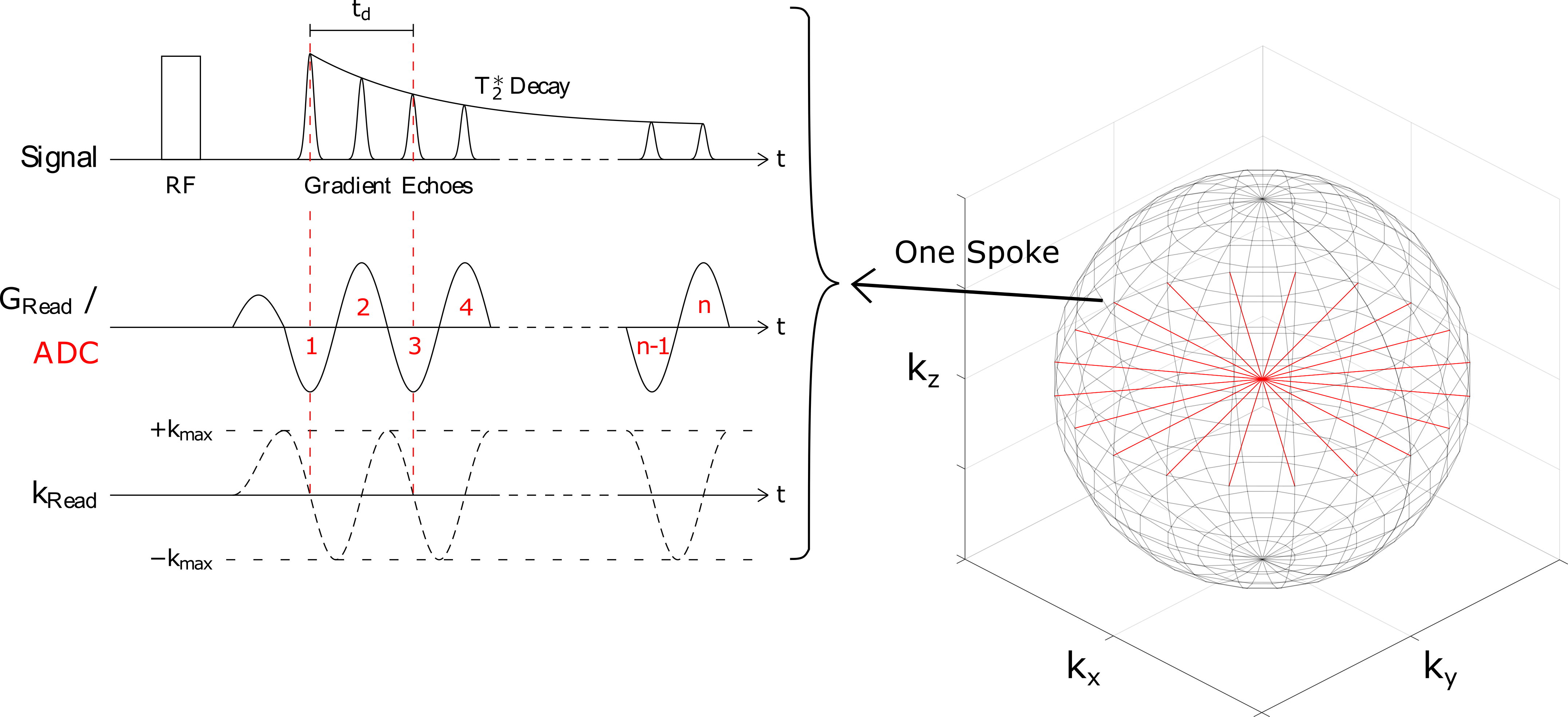
Figure 1: Sequence diagram of the implemented 3D radial EPSI. After a rectangular whole-volume RF excitation pulse and a prephaser gradient, an oscillating sinusoidal readout gradient GRead with n lobes is applied. After each spoke, the gradient is rotated around the sphere’s center to radially sample the k-space in all three dimensions. Note that the k-space center is sampled in every spoke.
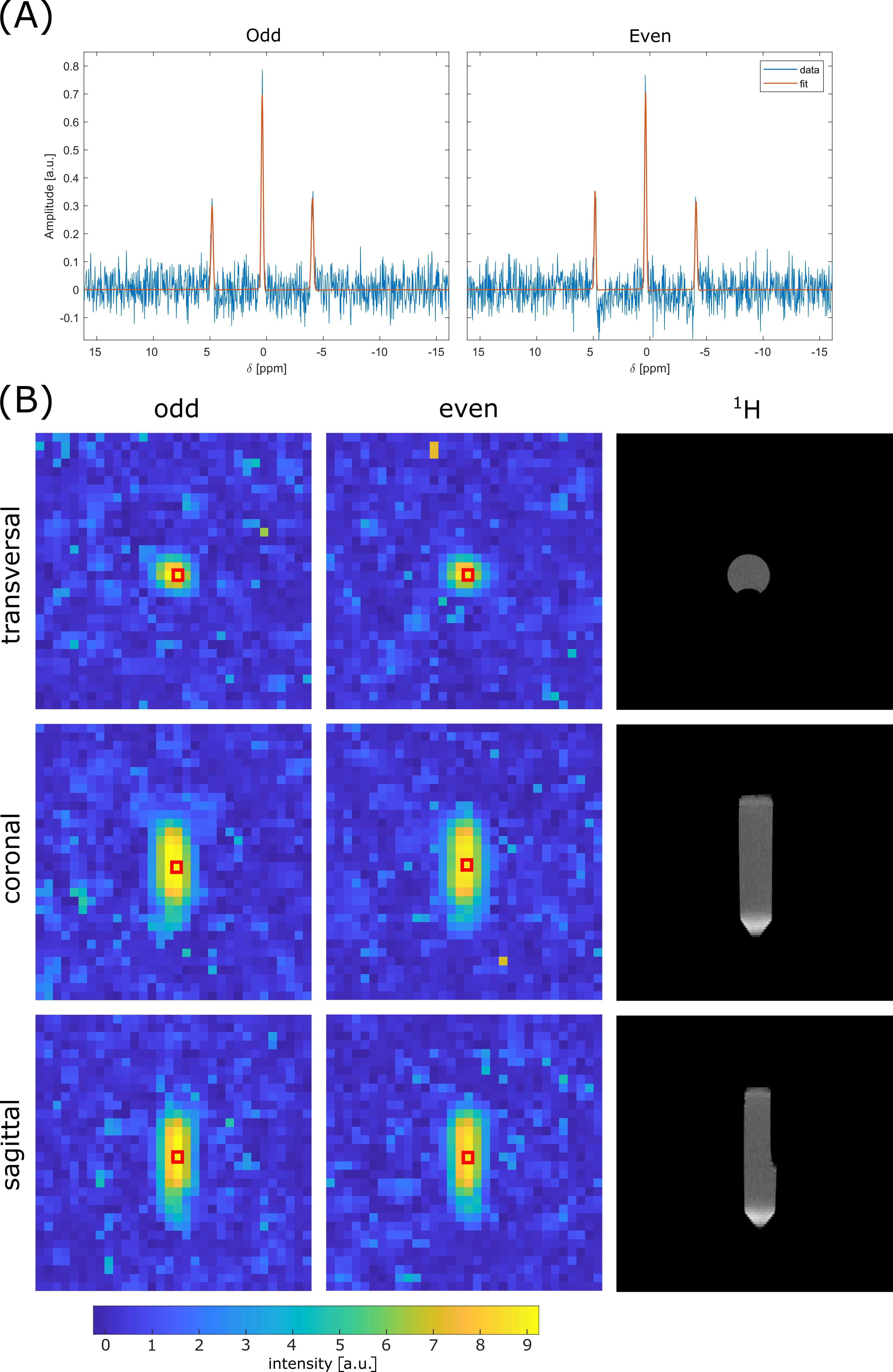
Figure 2: (A) Localized 13C spectra of thermally polarized ethylene glycol (EG) from a representative voxel (red square in (B)). The signals (blue line) from the odd (left) and even (right) dataset are corrected for zero- and first-order phases, and overlaid with the corresponding fits (orange line).
(B) 13C EG intensity maps of the odd (left column) and even (middle column) datasets, as well as 1H images of the phantom, are shown for all views with an interpolated voxel size of (6.25 mm)3. The intensity is determined by the fitted amplitude of the center peak of the EG triplet.
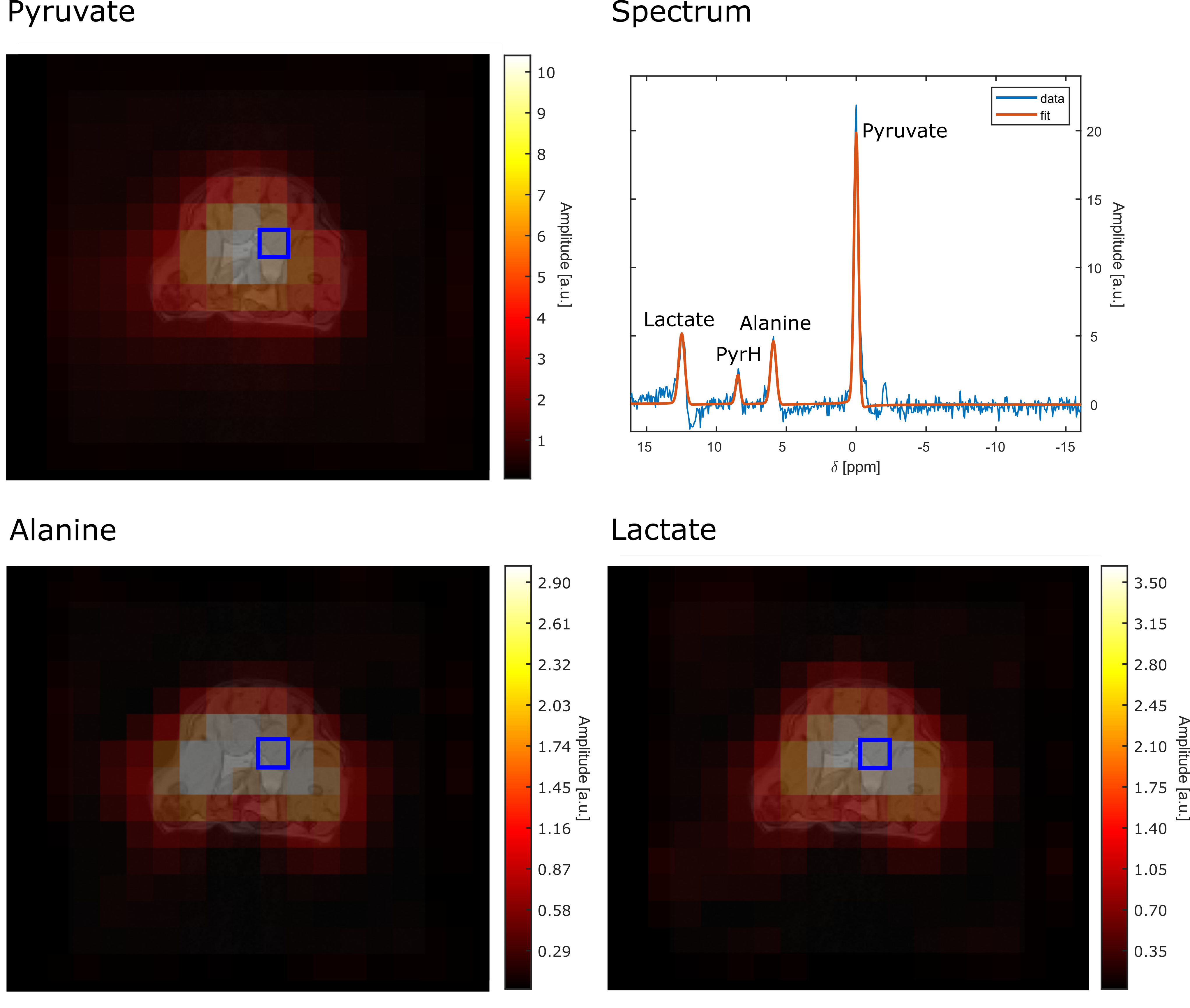
Figure 3: Hyperpolarized 13C maps from a rat acquired within a single 19.5-s acquisition block of rEPSI from the odd-echo dataset. A representative localized spectrum is displayed in the top right (blue square in the intensity maps). The signal (blue line) is corrected for zero- and first-order phases, and overlaid with the corresponding fit (orange line). The [1-13C]pyruvate (top left), [1-13C]lactate (bottom right) and [1-13C]alanine (bottom left) intensity maps are shown for one representative transversal slice with an interpolated voxel size of (7.5 mm)3.
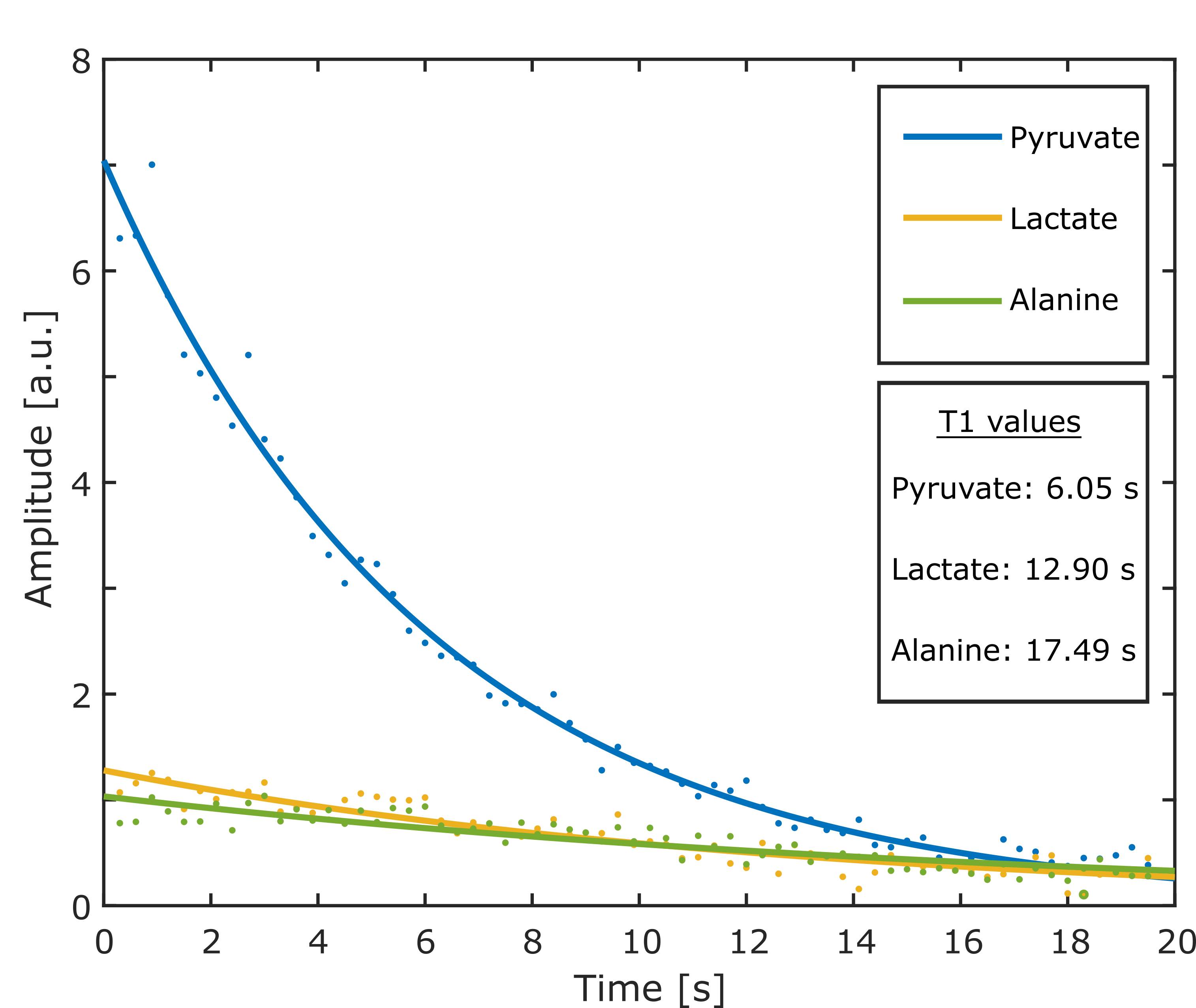
Figure 4: Global T1eff quantification for [1-13C]pyruvate (blue), [1-13C]lactate (yellow) and [1-13C]alanine (green). Shown is the fitted amplitude (dots) of the three metabolites within a single 19.5-s acquisition block extracted from the 65 acquired k-space centers. To determine the T1eff values, an exponential fit was applied to the data (continuous lines). Build-up effects due to the metabolic conversion were not taken into account.