4101
Assessment of Pulmonary Morphometry using Hyperpolarized 129Xe DWI with Variable-Sampling-Ratio Compressed Sensing Patterns1Key Laboratory of Magnetic Resonance in Biological Systems, State Key Laboratory of Magnetic Resonance and Atomic and Molecular Physics, National Center for Magnetic Resonance in Wuhan, Wuhan Institute of Physics and Mathematics, Innovation Academy for Precision Measurement Science and Technology, Chinese Academy of Sciences- Wuhan National Laboratory for Optoelectronics, Wuhan, China, 2University of Chinese Academy of Sciences, Beijing, China
Synopsis
Keywords: Data Acquisition, Hyperpolarized MR (Gas)
The long acquisition time of hyperpolarized 129Xe multiple b-values DWI made it hard to apply in patients with severe pulmonary diseases. Herein, we proposed a method of variable-sampling-ratio compressed sensing patterns for accelerating hyperpolarized 129Xe DWI. A four-fold reduction in acquisition time was achieved using the proposed method while preserving good image quality. Meanwhile, the method can be used for evaluating pulmonary injuries caused by cigarette smoking.Introduction
The technique of hyperpolarized (HP) 129Xe multiple b-values diffusion-weighted imaging (DWI) has been widely used in measuring the apparent diffusion coefficient (ADC) as well as the alveolar morphological parameters 1-3. Owing to the sensitivity of these morphological parameters to the alveolar microanatomical changes, they can be used to evaluate the physiological injuries in pulmonary microstructure caused by aging, smoking or lung diseases 1, 4, 5.Due to the long acquisition time, the application of HP 129Xe DWI is limited in patients with severe pulmonary diseases, who are unable to sustain long breath holds. In the previous studies, compressed sensing (CS) has been used for accelerating HP 129Xe DWI data acquisition 6, 7. Generally, a fixed acceleration factor (AF) was used 6. However, with this approach, obvious artifacts caused by k-space undersampling acquisition become inevitable when the AF increases, leading to high mean absolute error (MAE) values 8.
In this study, we proposed a method of variable-sampling-ratio compressed sensing (VCS) patterns for accelerating HP 129Xe DWI data acquisition while preserving good image reconstruction quality.
Methods
The VCS acquisition strategy put forward in the present study used various sampling ratios (the inverse of AF) and undersampling patterns for DWI data acquisition with different b-values. Optimal variable-sampling-ratios and corresponding k-space undersampling patterns for each b-value were obtained by retrospective simulations based on the fully sampled (FS) DWI dataset acquired from six young healthy volunteers. Then, the FS datasets were retrospectively undersampled using both VCS patterns and conventional CS pattern with a similar average AF. The quality of reconstructed images with retrospective VCS (rVCS) and CS (rCS) datasets were quantified using MAE and structural similarity (SSIM). In addition, prospective HP 129Xe VCS DWI datasets were acquired from fourteen cigarette smokers and thirteen healthy volunteers. The differences of lung morphological parameters obtained with the proposed method were compared between the groups using independent samples t-test. For the FS DWI experiments, the scanning parameters are as follows: TR/TE = 15.5 ms/12.5 ms; FOV = 380×380 mm2; matrix = 64×64; number of slices = 4; slice thickness = 30 mm; and scanning parameters for the prospective VCS acquisition are the same except the matrix of 64×(8/8/21/29).Results
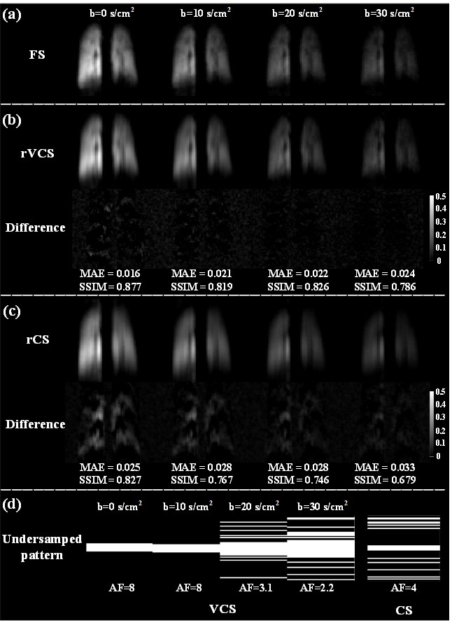
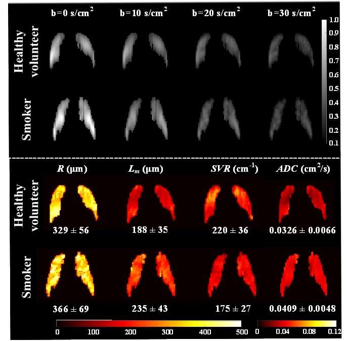
Figure 1 (a-c) showed the representative 129Xe DW images of the healthy volunteer acquired using the strategies of FS, rVCS and rCS. Obvious signal decay with increased b-values could be observed in all acquisition strategies. The rVCS images had better visual effect than the corresponding rCS images. When compared with FS images, images reconstructed with rVCS had lower MAE and higher SSIM than those with rCS. Meanwhile, increased MAE and decreased SSIM could be observed in the images with the larger b-values for both the methods. Figure 1 (d) showed the optimal k-space undersampling patterns used for VCS and CS simulations. Figure 2 showed the DW images obtained from a healthy volunteer and a cigarette smoker using prospective VCS acquisition. The pulmonary structure was preserved in both the healthy individual and the smoker. Compared to the healthy volunteer, heterogeneity and small ventilation defect regions could be observed in the lung of the cigarette smoker. Moreover, lower SVR and higher R, Lm, and ADC were found in morphometric parameter maps of the cigarette smoker. By using the VCS technique, significant differences were delineated between the pulmonary morphometric parameters of healthy volunteers and cigarette smokers (P < 0.001), while the acquisition time was reduced by four times.Discussion and Conclusion
A method employing VCS patterns for accelerating HP 129Xe DWI data acquisition was proposed. The optimal undersampling patterns of variable sampling ratios were obtained via retrospective simulations. Additionally, the proposed VCS method was also used for evaluating the pulmonary morphological changes caused by cigarette smoking in prospective study. Both the retrospective and prospective results demonstrated that the proposed VCS approach is capable of achieving comparable image quality and reliable pulmonary microstructural parameters with higher AFs.The proposed VCS method was able to accelerate the acquisition speed by four times while preserving good image details and quality. Our preliminary results demonstrated that the proposed method can be used for evaluating pulmonary injuries caused by cigarette smoking and may be helpful in diagnosing lung diseases in clinical practice.
Acknowledgements
This work is supported by National Natural Science Foundation of China (91859206, 21921004, 11905288, 81871321, 81930049, 82202119), National key Research and Development Project of China (2018YFA0704000), Key Research Program of Frontier Sciences (ZDBS-LYJSC004) and Scientific Instrument Developing Project of the Chinese Academy of Sciences (GJJSTD20200002, YJKYYQ20200067), CAS. Haidong Li acknowledges the support from Youth Innovation Promotion Association, CAS (2020330). Xin Zhou acknowledges the support from the Tencent Foundation through the XPLORER PRIZE.References
[1] Zhang H T, Xie J S, Xiao S, et al. Lung morphometry using hyperpolarized 129Xe multi-b diffusion MRI with compressed sensing in healthy subjects and patients with COPD. Med Phys, 2018, 45: 3097-3108.
[2] Li H D, Zhao X C, Wang Y J, et al. Damaged lung gas exchange function of discharged COVID-19 patients detected by hyperpolarized 129Xe MRI. Sci Adv, 2021, 7: eabc8180.
[3] Xie J S, Li H D, Zhang H T, et al. Single breath-hold measurement of pulmonary gas exchange and diffusion in humans with hyperpolarized 129Xe MR. NMR Biomed, 2019, 32: e4068.
[4] Kaushik S S, Cleveland Z I, Cofer G P, et al. Diffusion-weighted hyperpolarized 129Xe MRI in healthy volunteers and subjects with chronic obstructive pulmonary disease. Magn Reson Med, 2011, 65: 1154-1165.
[5] Bdaiwi A S, Niedbalski P J, Hossain M M, et al. Improving hyperpolarized 129Xe ADC mapping in pediatric and adult lungs with uncertainty propagation. NMR Biomed, 2022, 35: e4639.
[6] Ouriadov A, Guo F M, McCormack D G, et al. Accelerated 129Xe MRI morphometry of terminal airspace enlargement: Feasibility in volunteers and those with alpha-1 antitrypsin deficiency. Magn Reson Med, 2020, 84: 416-426.
[7] Chan H-F, Stewart N J, Parra-Robles J, et al. Whole lung morphometry with 3D multiple b-value hyperpolarized gas MRI and compressed sensing. Magn Reson Med, 2017, 77: 1916-1925.
[8] Chan H-F, Stewart N J, Norquay G, et al. 3D diffusion-weighted 129Xe MRI for whole lung morphometry. Magn Reson Med, 2018, 79: 2986-2995.
Figures