4097
Effects of patch size on 3D patch-based super-resolution reconstruction of hyperpolarized 13C cardiac MRI1Advanced Imaging Research Center, UT Southwestern Medical Center, Dallas, TX, United States, 2GE Healthcare, New York, NY, United States, 3Radiology, UT Southwestern Medical Center, Dallas, TX, United States
Synopsis
Keywords: Data Processing, Cardiomyopathy
The volumetric patch-based super-resolution method can reconstruct a single-slice low-resolution hyperpolarized 13C MRI to multiple 13C slices with enhanced spatial resolution by exploiting the corresponding high-resolution structural 1H MRI. In this study, the overall performance of this method and the effects of patch size were evaluated using a simulated digital phantom and an anthropomorphic cardiac 13C MR phantom. The optimal patch size for this reconstruction method was also applied to in-vivo human 13C cardiac images acquired with an injection of hyperpolarized [1-13C]pyruvate.Introduction
Carbon-13 (13C) MRI with hyperpolarized (HP) [1-13C]pyruvate captures key processes of glucose metabolism such as lactate fermentation and pyruvate oxidation of the heart1-4. However, the rapidly varying nature of HP signals and the quarter-size gyromagnetic ratio of 13C as compared to 1H restrict the spatial resolution of cardiac HP images to the order of centimeters, which is much larger than 1H cardiac images. Previously, we developed a two-dimensional5 and a volumetric6 patch-based super-resolution (PBSR) reconstruction algorithms to enhance the spatial resolutions of HP 13C MRI by exploiting high-resolution compartmental information collected from 1H patches. We noticed that the quality of the PBSR reconstructed images are sensitive to the patch size. In this study, we investigated the effects of patch size on 3D PBSR reconstruction for optimal reconstruction.Methods
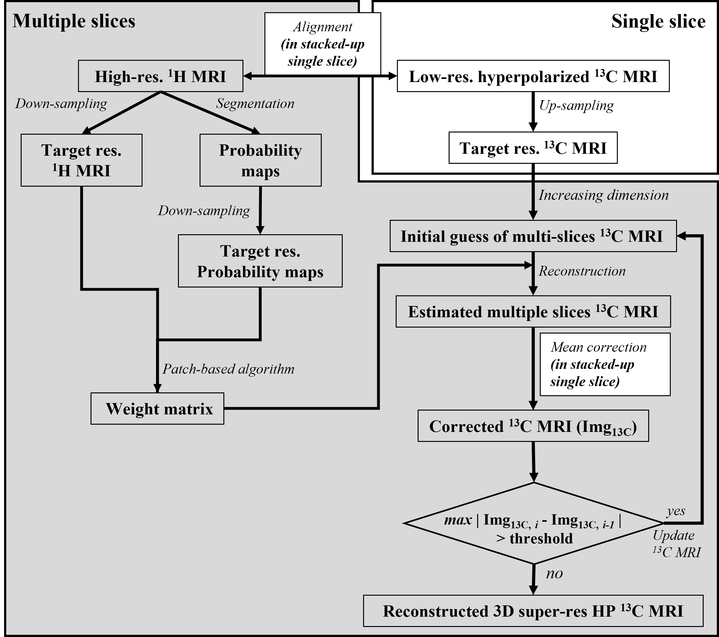
The reconstruction procedure is based on the previously presented 2D and 3D PBSR algorithms5, 6. Briefly, the process is composed of three steps as summarized in Figure 1. First, a patch-based weight matrix is calculated from the high-resolution segmented 1H MRI cardiac tissue compartments. Second, initial high-resolution multi-slice 13C images are estimated by interpolating the original 13C image. Finally, high-resolution multi-slice 13C images are iteratively updated by the guidance of the weight matrix and the subsequent mean correction until the incremental improvement is within a pre-determined threshold.The range of patch sizes was investigated from 3×3×3 to 19×19×9 of neighborhood voxels with an isotropic size increment of two. A digital phantom and an anthropomorphic MR phantom that contained [13C]bicarbonate were examined to identify a proper patch size, which was also applied to human HP 13C cardiac images.
a. Digital phantom
A numerical 3D digital phantom was generated with MATLAB (MathWorks, Natick, MA, USA)7. The phantom depicts a cross-sectional view of the left ventricle in the heart. Three major compartments are generated, including the ventricle, cardiac skeleton (myocardium), and its surrendering area in the chest. Both 1H (structural) and 13C (metabolic) images were created from this phantom.
b. 13C MR torso phantom
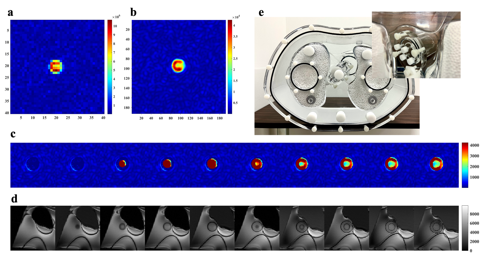
The torso phantom (Data Spectrum, Hillsborough, NC, USA) with a heart insert, filled with 0.4M [13C]bicarbonate. The 1H/13C-integrated imaging protocol included a 1H multi-slice 2D T1-weighted gradient echo (TE/TR=1.712/4.442ms, FOV=40×40cm2, matrix size=512×512, slices=3mm×10) and a 2D 13C single-slice metabolite-selective multi-echo spiral imaging (TE/TR=18.42/5000ms, FOV=40×40cm2, matrix=40×40, slice=30mm×1) along the short-axis view plane8.
c. In-vivo human HP cardiac 13C MRI
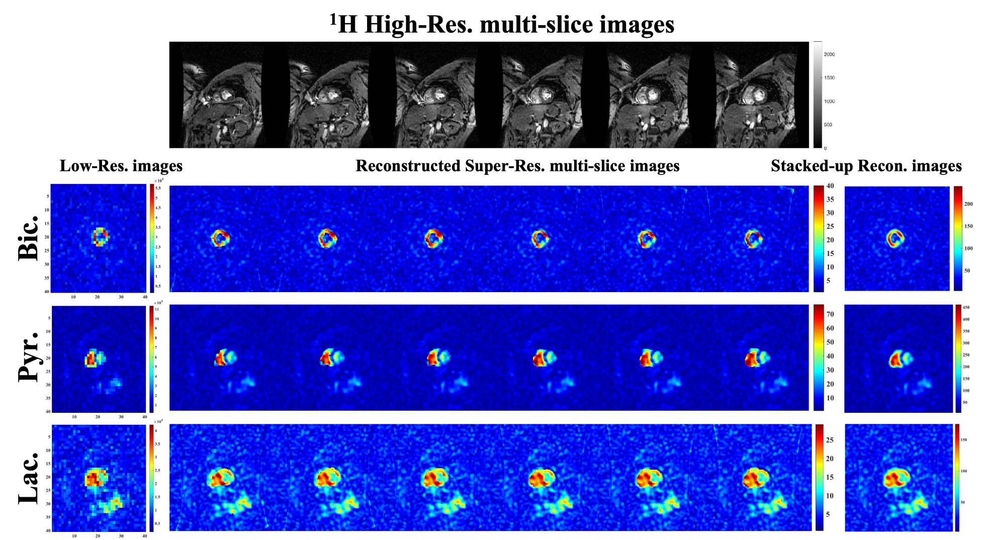
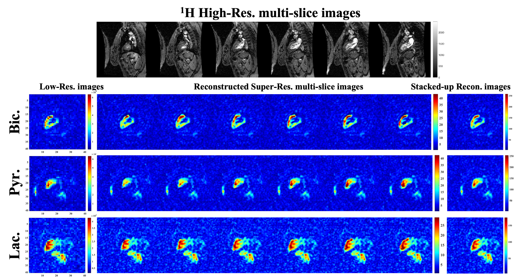
A set of cardiac images were acquired from a healthy volunteer (50 year old, female). For 1H imaging, the T1-weighted FIESTA was acquired with short-axis and long-axis views (TE/TR/IR=1.364/3.504/250ms, FOV=40×40cm2, matrix=256×256, slices=5mm×6). For 13C images, single-slice HP images of [13C]bicarbonate, [1-13C]lactate, and [1-13C]pyruvate were acquired 6s after an injection of 250mM HP [1-13C]pyruvate solution using a multi-echo spiral imaging sequence (single shot, FOV=40×40cm2, spatial resolution=1×1cm2, slice=30mm×1, flip angle=90° for bicarbonate, 90° for lactate, and 5° for pyruvate) in an interleaved spiral readouts every two R-R intervals8.
All MR images were acquired from a clinical 3T MRI scanner (GE Healthcare, Waukesha, WI, USA). The body coil was used for 1H RF excitation and data acquisition. A two-loop 13C transmit/receive Helmholtz coil was used for 13C (PulseTeq Limited, Chobham, Surrey, UK).
Results
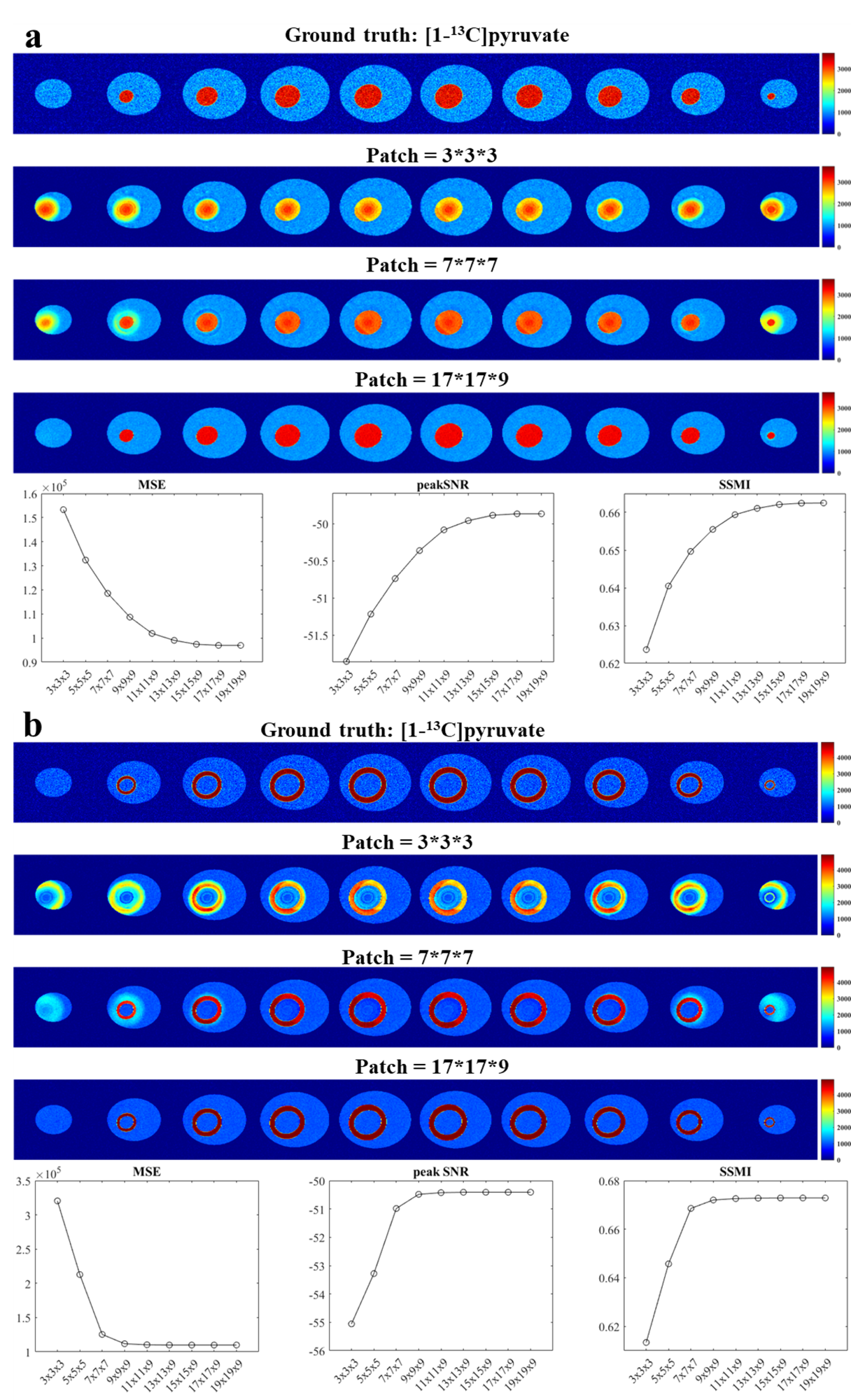
In the digital phantom, the reconstruction accuracy asymptotically improved as the patch size increased and was saturated with the patch size of 17×17×9 (Figure 2). The reconstructed images were evaluated using image quality metrics (MSE, SNRpeak, and SSMI) between the ground truths and the reconstructed images. All three indices showed a similar trend of gradual changes along with the increasing patch size and reached a stable level while the patch size was larger than 17×17×9 for pyruvate, and 9×9×9 for bicarbonate images. The reconstructed images from the torso phantom showed the correct structural distribution with the patch size of 5×5×5 (Figure 3). HP [13C]bicarbonate, [1-13C]pyruvate, and [1-13C]lactate metabolic maps of the human heart in the short axis (Figure 4) and the long axis planes (Figure 5) were also reconstructed using the PBSR with a 5×5×5 patch. High structural similarity in reconstructed 13C images in torso phantom images (SSIM = 0.9943) and human cardiac images (SSIM>0.97 for all metabolites and view planes) when compared to low-resolution 13C images. The entropy of 13C images was higher in the digital phantom (6.7557) than in the torso phantom images (4.6309) and cardiac images (5.2492).Discussion
A larger patch reconstructs more accurate images since the PBSR algorithm considers the signal continuity and redundancy between two neighboring pixels, and it includes more neighborhood information9. In addition, the saturating patch size depends on the dimensions of input image and reconstructed image. A large ratio of reconstruction-to-input size requires high computation complexity and therefore a large patch size10. The digital phantom required a 17×17×9 patch for the ratio of 1.6 (256/160), while a 5×5×5 patch was sufficient for the torso phantom and human cardiac images whose ratio was 1.2 (192/160). In addition, the higher entropy of the low-resolution image for the PBSR might require the larger patch because of the image complexity.Conclusion
The current investigation demonstrated the optimal patch size to the PBSR would depend on the ratio of reconstruction-to-input size.Acknowledgements
National Institute of Health: R01NS107409, R21EB030765, P30DK127984, P41EB015908 Department of Defense: W81XWH2210485 The Welch Foundation: I-2009-20190330.References
1. Cunningham CH, Lau JY, Chen AP, Geraghty BJ, Perks WJ, Roifman I, et al. Hyperpolarized 13C Metabolic MRI of the Human Heart: Initial Experience. Circ Res. 2016;119(11):1177-82.
2. Rider OJ, Apps A, Miller J, Lau JYC, Lewis AJM, Peterzan MA, et al. Noninvasive In Vivo Assessment of Cardiac Metabolism in the Healthy and Diabetic Human Heart Using Hyperpolarized (13)C MRI. Circ Res. 2020;126(6):725-36.
3. Apps A, Lau JYC, Miller J, Tyler A, Young LAJ, Lewis AJM, et al. Proof-of-Principle Demonstration of Direct Metabolic Imaging Following Myocardial Infarction Using Hyperpolarized 13C CMR. JACC Cardiovasc Imaging. 2021;14(6):1285-8.
4. Park JM, Reed GD, Liticker J, Putnam WC, Chandra A, Yaros K, et al. Effect of Doxorubicin on Myocardial Bicarbonate Production From Pyruvate Dehydrogenase in Women With Breast Cancer. Circ Res. 2020;127(12):1568-70.
5. Ma J, Park JM. Super-Resolution Hyperpolarized (13)C Imaging of Human Brain Using Patch-Based Algorithm. Tomography. 2020;6(4):343-55.
6. Sung-Han Lin JJM, Jae Mo Park, editor Volumetric patch-based image reconstruction for enhancing hyperpolarized 13C MRI with hybrid image guidance. ISMRM Annal Meeting 2022 07-12 May 2022; London, UK.
7. Schabel M. 3D Shepp-Logan phantom: MATLAB Central File Exchange; 2022 [Retrieved October 12, 2022.]. Available from: (https://www.mathworks.com/matlabcentral/fileexchange/9416-3d-shepp-logan-phantom),.
8. Ma J, Chen J, Reed GD, Hackett EP, Harrison CE, Ratnakar J, et al. Cardiac T 2 * measurement of hyperpolarized (13) C metabolites using metabolite-selective multi-echo spiral imaging. Magn Reson Med. 2021;86(3):1494-504.
9. Rousseau F. Brain Hallucination. Springer Berlin Heidelberg; 2008. p. 497-508.
10. Zhao Y, Wang R, Jia W, Yang J, Wang W, Gao W. Local patch encoding-based method for single image super-resolution. Information Sciences. 2018;433-434:292-305.
Figures