4094
3D Pulmonary Dynamic Ventilation Imaging with High Spatial and Temporal Resolution Using Hyperpolarized 129Xe MRI1Key Laboratory of Magnetic Resonance in Biological Systems, State Key Laboratory of Magnetic Resonance and Atomic and Molecular Physics, National Center for Magnetic Resonance in Wuhan, Wuhan Institute of Physics and Mathematics, Innovation Academy for Precision Measurement Science and Technology, Chinese Academy of Sciences- Wuhan National Laboratory for Optoelectronics, Wuhan, China, 2University of Chinese Academy of Sciences, Beijing, China
Synopsis
Keywords: Pulse Sequence Design, Hyperpolarized MR (Gas)
The mutual restriction of temporal and spatial resolution is a challenge for hyperpolarized gas dynamic MRI, especially for 3D acquisition. Herein, we proposed a methodology to image the pulmonary dynamic ventilation with high temporal and spatial resolution using hyperpolarized gas MRI based on a 3D gradient echo sequence. Furthermore, we observed the difference of pulmonary dynamic ventilation in the posterior-anterior direction.Introduction
Abnormal pulmonary ventilation is closely associated with pathological changes of the lung. Noninvasively imaging dynamic ventilation of the lung with both high temporal and spatial resolution is of great demand for diagnosing lung diseases that related to obstructive ventilation. Hyperpolarized (HP) 129Xe gas MRI is a promising and valuable tool for evaluation structure and function changes with lung diseases without invasion and ionizing radiation1-3. Unfortunately, it is a challenge to obtain dynamic images with high temporal and spatial resolution due to the short respiratory cycle. Moreover, the unrecoverable magnetization of HP gas, the depolarized by continuous RF pulses and the longitudinal relaxation in the lung make it difficult to acquire enough images with high quality in the limited respiratory cycle. In this study, we presented a multi-respiration acquisition scheme for 3D imaging of the pulmonary dynamic ventilation that enables to enhance the temporal resolution up to 44 ms with spatial resolution of 0.5 × 0.8 × 1.9 mm3.Methods
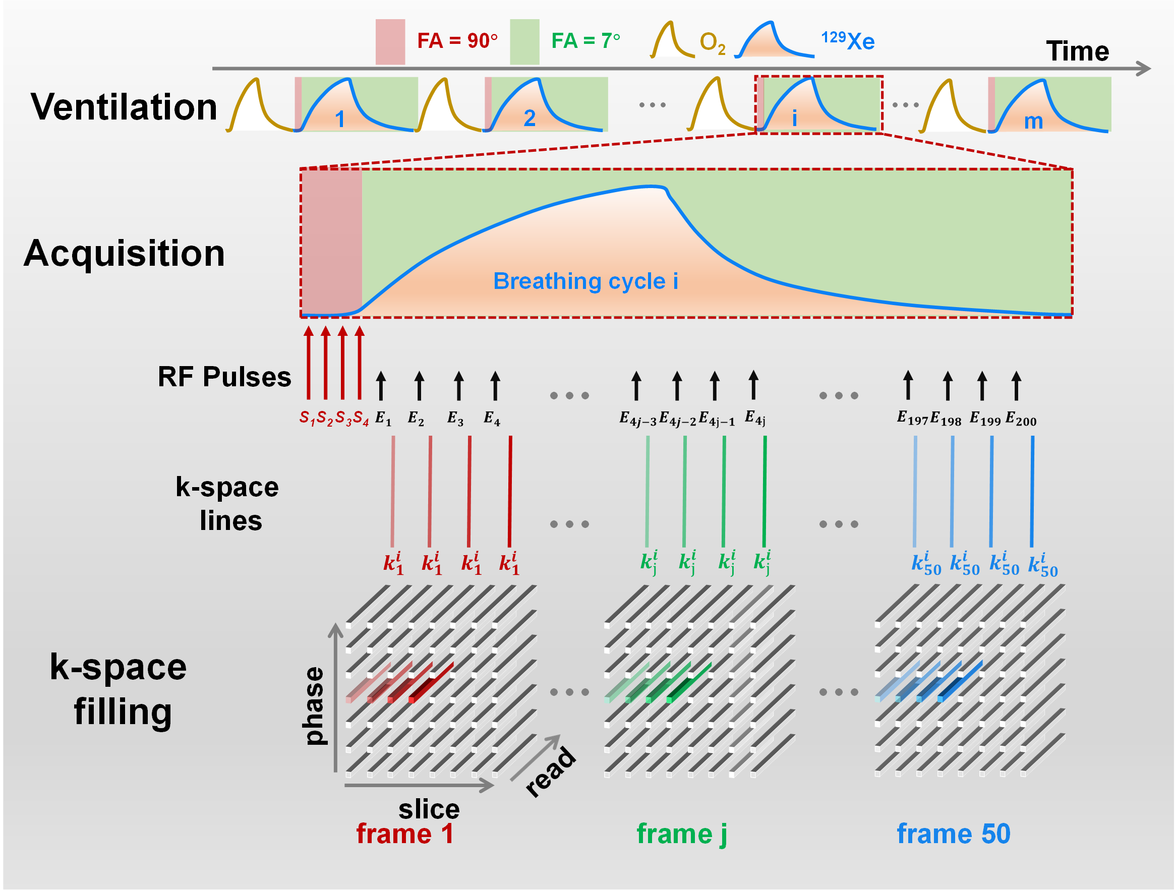
The specific multi-respiration acquisition scheme, including ventilation, acquisition and k-space filling strategies, were shown in Figure 1. We exploited a modified gradient echo sequence that allowed exchange the number and the order of repetition and phase-encoding loops. The main idea of the proposed method is that repeatedly acquiring partial k-space lines in one respiratory cycle (eight lines in experiment) and acquiring the other k-space lines in other respiratory cycles. To maintain normal physiological state, one oxygen gas breath is used between every two 129Xe breathes. Before the data acquisition for each respiratory cycle, four 90° saturation RF pulses were applied to destroy the residual HP gas magnetization in the last adjacent respiratory cycle. Then, all the k-space data were filled into the corresponding frames according to the period in one respiratory cycle as shown in Figure 1, and the dynamic images were obtained with high temporal resolution, which equals to four TRs. For the 3D MRI experiments, the following parameters were used: number of frames (number of repetitions) = 50, TR/TE = 5.5/1.5ms, gauss RF pulse duration = 0.5 ms, flip angle = 7°, matrix = 96 × 64 × 16, FOV = 50 × 50 × 30 mm3, bandwidth = 50 kHz.Results
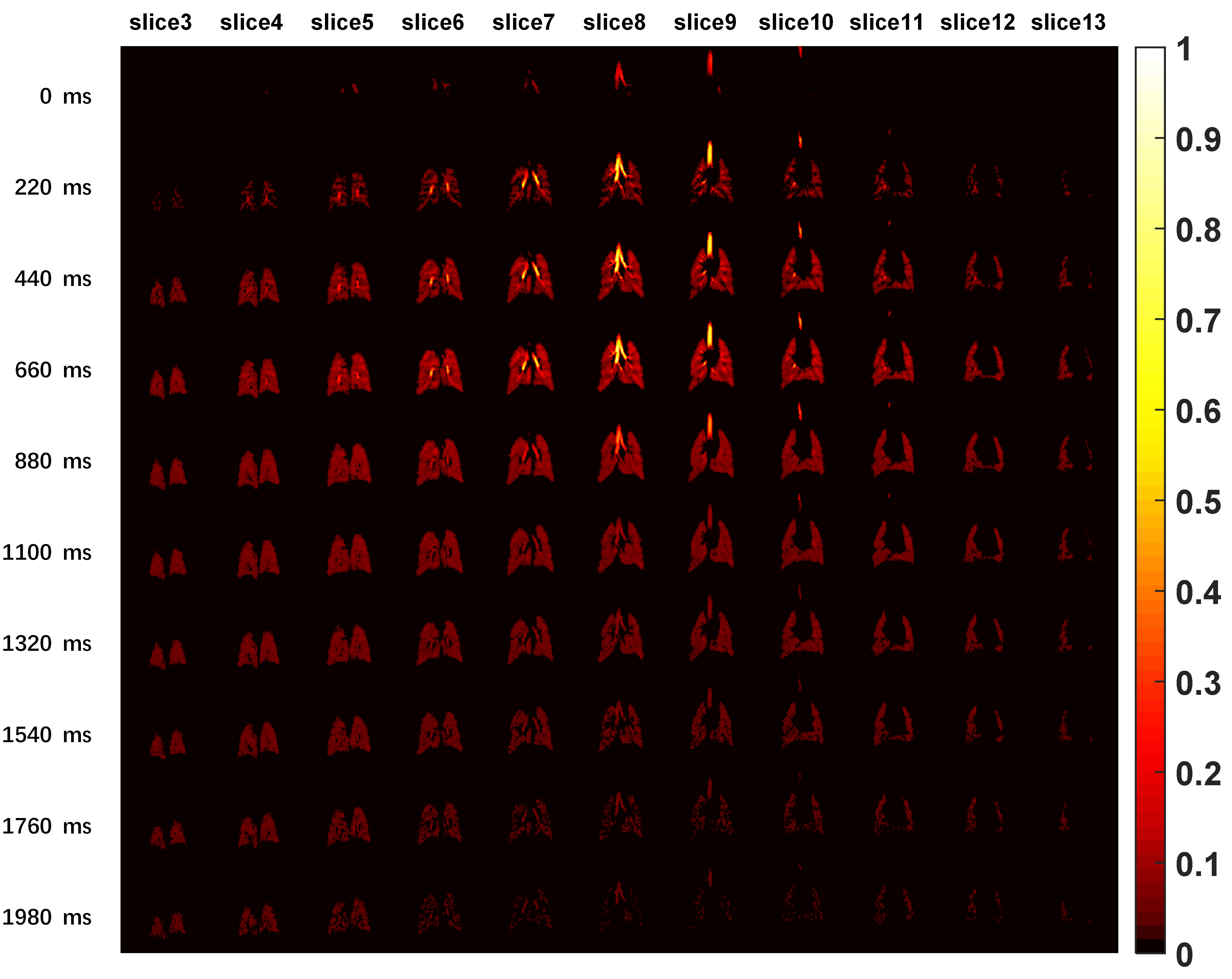
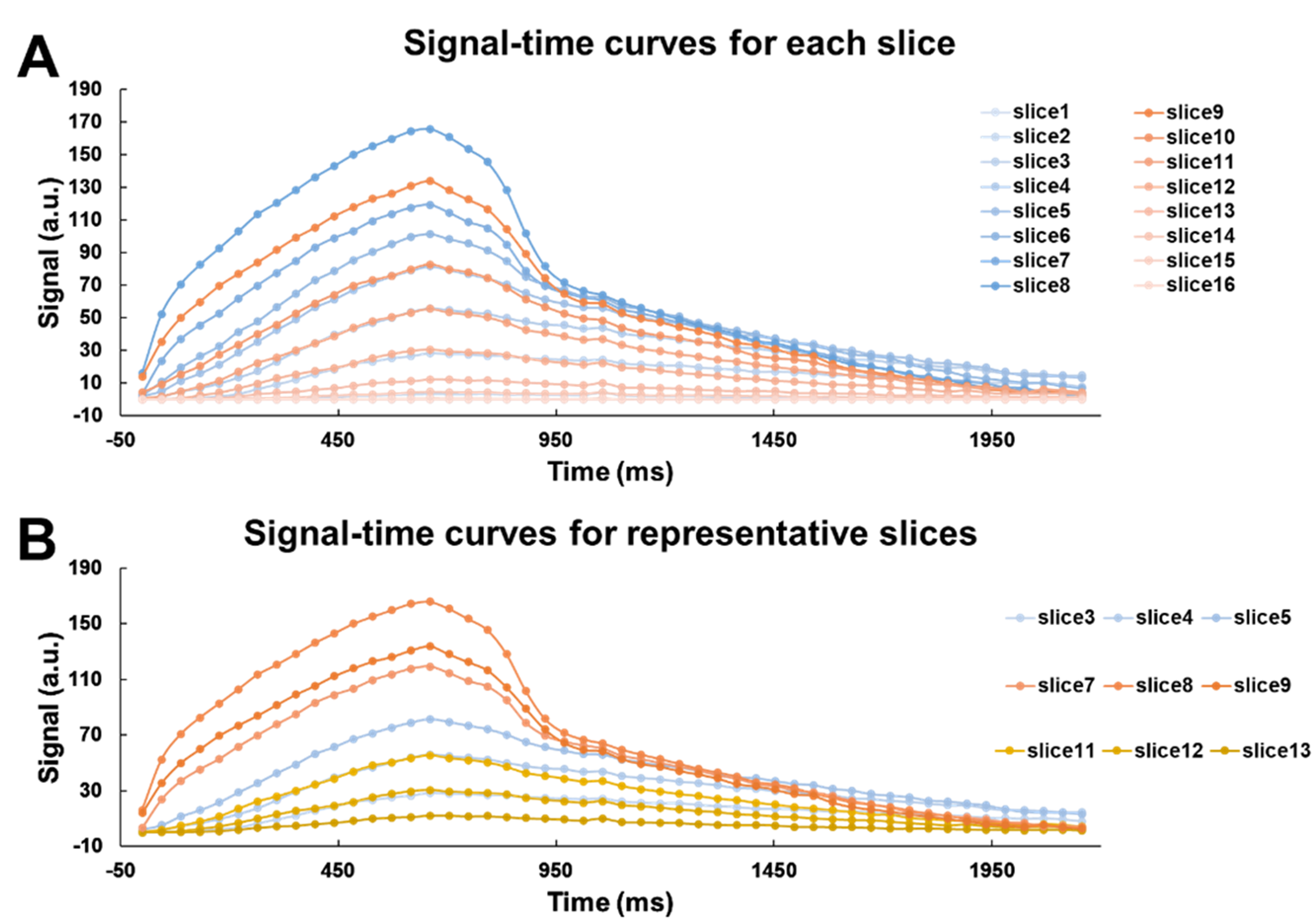
Figure 2 showed the representative 3D dynamic ventilation images obtained with the proposed method from a healthy rat. Benefiting from the high temporal and spatial resolution (44 ms and 0.5 × 0.8 × 1.9 mm3, respectively), we could observe the dynamic ventilation information slice by slice. It was clearly found that the signal intensity in the trachea and the bronchi was much higher than that in the lung parenchyma during the inspiration, but opposite trends were observed during the expiration. Figure 3A showed the change of the signal intensity over time for all slices. We could find that the airflow rate of the center slices (slice7~9) was faster than that in the anterior slices (slice11~13) and posterior slices (slice3-5), as shown in Figure 3B.Discussion and Conclusion
A method for directly imaging the pulmonary dynamic ventilation using HP 129Xe MRI was proposed, and 3D dynamic ventilation images with a temporal resolution of 44 ms and a spatial resolution of 0.5 × 0.8 × 1.9 mm3 were obtained. The difference of pulmonary dynamic ventilation in the posterior-anterior direction was probably because of the supine position and the dependence of gravity gradient. Our preliminary results demonstrated the feasibility of the proposed method for imaging pulmonary dynamic ventilation with high temporal-spatial resolution using HP gas MRI in vivo, which is promising for early diagnosis of lung diseases that related to dynamic ventilation abnormalities, for instance COPD4 and asthma5.Acknowledgements
This work is supported by National Natural Science Foundation of China (91859206, 21921004, 11905288, 81871321, 81930049, 82202119), National key Research and Development Project of China (2018YFA0704000), Key Research Program of Frontier Sciences (ZDBS-LYJSC004) and Scientific Instrument Developing Project of the Chinese Academy of Sciences (GJJSTD20200002, YJKYYQ20200067), CAS. Haidong Li acknowledges the support from Youth Innovation Promotion Association, CAS (2020330). Xin Zhou acknowledges the support from the Tencent Foundation through the XPLORER PRIZE.
References
1. Zhang M, Li HD, Li HC, Zhao XC, Zhou Q, Rao QC, et al. Quantitative evaluation of lung injury caused by PM2.5 using hyperpolarized gas magnetic resonance. Magn Reson Med. 2020;84(2):569-78.
2. Li HD, Zhang ZY, Zhao XC, Han YQ, Sun XP, Ye CH, et al. Quantitative evaluation of pulmonary gas-exchange function using hyperpolarized 129Xe CEST MRS and MRI. NMR Biomed. 2018;31(9):e3961.
3. Li HD, Zhang ZY, Zhao XC, Sun XP, Ye CH, Zhou X. Quantitative Evaluation of Radiation-Induced Lung Injury with Hyperpolarized Xenon Magnetic Resonance. Magn Reson Med. 2016;76(2):408-16.
4. Zhang HT, Xie JS, Xiao S, Zhao XC, Zhang M, Shi L, et al. Lung morphometry using hyperpolarized 129Xe multi-b diffusion MRI with compressed sensing in healthy subjects and patients with COPD. Med Phys. 2018;45(7):3097-108.
5. McIntosh MJ, Kooner HK, Eddy RL, Jeimy S, Licskai C, Mackenzie CA, et al. Asthma control, airway mucus, and 129Xe MRI ventilation after a single benralizumab dose. Chest. 2022.
Figures