4090
Susceptibility Managed Optimization (SUMO) : Very HIgh order shimming for Single Voxel Spectroscopy at 7T1Resonance Research Inc., Billerica, MA, United States, 2Radiology, University of Missouri, Columbia, MO, United States
Synopsis
Keywords: Shims, Shims
B0 shimming for single voxel spectroscopy (SVS) is of importance due to its role in determining linewidth, accuracy of the analysis and interpretation. However, for difficult brain regions, such as the prefrontal cortex, where shifts of 3-6mm in voxel position can dramatically alter the achieved linewidth in SVS, the lack of a reliable method to predict the achievable homogeneity results in failed studies and/or increased study times. In this work we demonstrate an objective and non-iterative method (SUMO) for voxel positioning that predicts and ensures adequate B0 homogeneity is achievable.Introduction:
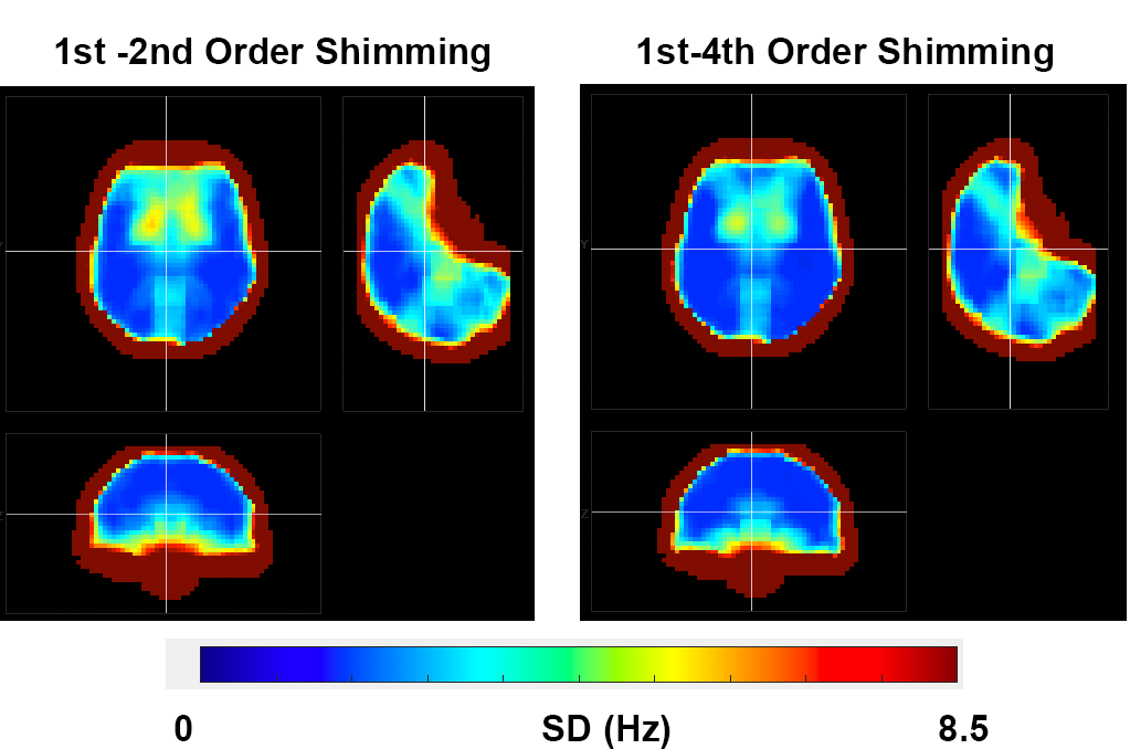
Introduction: B0 shimming for single voxel spectroscopy (SVS) is of importance due to its role in determining linewidth, accuracy of the analysis and interpretation. With relatively small volumes, ~8cc, B0 variation is expected to be small and shimming with 2nd order spherical harmonics is generally thought to be sufficient. However, this assumption may not be valid in the anterior and inferior portions of the frontal and temporal lobes and some regions adjacent to the skull (Fig. 1). Methods to map the field in such regions with rapidly spatially varying B0 fields have also been thought to be problematic due to limited accuracy of many 3D B0 mapping methods, limiting the predictability of shimming. On a practical basis in difficult neocortical regions without clear landmarks and the probabilistic methods used in determining neocortical regions of interest (such as the prefrontal cortex, PFC), repositioning the voxel by a few mm can significantly improve B0 homogeneity. With the alternative being discarding poor spectral quality data altogether, shifting the voxel position is the best option although costly insofar as the imaging time needed for the repositioning, shimming and reacquiring the data. However, we hypothesize that after collection of a single B0 map of sufficient accuracy spanning the entire brain, the achievable B0 homogeneity for a voxel of arbitrary size, shape and location can be predicted and will ensure that adequate homogeneity is achieved.The goals of this work were to: 1) accurately predict and achieve higher order shimming (1st-4th order) for SVS studies; 2) objectively and efficiently position the voxel to achieve a target B0 homogeneity and 3) validate these methods in the PFC using LC modeling1 of SVS measurements at 7T in the human brain.
Methods:
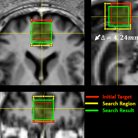
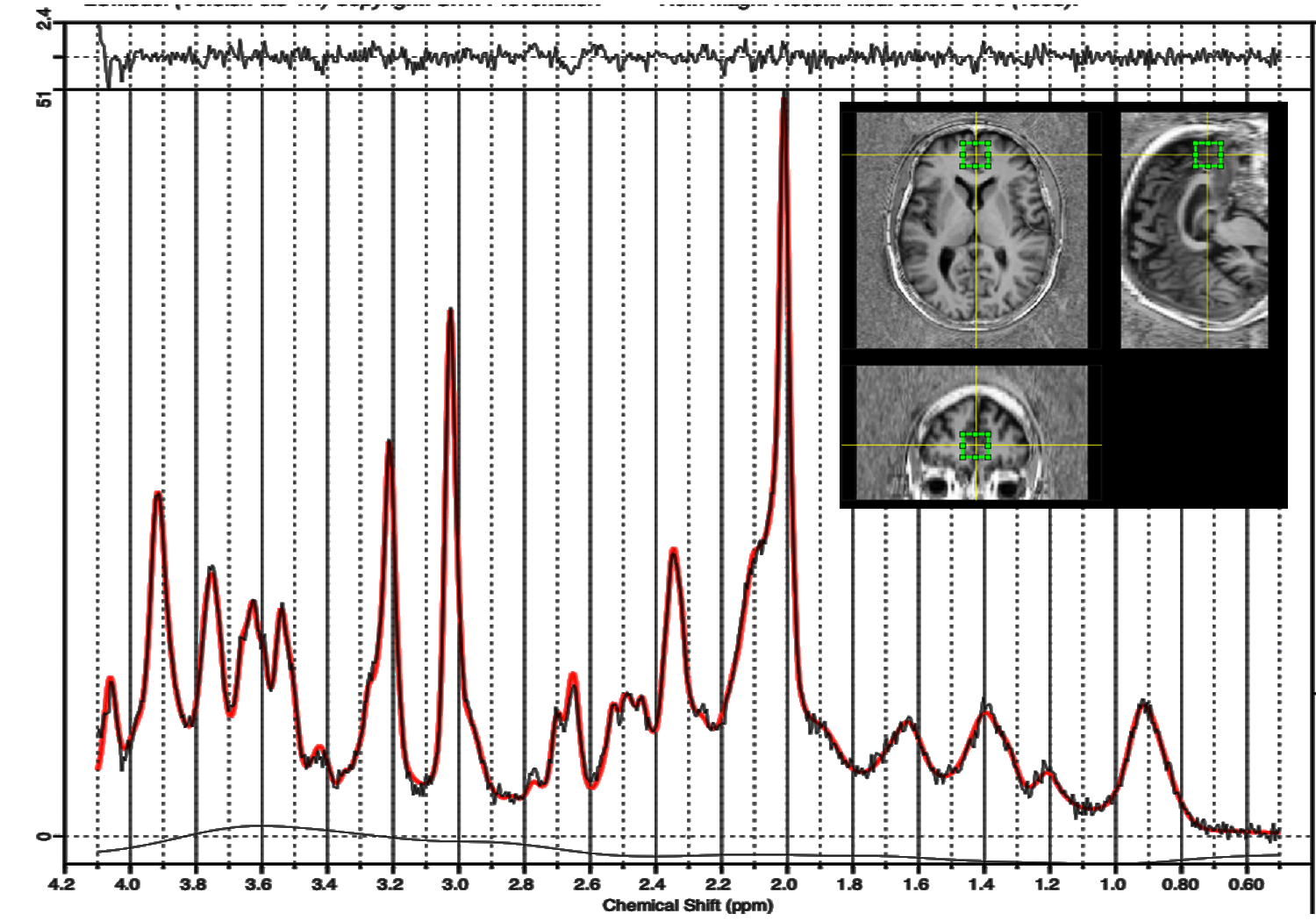
Data was acquired from 6 healthy subjects using a Siemens 7T Terra with a 16-channel transceiver array, and a 3rd and 4th order shim insert from Resonance Research Inc.2-4. For anatomical visualization and positioning, a 1mm isotropic MP2RAGE image was acquired. Whole brain B0 field maps at 3mm isotropic resolution (216x216x123mm3 FOV) were acquired using BOLERO5. This B0 field map enables prediction of the achievable field homogeneity after localized shimming for arbitrary voxel sizes and positions over the entire brain. For the PFC, where small displacements on the order of 3-6mm can have significant effects on the achievable linewidth after shimming, the voxel position was optimized using Susceptibility Managed Optimization, SUMO. This process selects the voxel location by 1) manually selecting an initial target location (Fig. 2 red box), 2) defining a grid of potential voxel locations about the target location (Fig. 2 yellow box), 3) calculating the achievable homogeneity over that grid and 4) automatically selecting the nearest location to the target that achieves the user-determined homogeneity threshold (Fig. 2, green box). In this work a threshold of 5Hz SD was used. The location was shimmed (1st-4th order), SVS data acquired, and verification B0 maps were acquired to document the achieved homogeneity. SVS data (20x20x20mm3) was acquired using a STEAM sequence6 (TR/TE/TM/NA of 6sec/8ms/20ms/32 and analyzed with LC Model1.Results:
Displayed in Fig.1 are color-coded B0 (3mm isotropic resolution) images showing the predicted SD after 1st-2nd and 1st-4th order shimming (voxel 20x20x20mm3). As described, small displacements of a single or several B0 map pixels (3mm) can decrease the linewidth by 50% or more (~10Hz reduction in FWHM). To demonstrate this effect, we evaluated the PFC with a 3x3 search grid in the AP and HF directions (Fig. 2). The RL position was fixed so that the voxel remained along the mid-line. Across 6 subjects, the predicted SD from the initial target location was 8.9+3.8Hz using 1st-4th order shimming. Application of SUMO using a threshold of 5Hz SD for 1st-4th order shimming, identified at least one acceptable voxel of <5Hz SD in 5 of 6 subjects (in the remaining subject the minimum SD within the search grid was 5.3Hz over the target ROI, which was used). The average distance between the initial target and the closest voxel meeting the threshold SD was 4.2±2.3mm. The measured SD after 1st-4th order shimming was 4.0+0.9Hz in excellent agreement with the predicted SD, 4.5+0.9Hz, calculated prior to shimming, demonstrating the accuracy of the B0 maps. The predicted SD after 1st-2nd order shimming from this location was 7.3+2.5Hz, indicating the presence of inhomogeneities >2nd order in the SUMO selected voxel. LC Model analysis of the acquired spectra yielded a mean FWHM linewidth of 10.7+1.5Hz. Assuming a gaussian line-shape, this is equivalent to an SD of 4.6+0.6Hz (Fig. 3). The predicted and achieved linewidths for 1st-4th order shimming calculated from the B0 maps, assuming gaussian line shapes, are 10.5+2.1 and 9.5+2.1 respectively, which are in excellent agreement with the LC Model calculated linewidth from the spectra, 10.7+1.5Hz.Discussion/Conclusions:
We have demonstrated that small spatial displacements (3-6mm) in difficult brain regions such as the PFC can have dramatic effects on shimming. BOLERO B0 maps can accurately predict the achievable homogeneity which is in good agreement the measured linewidths in the acquired spectra. Thus, SUMO can provide an objective and non-iterative method for voxel positioning to ensure spectral quality.Acknowledgements
NIH R01 – EB024408References
1. Provencher SW. Automatic quantitation of localized in vivo 1H spectra with LCModel. NMR in biomedicine. 2001;14(4):260-4.2.
2. Pan JW, Lo KM, Hetherington HP. Role of very high order and degree B0 shimming for spectroscopic imaging of the human brain at 7 tesla. Magnetic resonance in medicine. 2012;68(4):1007-17.3.
3. Avdievich NI. Transceiver-Phased Arrays for Human Brain Studies at 7 T. Applied magnetic resonance. 2011;41(2-4):483-506.4.
4. Li X, Pan JW, Avdievich NI, Hetherington HP, Rispoli JV. Electromagnetic simulation of a 16-channel head transceiver at 7 T using circuit-spatial optimization. Magnetic resonance in medicine. 2021;85(6):3463-78.5.
5. Hetherington HP, Chu WJ, Gonen O, Pan JW. Robust fully automated shimming of the human brain for high-field 1H spectroscopic imaging. Magnetic resonance in medicine. 2006;56(1):26-33.6.
6. Li Y, Xu D, Ozturk-Isik E, Lupo JM, Chen A, Vigneron D, et al. T1 and T2 Metabolite Relaxation Times in Normal Brain at 3T and 7T. J Mol Imaging Dynam. 2012;S1:002.:5.
Figures