4074
Investigating the impact of head position on the spatial distribution of B1+ field for a 60-ch Tx coil at 7T1University of Pittsburgh, Pittsburgh, PA, United States
Synopsis
Keywords: High-Field MRI, Simulations, Head position, B1+ field, 7T MRI
In this work, we computationally and experimentally evaluated the B1+ field and SAR generated by a 60-channel Tic-Tac-Toc RF coil at 7T MRI scanner on a variety of participants, head models and head positions. On three anatomically detailed human head models, we found slight changes in the intensity and the distribution of B1+ field and SAR over the whole brain when the head was moved out from the top of the coil. This was confirmed in both simulation and experimental B1+ maps. Our results highlighted the impact of imaging setup on imaging quality.Introduction
To improve the imaging quality under ultra-high field, the Tic-Tac-Toe (TTT) radiofrequency (RF) head coil design for 7T MRI have been devised to increase B1+ homogeneity and have demonstrated load insensitivity 1-3. This has been further improved with the new 60-channel double-row TTT coil system4. However, the impact of imaging setup, such as the spatial position of the head, on imaging quality is still under investigation. Thus, in this study, we assessed the impact of the head position on the B1+ field distribution/intensity, attempting to provide better understanding of imaging strategy and guidance of examination setup.Methods
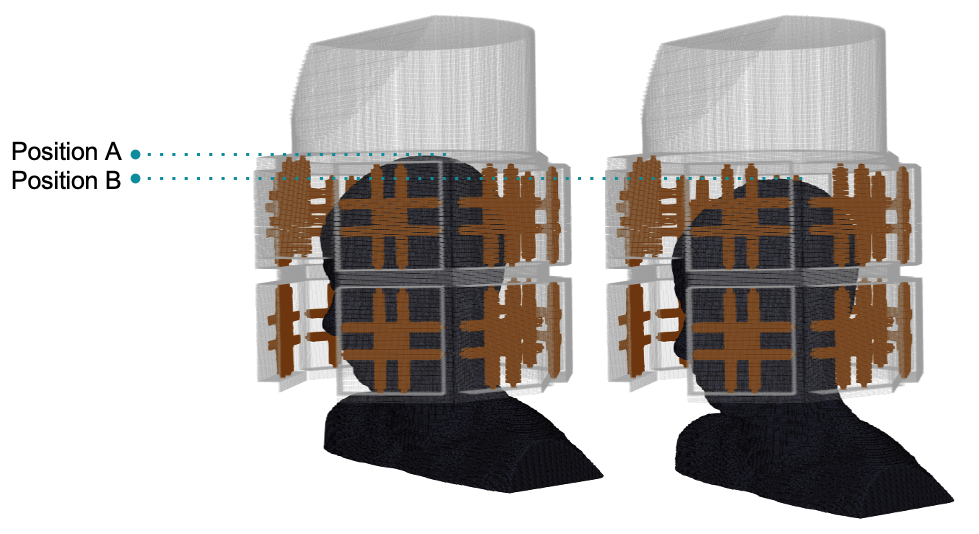
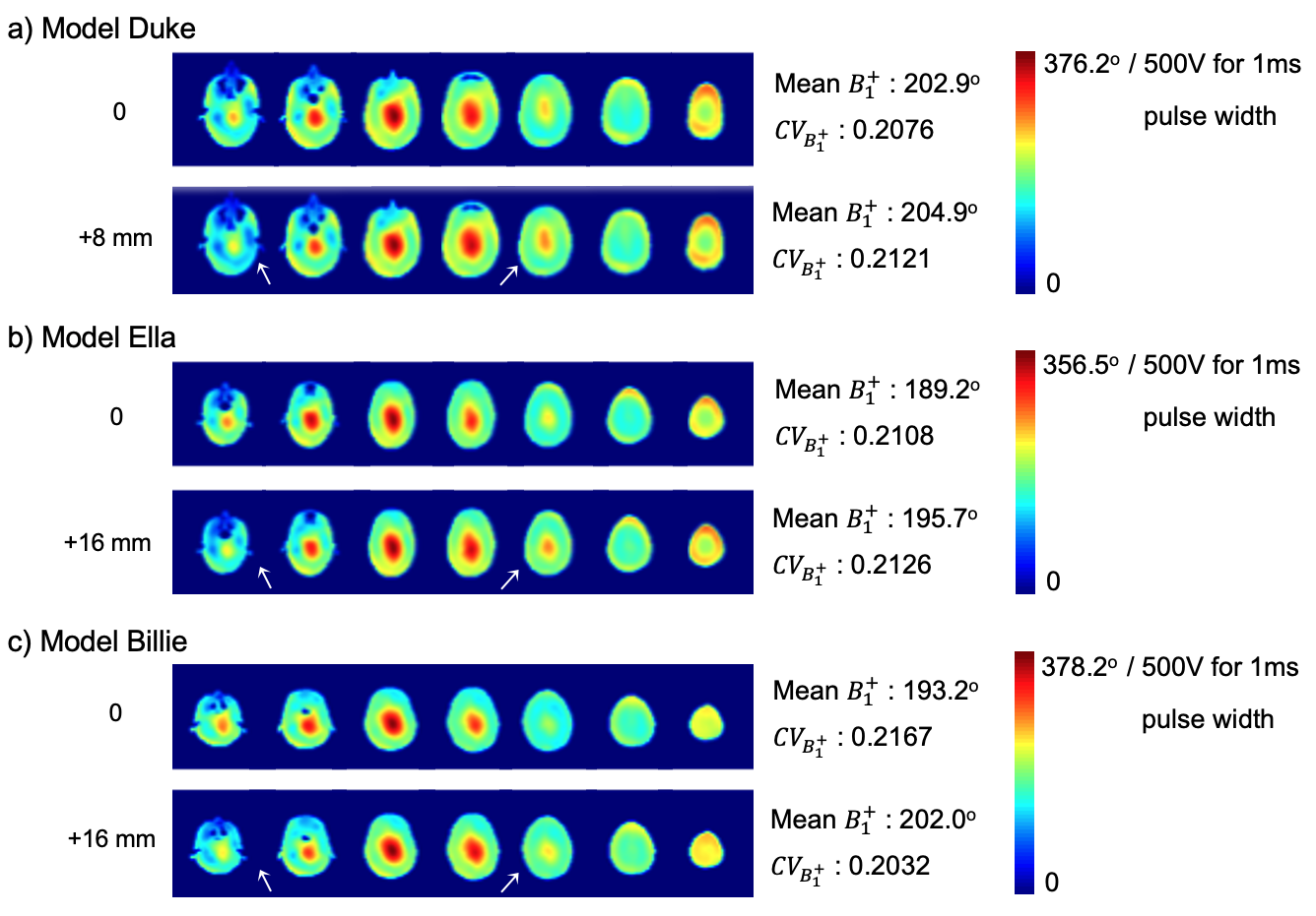
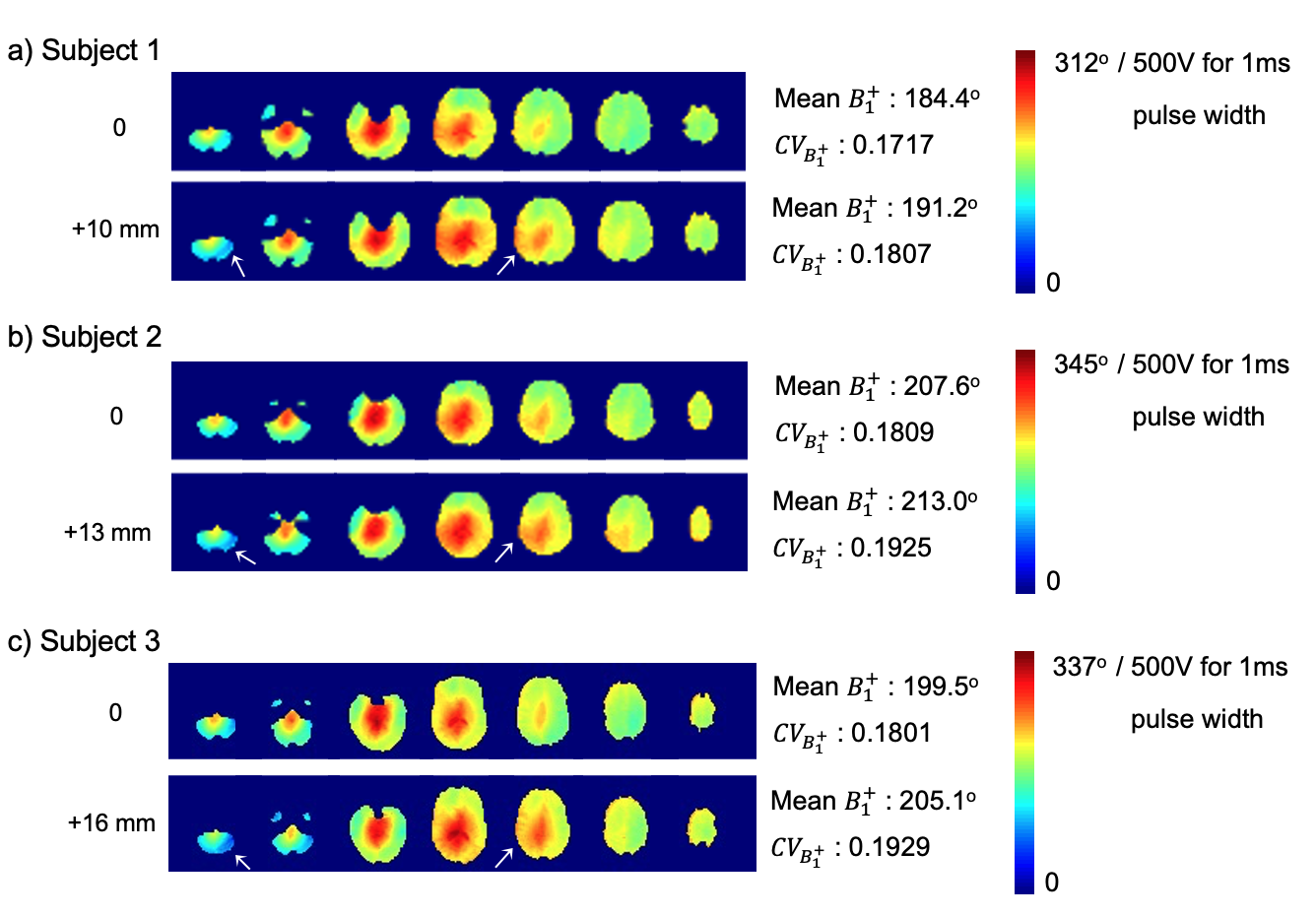
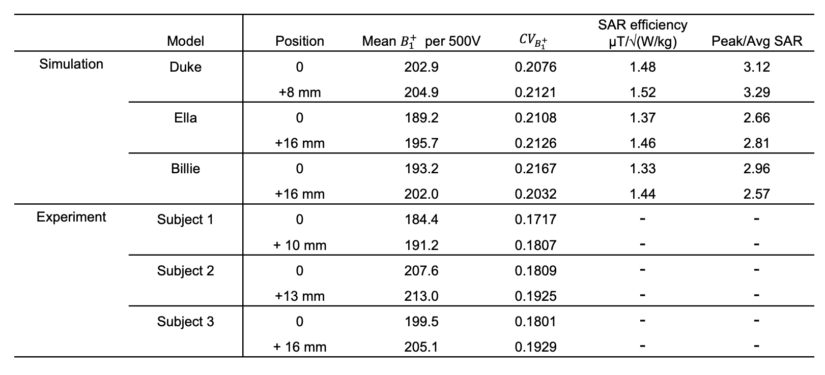
To computationally simulate the influence of head position on the B1+ field, electromagnetic fields produced on the geometry of the 60-channel octagonal Tic-Tac-Toc RF head coil were simulated with an in-house Finite-Difference Time-Domain (FDTD) algorithm. The simulations were performed by using three different anatomically detailed head models including the Duke (34 years old male, 155 lbs), Ella (26 years old female, 126 lbs), and Billie (11 years old female, 75 lbs) models from the Virtual Family5. We adopted two different head positions (Figure 1). For the Duke model, we moved out 8 mm from the reference position (i.e. the position very close to the coil). For the Ella and Billie models, we moved out 16mm from the reference position. To analyze and compare B1+ field and SAR distributions, we calculated mean B1+ value, coefficients of variation(CV) of B1+ field, specific absorption rate (SAR) efficiency and peak/average SAR over the whole brain including the cerebellum. Experimentally, a 12-minute Turbo-FLASH sequence (TR/TE = 2000/1.16ms, flip angle from 0° to 90° in 18° increments, 3.2mm isotropic resolution) was used to obtain in vivo B1+ maps, which were acquired from three participants with the similar experimental position to the simulation to evaluate the real-world scenario.Results
The simulated B1+ fields of three head models were visualized in Figure 2. We observed that all three models had a higher average B1+ field over the whole brain when the head was moved out from the top of the coil (position B in Figure 1). Regionally speaking, there existed a higher B1+ field at the superior region of the brain and lower B1+ field in the cerebellum when moving out the head. Also, it showed that the B1+ field became slightly less homogeneous when the position was moved out, which was indicated by higher coefficients of variation of B1+ field (Figure 2). Besides, the SAR efficiency and peak/average SAR of simulations were assessed (Table 1); the outer position provided better SAR efficiency compared to the reference position. Moreover, the experimental in vivo B1+ maps demonstrated consistent findings with the simulated outcomes (Figure 3 & Table 1), confirming the effect of head position under the scanning of ultra-high fields.Discussion and Conclusion
In this study, we found that the head position would slightly affect the behavior of B1+ field at 7T MRI; the head at an outer position would have a relatively higher average B1+ value, but it would be less homogeneous. Also, the regional difference of B1+ field would be also affected by the position. Our result suggested that the position of scanning objects had a certain impact on imaging quality at 7T MRI. The examination setup should consider the regions of interest to optimize imaging strategies under ultra-high field, especially for repeated measures in a longitudinal study.Acknowledgements
This work was supported by NIH R01MH111265, R01AG063525, R56AG074467, and T32MH119168. This research was also supported in part by the University of Pittsburgh Center for Research Computing (CRC) through the resources provided.References
[1] Krishnamurthy, N. et al. Computational and experimental evaluation of the Tic-Tac-Toe RF coil for 7 Tesla MRI. PLOS ONE 14, e0209663 (2019).
[2] Santini, T. et al. 64-channel Double-Octagon Tx Head Coil for 7T Imaging. The International Society of Magnetic Resonance in Medicine (ISMRM) 2017 Annual Meeting. https://archive.ismrm.org/2017/4308.html.
[3] Sajewski, A. N. et al. Impact of Coupling on the B1+ Field Produced by a 15-panel TicTac-Toe RF Array. The International Society of Magnetic Resonance in Medicine (ISMRM) 2022 Annual Meeting. https://archive.ismrm.org/2022/1544.html
[4] Sajewski, A. N. et al. Anti-claustrophobic 7T Tic-Tac-Toe RF coil system with 60/32 Transmit/Receive channels. 2022 In Vivo Magnetic Resonance Gordon Research Conference
[5] Christ, A. et al. The Virtual Family--development of surface-based anatomical models of two adults and two children for dosimetric simulations. Phys. Med. Biol. 55, N23-38 (2010).
Figures