4073
Simultaneous multi-slice 23Na imaging using radial CAIPIRINHA1Medical Physics in Radiology, German Cancer Research Center (DKFZ), Heidelberg, Germany, 2Faculty of Physics and Astronomy, University of Heidelberg, Heidelberg, Germany, 3University Hospital Erlangen, Institute for Radiology, Friedrich‐Alexander‐Universität Erlangen‐Nürnberg (FAU), Erlangen, Germany, 4Physikalisch-Technische Bundesanstalt (PTB), Braunschweig and Berlin, Germany, 5Faculty of Medicine, University of Heidelberg, Heidelberg, Germany
Synopsis
Keywords: High-Field MRI, Non-Proton, 23Na,SMS,radial CAIPIRINHA
In this work, a simultaneous multi-slice (SMS) approach using radial CAIPIRINHA was implemented for sodium MRI. Initial experiments were confined to two simultaneously excited slices. Here, simulated and measured slice profiles agreed well with expectations. Further, the SNR of the acquired SMS images was analyzed and compared to reference measurements of the individual slices. An SNR increase of up to 38% when maintaining flip angle and up to 13% for constant SAR was observed. Lastly, in-vivo head images (2.9x2.9x12mm3) were obtained showing the feasibility of 23Na SMS MRI.Introduction
Sodium (23Na) MRI has evolved into a valuable tool for various biomedical research applications1. Commonly, 3D measurements are conducted in 23Na MRI since the accumulation of sufficient SNR is often the bottleneck rather than spatial encoding. However, certain applications benefit from 2D imaging, such as cardiac MRI with higher in-plane resolutions2 or measurements in prolonged structures such as calf muscle3. The acquisition of multiple 2D slices is, however, often needed to provide relevant information required for diagnostics. Instead of sequential measurements of multiple slices, this work proposes simultaneous multi-slice (SMS) imaging for sodium MRI using a radial CAIPIRINHA approach4. For proof of concept, double-slice imaging is targeted in this work.Methods
SMS pulses were generated by processing a sinc excitation pulse with a CAIPIRINHA5 routine that modulates the pulse envelope corresponding to the superposition of two slice-selective RF pulses with base frequencies of the targeted slices. Here, a phase offset of π was applied to every second excitation pulse of the second slice, resulting in two different excitation pulses as shown in Figure 1b) and 1c). This allows specific image reconstruction of slice1 (slice2) by modulating the phase in every second k-space projections by 0 (π) during reconstruction, which was performed using a NUFFT operator6.The SMS pulses were implemented in a spoiled 2D radial sequence with density adapted readout gradients7 (Figure 1d), thus allowing image acquisition following the approach of radial CAIPIRINHA4.
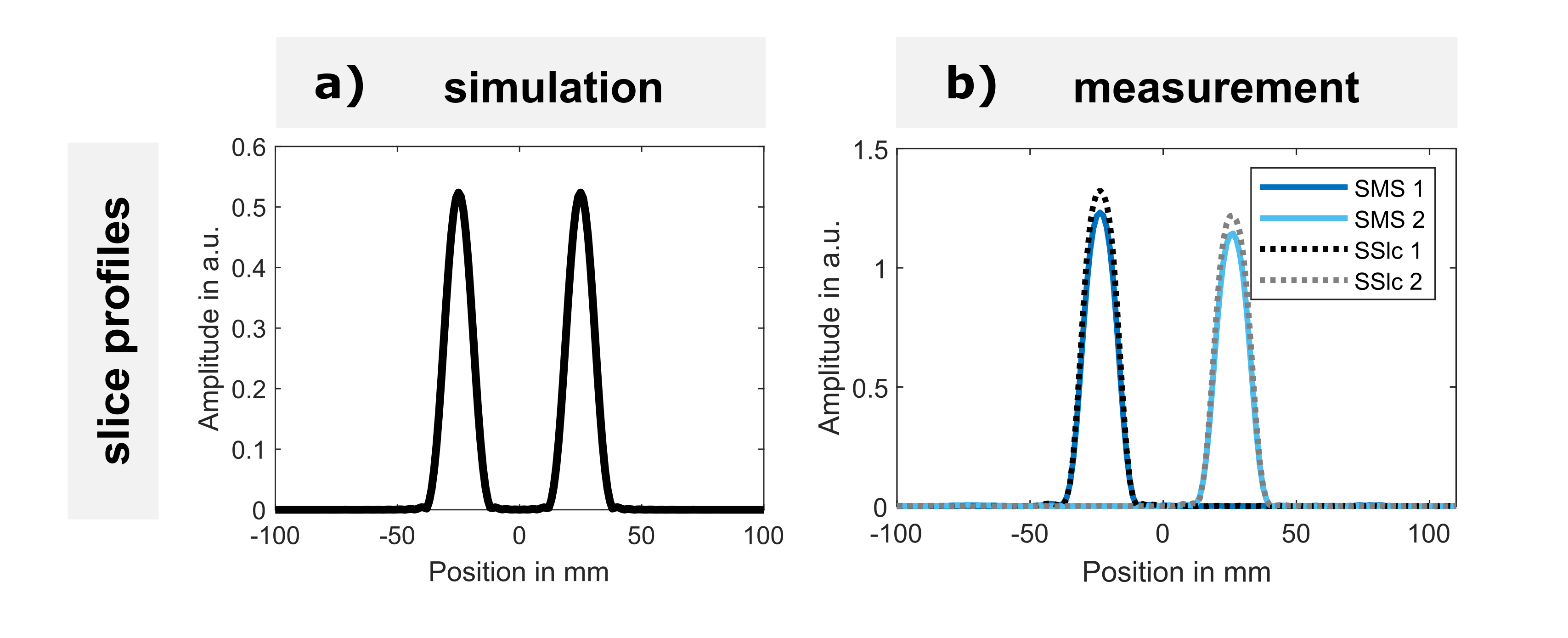
Bloch simulations were used to validate the resulting slice profiles of the SMS pulses, the results of which are shown in Figure 2a) (τ=1024µs, bandwidth-time-product (BWTP)=2.7, T1=64ms, T2*=55ms, FA=35°, slice thickness=12mm, slice distance=50mm).
Measurements were obtained on a 7T research system (Magnetom 7T, Siemens Healthcare) using a cylindrical phantom filled with 0.9% saline solution and equipped with vials of varying agar concentrations between 2% and 7%, providing relaxation times of T1=35-50ms, T2s*=4-11ms, and T2l*=25-40ms8. The slice profiles were measured using a gradient echo sequence with readout in z-direction (FA=35°, TE=5ms, TR=20ms, BWTP=2.7, slice thickness=12mm, slice distance=50mm). Equivalent slice profiles using single-slice excitation of the targeted slices were obtained for comparison.
Testing the general applicability of the SMS approach, two-slice images were acquired in the phantom next to a saline bottle, which provides a visual difference between the acquired slices, shown in Figure 3a).
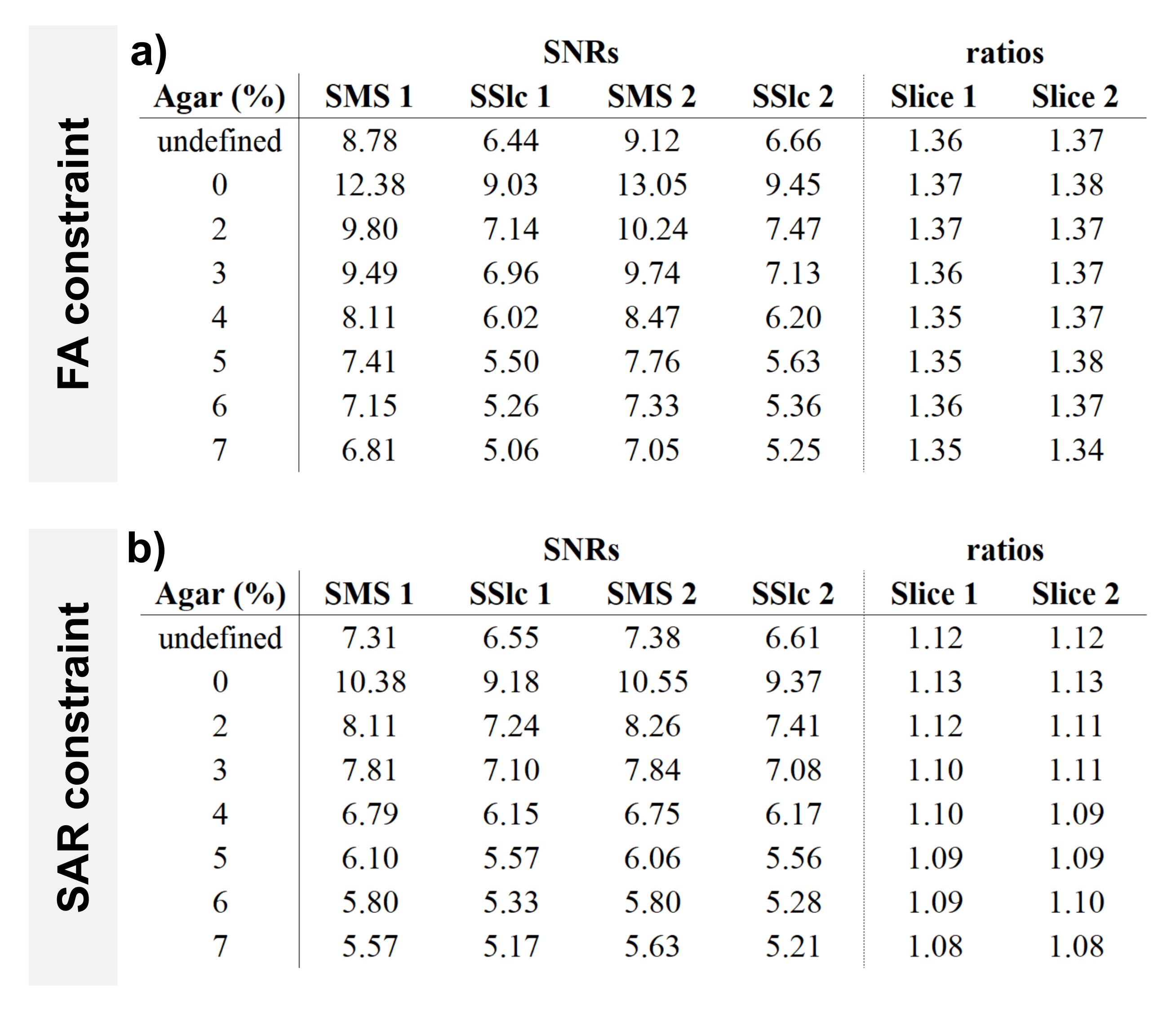
In addition, SMS and single-slice phantom measurements were acquired for SNR comparison with the constraint of constant acquisition time for the total measurement of both slices. Furthermore, in a first experiment the FA was held constant for the SMS and single-slice measurements (FA=34°, TE=5ms, TR=20ms, BWTP=2.7, slice thickness=12mm, slice distance=50mm). This measurement represents the case when the target FA is not restricted by SAR. Secondly, for the case of SAR restrictions, measurements with constant SAR between SMS and single-slice excitation were performed. This was ensured by normalizing the integral of the squared RF pulses, leading to a reduced FA=24° for SMS excitation. Subsequently, SNR was determined in all measurements on the unfiltered images using the ROI method9,10:
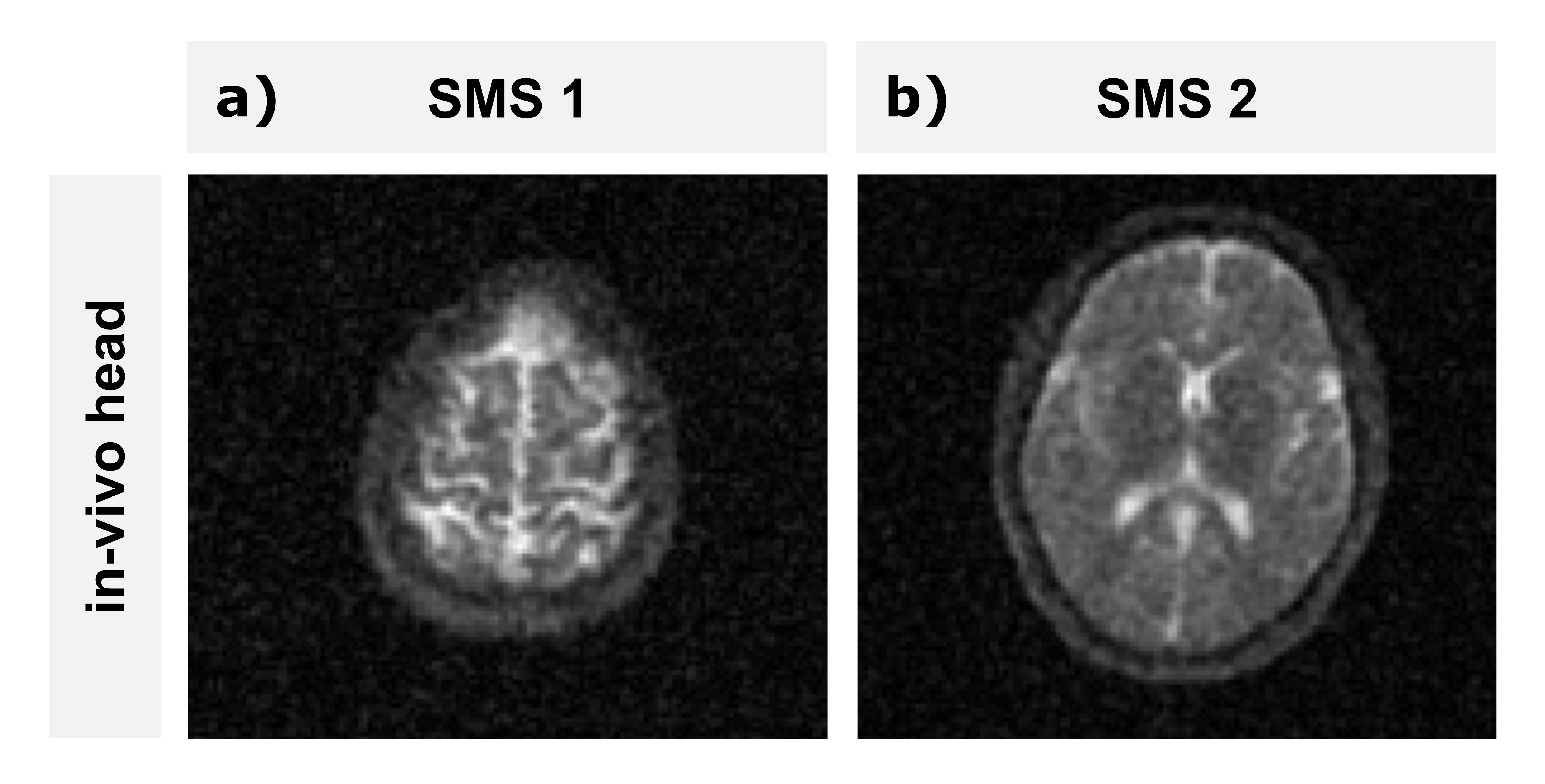
Finally, an SMS measurement was performed in the head of a healthy volunteer to validate the applicability in-vivo.
Results
The simulated SMS slice profile agrees well with targeted slice thickness and inter-slice distance. Measurements of SMS slice profiles agree well with single-slice excitation profiles (Figure 2). The peak-peak distance agrees within 1mm for simulation and measurement with targeted distance of 50mm. Full width at half maximums (FWHMs) of simulation agree within 1mm with the targeted slice thickness of 12mm and FWHMs of measurement within 2.5mm. The peak amplitudes of SMS slice profiles are 7% lower than corresponding single-slice profiles. In both measurements the signal amplitude of the right slice was found to be lower than for the left slice (Figure 2).SMS phantom measurements demonstrate acquisition of two distant slices (Figure 3). Acquisitions with constant FA led to an SNR increase of up to 38% compared to single-slice acquisition, with similar SNR benefits for all compartments (Figure 4a). The constraint of equal SAR led to an SNR increase of up to 13% with a trend of decreasing SNR benefit for increasing agar concentration or decreasing relaxation time (agar 0%: SNR benefit 13%, agar 7%: SNR benefit 8%; Figure 4b).
Figure 5 demonstrates the applicability of SMS acquisition for 23Na in-vivo imaging, where the two slices are clearly distinguished.
Discussion & Conclusion
A first investigation of 23Na SMS imaging was performed by implementing multi-band excitation pulses in a 2D spoiled GRE sequence to enable double-slice imaging according to radial CAIPIRINHA. Measured SMS slice profiles were in good agreement with reference measurements. The signal amplitude reduction of the right slice in both measurements (Figure 2) can be explained by reduced B1 amplitude. SNR evaluations of SMS and corresponding single-slice acquisitions indicate an SNR increase for acquisitions with both FA and SAR constraints. SMS in-vivo images of the human head were possible and of good quality. The application of SMS can thus be considered generally feasible when measuring two slices in 23Na MRI.Acknowledgements
No acknowledgement found.References
1 Madelin G, Regatte RR. Biomedical applications of sodium MRI in vivo. J Magn Reson Imaging. 2013 Sep;38(3):511-29. doi: 10.1002/jmri.24168. Epub 2013 May 30. PMID: 23722972; PMCID: PMC3759542.
2 Konstandin S, Schad LR. Two-Dimensional Radial Sodium Heart MRI Using Variable-Rate Selective Excitation and Retrospective Electrocardiogram Gating with Golden Angle Increments. Magn Reson Med. 2013; 70:791–799
3 Utzschneider M, Behl NGR, Lachner S, Gast LV, Maier A, Uder M, Nagel AM. Accelerated quantification of tissue sodium concentration in skeletal muscle tissue: quantitative capability of dictionary learning compressed sensing. MAGMA. 2020 Aug;33(4):495-505. doi: 10.1007/s10334-019-00819-2. Epub 2020 Jan 16. Erratum in: MAGMA. 2020 Feb 12;: PMID: 31950390.
4 Yutzy, S. R., Seiberlich, N., Duerk, J. L., & Griswold, M. A. (2011). Improvements in multislice parallel imaging using radial CAIPIRINHA. Magnetic resonance in medicine, 65(6), 1630-1637.
5 Breuer, F. A., Blaimer, M., Heidemann, R. M., Mueller, M. F., Griswold, M. A., & Jakob, P. M. (2005). Controlled aliasing in parallel imaging results in higher acceleration (CAIPIRINHA) for multi‐slice imaging. Magnetic Resonance in Medicine: An Official Journal of the International Society for Magnetic Resonance in Medicine, 53(3), 684-691.
6 Fessler JA, Sutton BP. Nonuniform Fast Fourier Transforms Using Min-Max Interpolation. IEEE Transactions on Signal Processing 2003;51:560-574.
7 Nagel AM, Heiler PM, Schad LR. Two-Dimensional Radial Acquisition Technique With Density Adaption in Sodium MRI. Magn Reson Med. 2011; 65:1091–1097
8 Kratzer, F. J., Flassbeck, S., Schmitter, S., Wilferth, T., Magill, A. W., Knowles, B. R., ... & Nagel, A. M. (2021). 3D sodium (23Na) magnetic resonance fingerprinting for time‐efficient relaxometric mapping. Magnetic resonance in medicine, 86(5), 2412-2425.
9 Gudbjartsson, H., & Patz, S. (1995). The Rician distribution of noisy MRI data. Magnetic resonance in medicine, 34(6), 910-914.
10 Cárdenas‐Blanco, A., Tejos, C., Irarrazaval, P., & Cameron, I. (2008). Noise in magnitude magnetic resonance images. Concepts in Magnetic Resonance Part A: An Educational Journal, 32(6), 409-416.
Figures
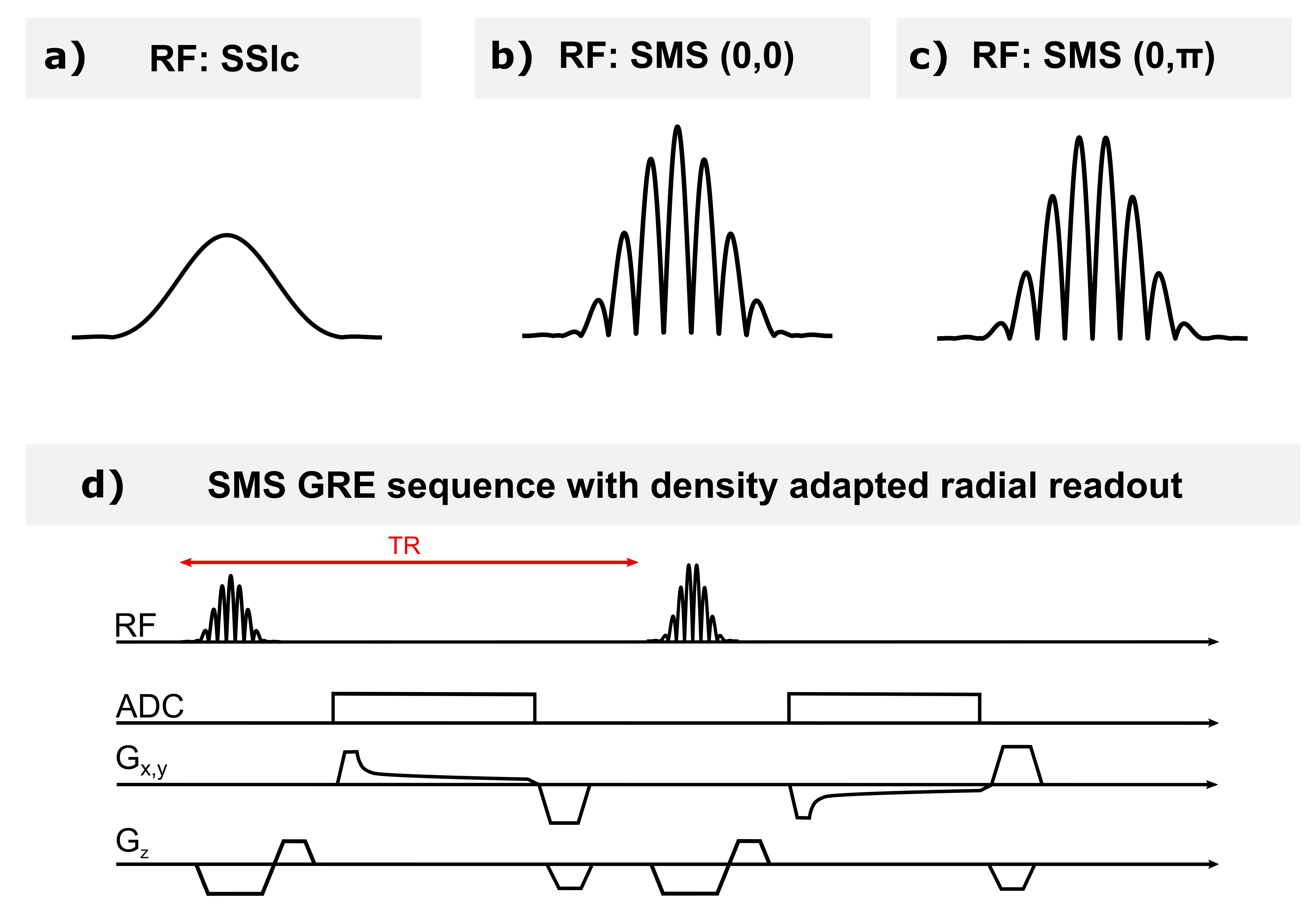
Figure 1: RF pulses for a) single-slice (SSlc) and b), c) SMS excitation. Single-slice excitation is performed by the application of a conventional sinc pulse, while SMS excitation uses CAIPIRINHA pulses. CAIPIRINHA pulses are obtained by the summation of the respective sinc excitation pulses of the two target slices modulated with corresponding local Larmor frequencies. Two relative phases between target slices lead to different pulse envelopes: the same phase for both pulses is shown in b) and a phase difference of π in c). d) shows the SMS implementation in a spoiled FLASH sequence.