4066
lower limbs skeletal muscle perfusion evaluation with the Fourier decomposition technique
Jianxun Qu1 and Tianye Lin2
1SIEMENS Healthineers, Beijing, China, 2Radiology Department, Peking University Cancer Hospital & Institute, Beijing, China
1SIEMENS Healthineers, Beijing, China, 2Radiology Department, Peking University Cancer Hospital & Institute, Beijing, China
Synopsis
Keywords: Muscle, Perfusion
This study incorporated the Fourier decomposition (FD) technique to the lower extremity blood oxygen level-dependent, or reactive hyperemia, technique to evaluate the perfusion level of lower limbs. A periodically repeated ischemia-reperfusion paradigm was used to support the frequency analysis in FD. A characteristic frequency intensity corresponding to the ischemia-reperfusion cycle frequency was observed. The feasibility of the proposed method was demonstrated on healthy subjects.Introduction
Fourier decomposition (FD) is a novel perfusion imaging technique that has been successfully introduced in pulmonary perfusion imaging [1] and also has been practised in other regions [2]. The perfusion level of the interested organ could be extracted from the frequency analysis of the time-resolved measurement. This study explores the feasibility of practising the FD technique in lower extremity perfusion imaging. However, due to the intrinsic low perfusion level of the skeletal muscle at the resting state, the direct application of FD might still be challenging. To increase the perfusion contrast, the reactive hyperemia technique, also termed blood oxygen level-dependent (BOLD), was employed to provoke measurable changes [3]. Conventionally, the BOLD protocol consists of a cuff-induced ischemia session lasting for several minutes, followed by a reperfusion session after the pressure is released. This work modified the ischemia-reperfusion protocol to a periodically repeated paradigm to support the frequency analysis in FD.Methods
This study recruited two healthy subjects, with informed consent acquired before the experiment. The reactive hyperemia experiment was performed on a 3T whole-body scanner equipped with an 18-channel phased array Ultraflex coil. The subject positioning was the same as in our previous study [4]. The MRI scan had two sessions, a sixty-second resting session and then the ischemia-reperfusion session, which consisted of fifteen repeated blocks, as shown in Figure 1A. Each block contained a 10-second ischemia state when the cuff pressure was exerted and a 10-second reperfusion state when the pressure was released. The ischemia-reperfusion frequency was 1/20-second or 0.05 Hz. The entire ischemia-reperfusion session lasts for 300 seconds. Gradient echo (GRE) echo planar imaging (EPI) was used to sample the signal change along the whole process, with the following parameters: TR/TE 1000/15 ms, the field of view (FOV) 154 * 154 mm2, resolution 96 * 96, slice thickness 6 mm, number of slices 24, and voxel size 1.6 * 1.6 * 6.0 mm3. The acceleration factor in the phase encoding direction is 2, and in the slice direction is 2. An excitation flip angle of 45 degrees was used to reduce the influence of T1 relaxation. In total, 360 measurements were acquired. In the post-processing, ANTs (Advanced Normalization Tools) was used for the non-rigid registration. A pixel-wise Fourier transform was then performed. The frequency spectrum of the imaging volume was calculated for both the resting session and the ischemia-reperfusion session. The frequency intensity at 0.05 Hz was regarded as the metric to assess perfusion level (Figure 1B).Results:
Figure. 2 shows the frequency analysis of the BOLD measurement time series. No characteristic frequency was observed from the resting state data (Figure 2A). As a comparison, for the ischemia-reperfusion data, the frequency spectrum showed an apparent peak at 0.05 Hz, as indicated by the arrow. A pixel-wised frequency spectrum intensity, at 0.05Hz, is shown in Figure. 3B. Compared to the anatomical images, there is a conspicuous muscle group wise distribution. The frequency intensity maps of the adjacent frequencies are shown in Figure 4. The intensity is not comparable to that of the 0.05Hz.Discussion and Conclusion:
This is the first work incorporating the Fourier decomposition technique and the ischemia hyperemia technique in the perfusion evaluation of lower limbs. A periodically exerted ischemia-reperfusion block replaced the long-lasting ischemia session in the conventional skeletal muscle BOLD experiment. The FD method markedly reduced the total ischemia duration, which mitigates the discomfort and could potentially reduce the side effect of long-lasting ischemia, especially for subjects vulnerable to perfusion deficiency. As a preliminary study, the spectrum intensity of the characteristic frequency, 0.05Hz in this study, was used directly to evaluate the perfusion level. A temporal resolution of 1 second was used. The temporal resolution could be increased further to incorporate other physiological movements, for instance, pulsation. As a limitation, this work only applied the FD technique to healthy subjects. Its effectiveness wait to be further tested for subjects with altered perfusion status.Acknowledgements
No acknowledgement found.References
[1] Andreas Voskrebenzev, et al. Feasibility of Quantitative Regional Ventilation and Perfusion Mapping With Phase-Resolved Functional Lung (PREFUL) MRI in Healthy Volunteers and COPD, CTEPH, and CF Patients, MRM, 2017 [2] Julian Glandorf, et al. Feasibility of Flow-related Enhancement Brain Perfusion MRI, ISMRM, 2022 [3] Shiteng Suo, Lan Zhang, et al., Evaluation of skeletal muscle microvascular perfusion of lower extremities by cardiovascular magnetic resonance arterial spin labeling, blood oxygenation level-dependent, and intravoxel incoherent motion techniques, Journal of Cardiovascular Magnetic Resonance, 2018 [4] Xiaoxi Yu, Zhaoxi Liu, Jianxun Qu, et al. Blood Oxygen Level-Dependent Imaging of Lower Limbs: Perfusion and Clinical Assessment of Peripheral Artery Disease, 2022, ISMRMFigures
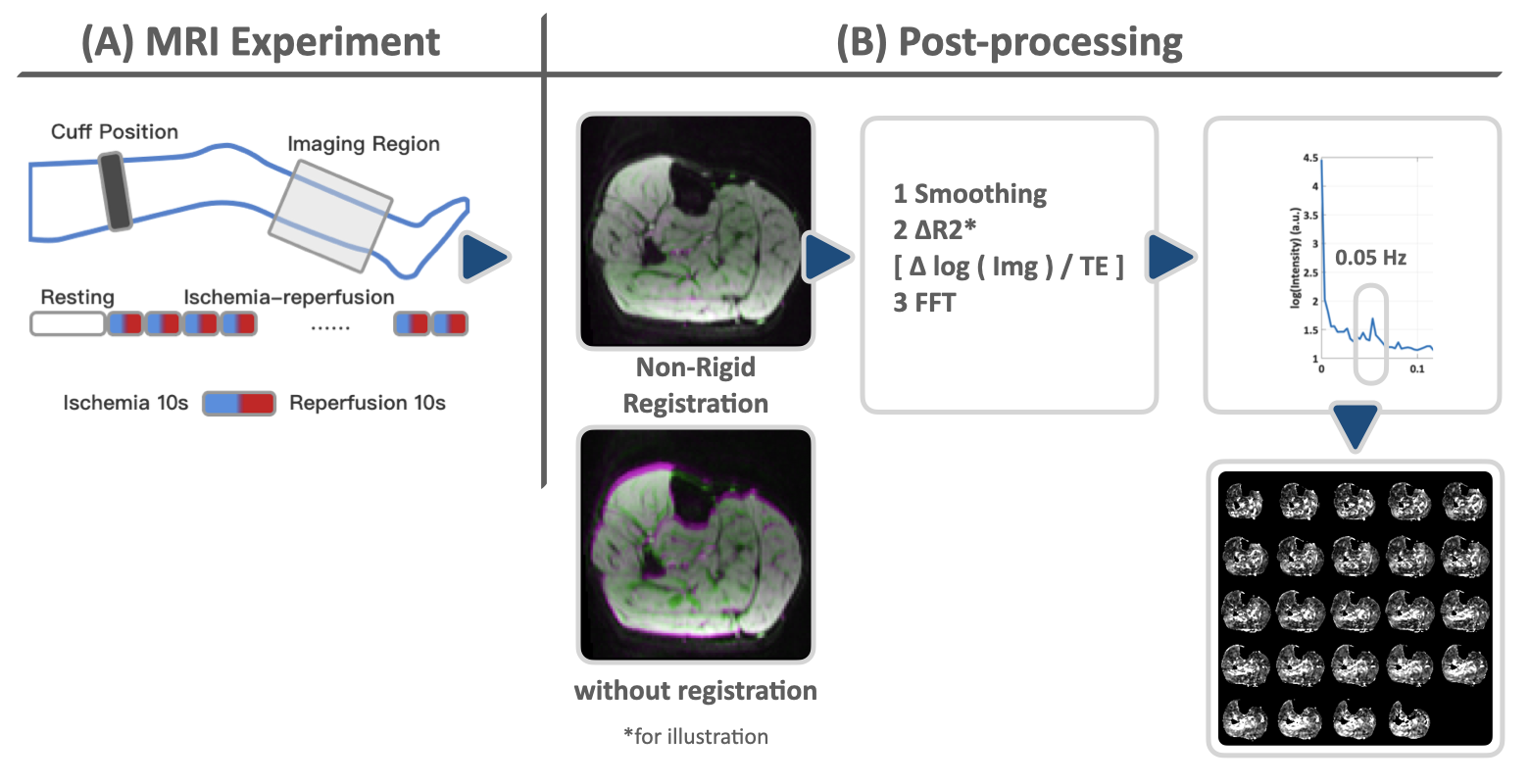
Figure 1, The MRI experiment setting (A) and the post-processing pipeline (B).
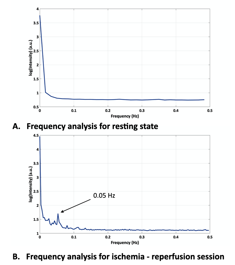
Figure 2, The frequency analysis of the resting session (A) and the ischemia - reperfusion session (B). There's a conspicuous peak at 0.05 Hz.
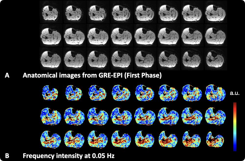
Figure 3, The anatomical images (A) and the frequency intensity map at 0.05Hz (B).
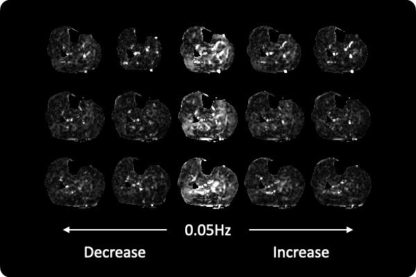
Figure 4, The frequency intensity map from the adjacent frequencies. The 0.05 Hz map showed stronger intensity compared to the adjacent frequencies
DOI: https://doi.org/10.58530/2023/4066