4059
Evaluation of viscoelastic changes of the psoas major muscle after exercise using MR elastography1Department of Radiological Sciences, Graduate School of Human Health Sciences, Tokyo Metropolitan University, Tokyo, Japan, 2Office of Radiation Technology, Keio University Hospital, Tokyo, Japan, 3Human Technology Research Insutitue, National Institute of Advanced Industrial Science and Techonology, Ibaraki, Japan, 4Department of Sport and Health Sciences, Faculty of Human Sciences, University of East Asia, Yamaguchi, Japan, 5System Emotional Science, Faculty of Medicine, University of Toyama, Toyama, Japan
Synopsis
Keywords: Muscle, Elastography
The shear modulus of the psoas major muscle (PM) after exercise has been evaluated using magnetic resonance elastography (MRE) previously, however, a separate evaluation of elasticity and viscosity has not been performed. We evaluate storage modulus representing elasticity and loss modulus representing viscosity of the PM using MRE after exercise in this study.
Comparing the before and immediately after exercise, the loss modulus showed a greater rate of decrease than the storage modulus, suggesting that the loss modulus, which reflects viscosity, is more useful for detecting changes of muscle tissue immediately after exercise.
Introduction
Stiffness is one of the important parameters of muscle function and is easily influenced by muscle exercise1. Magnetic resonance elastography (MRE) is a phase-contrast technique that allows measurement of the shear modulus of tissues, noninvasively and quantitatively even in deep lying tissues2,3. The psoas major muscle (PM) is one of the lumbar back muscles that are deeply located in the human body. The previous studies have evaluated the time-course of physical properties of the PM by shear modulus, T2 values, and apparent diffusion coefficient4. This previous study suggested that the shear modulus of the PM significantly decreased from 5.5 min after exercise to 31.5 min after exercise, and significantly increased from 96.5 min after exercise to 109.5 min after exercise. Although not a significant change, there was a trend toward an increase between 31.5 min to 96.5 min after exercise. It has been suggested that these changes may be influenced by changes in viscosity, however, this previous study used local frequency estimation (LFE) for the estimation of the shear modulus and was not able to evaluate elasticity and viscosity separately. Algebraic Inversion of the Differential Equation (AIDE) is one of the methods for shear modulus estimation and it is able to estimate storage modulus representing elasticity and loss modulus representing viscosity5. The objective of this study was to evaluate elastic and viscous properties of the PM in the shear modulus change after exercise.Mehod
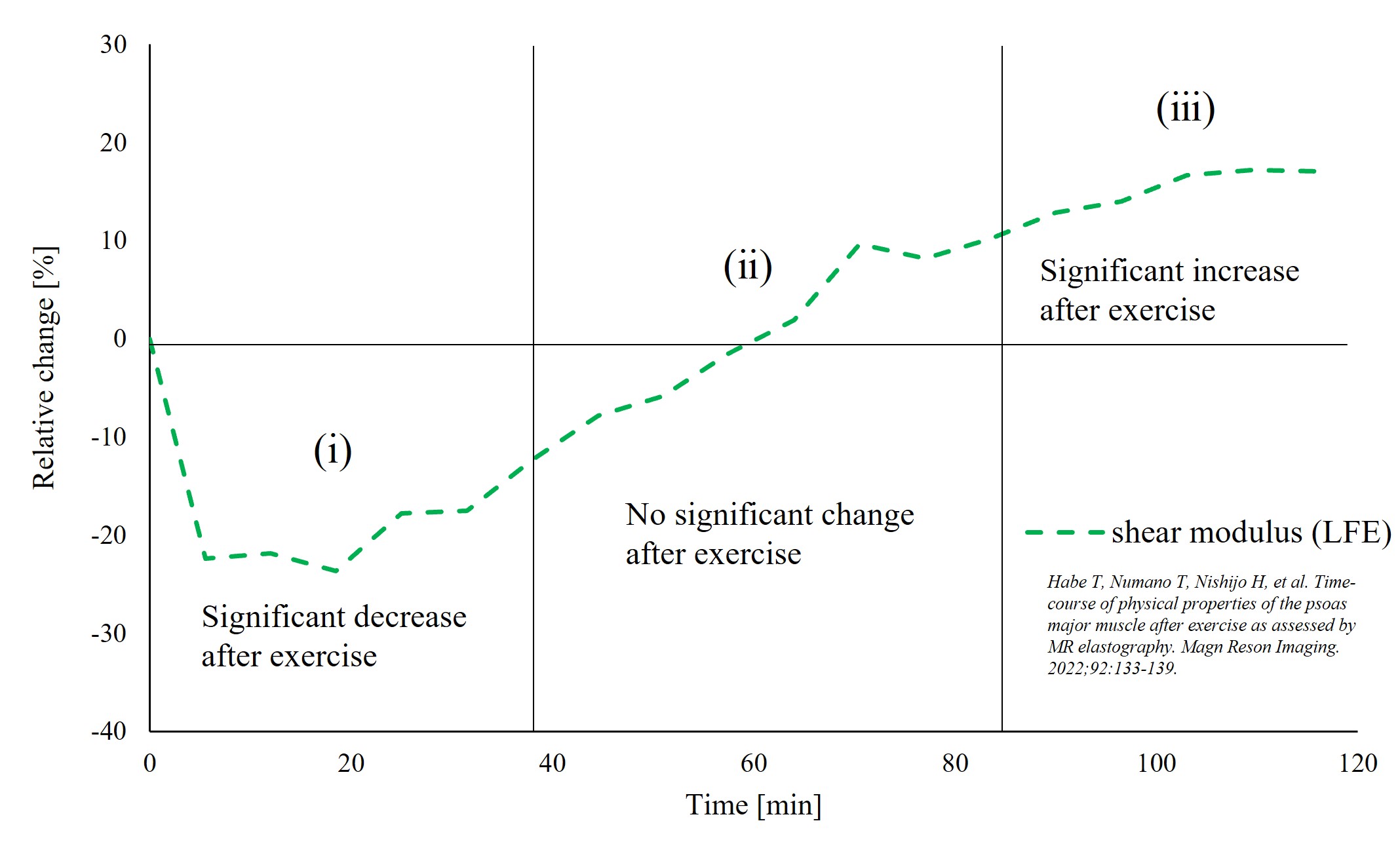
Volunteers with no history of low back pain were enrolled in this MRE study. Nine male volunteers participated this study (mean age, 23.1 ± 2.1 years; height, 175.2 ± 5.9 cm; body weight, 64.2 ± 6.8 kg). MRE experiments were performed on a clinical MR imager (Achieva 3.0T, Philips Healthcare, Best, The Netherlands) using a SENSE Torso coil (Philips Healthcare, Best, The Netherlands). MRE was performed with a gradient-echo type multi-echo MRE sequence6. The experimental system and acquisition parameters for PM MRE was similar to that of a previously reported7,8. The previous study showed that time points after exercise can separate 3 groups from the perspective of the changes of shear modulus: (ⅰ) significant decrease, (ⅱ) no significant change, and (ⅲ) significant increase (Fig. 1)4. We estimated storage modulus, loss modulus, and shear modulus magnitude from the representative 2 data from each of (ⅰ)-(ⅲ) using AIDE algorithm in this study. The data of the acquired parameters were normalized as relative changes to the pre-exercise values (Relative change (%) = 100(pre-exercise – post-exercise)/pre-exercise). To compare post-exercise data at each time-point with pre-exercise control data, post-hoc analyses with Durbin-Conover test were conducted with a Holm correction applied. Statistical significance was set at p < 0.05 for all analyses.Result
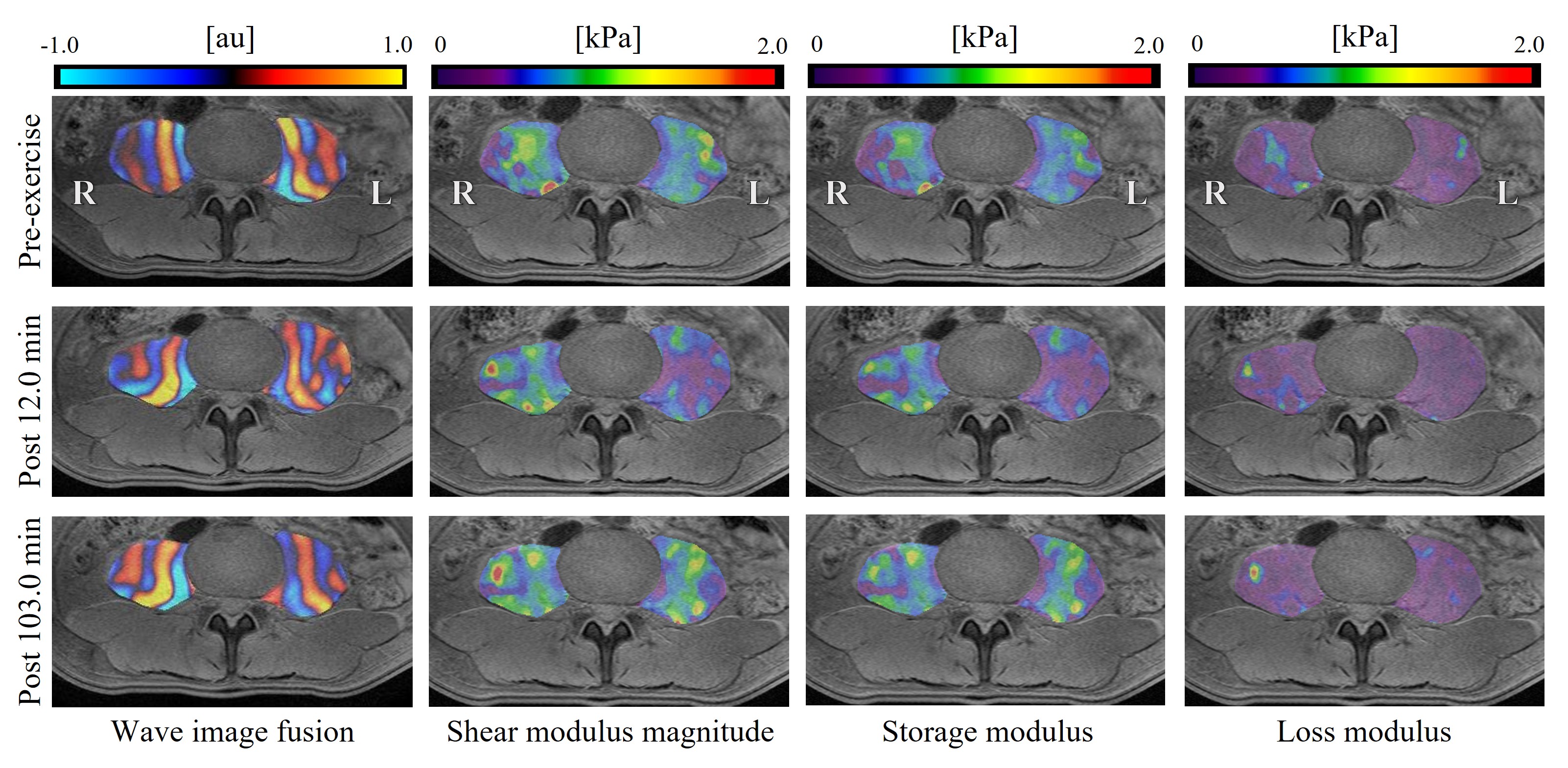
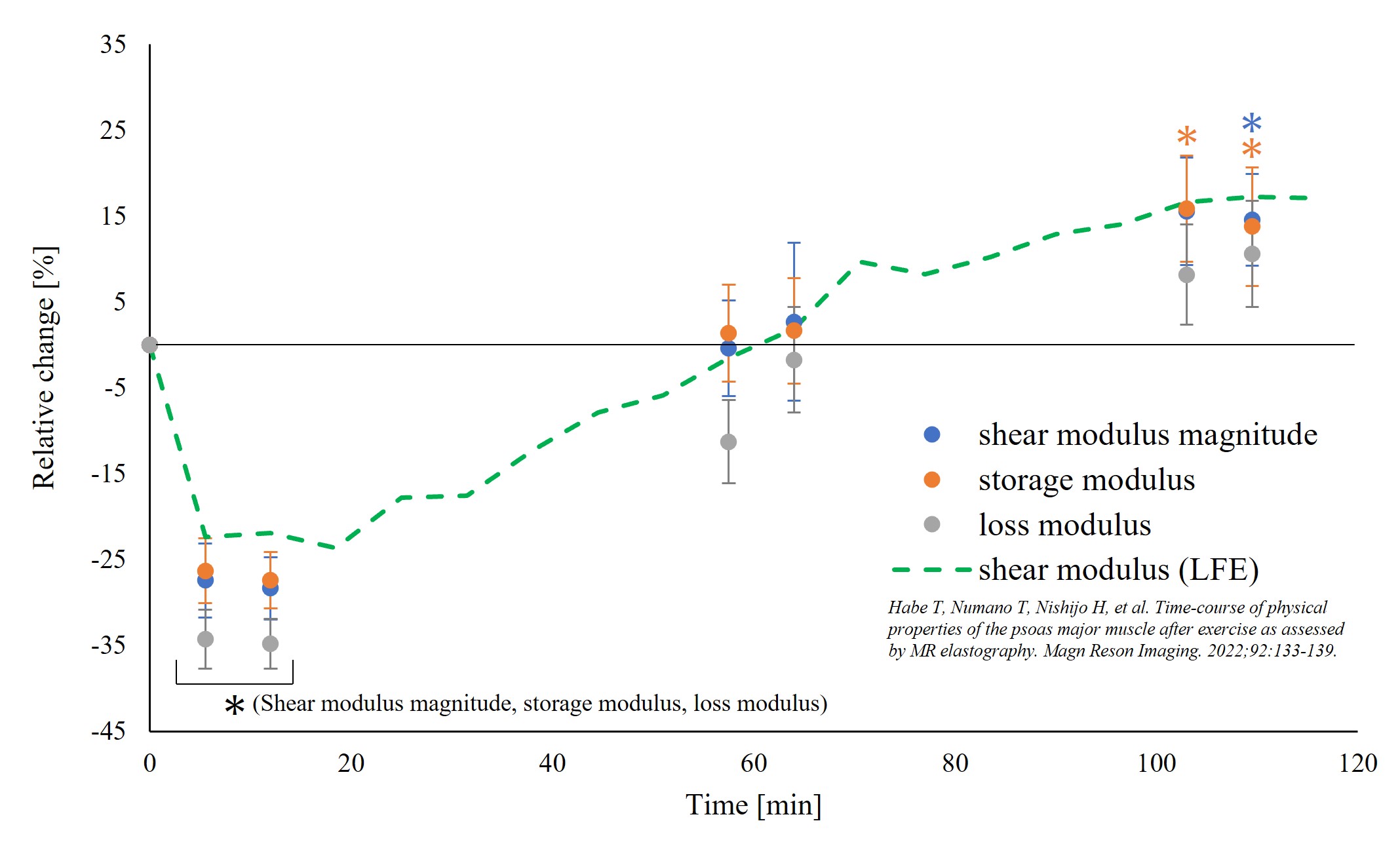
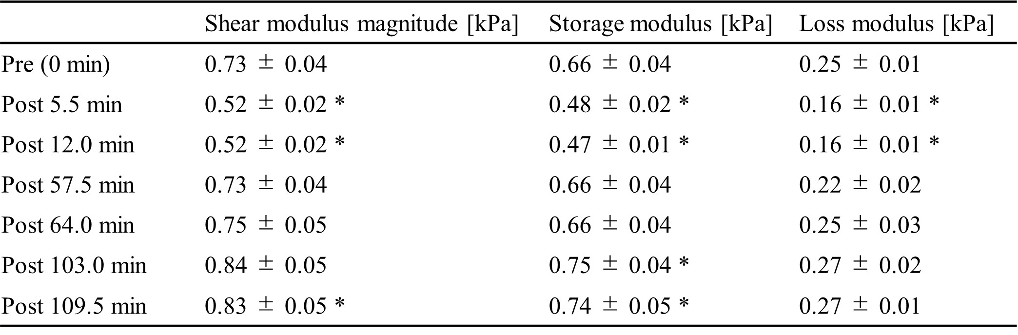
Figure 2 shows representative MRE images of the PM. Fig. 3 shows the relative changes of each modulus and Fig. 4 shows non-normalized data of each modulus. The storage modulus, loss modulus, and shear modulus magnitude showed significant decrease at 5.5 min and 12.0 min after exercise and loss modulus showed a larger relative change than storage modulus and shear modulus magnitude. The storage modulus showed significant increase at 103.0 min and 109.5 min after exercise and shear modulus magnitude showed significant increase at 109.5 min after exercise.Discussion
In this study, relative change of loss modulus was larger than that of storage modulus and shear modulus magnitude at 5.5 min and 12.0 min after exercise. Water retention and inflammation or changes in the myocytic structures occurred in muscle tissue after exercise and it led to the decrease of viscosity9. In the previous studies4, the decrease in viscoelastic modulus of the PM coincided with an increase in T2 value and apparent diffusion coefficient, suggesting that changes in water content caused a decrease in viscosity, which in turn caused a decrease in viscoelastic modulus. The result of this study suggests that changes in loss modulus (viscosity) of the PM immediately after exercise are more sensitive to detecting changes of muscle tissues than changes in storage modulus (elasticity). As for studies on other organs, it has been suggested that viscosity is useful for grading hepatic necro-inflammation in the liver10.The storage modulus increased significantly at 103.0 min and 109.5 min after exercise, while the loss modulus did not change significantly. Previous studies4 have suggested that an increase in intramuscular Ca2+ concentration after exercise contributes to the increase in viscoelastic modulus, suggesting that viscous effects play little role in the increase in viscoelastic modulus.
Conclusion
This study indicates that the loss modulus, which represents viscosity, facilitate the detection of tissue changes immediately after exercise. The present study also suggests that the increase in viscoelastic modulus of the PM after exercise is largely due to increased elasticity.Acknowledgements
References
1. Hug F, Tucker K, Gennisson JL, Tanter M, Nordez A. Elastography for muscle biomechanics: toward the estimation of individual muscle force. Exerc Sport Sci Rev 2015;43(3):125–33.
2. Muthupillai R, Lomas DJ, Rossman PJ, et al. Magnetic resonance elastography by direct visualization of propagating acoustic strain waves. Science (New York, NY) 1995;269(5232):1854-7.
3. Muthupillai R, Ehman RL. Magnetic resonance elastography. Nature Medicine 1996;2(5):601-603.
4. Habe T, Numano T, Nishijo H, et al. Time-course of physical properties of the psoas major muscle after exercise as assessed by MR elastography. Magn Reson Imaging. 2022;92:133-139.
5. Oliphant TE, Manduca A, Ehman RL, Greenleaf JF. Complex-valued stiffness reconstruction for magnetic resonance elastography by algebraic inversion of the differential equation. Magn Reson Med. 2001;45(2):299-310.
6. Numano T, Mizuhara K, Hata J, et al. A simple method for MR elastography: a gradient-echo type multi-echo sequence. Magn Reson Imaging 2015;33(1):31-7.
7. Numano T, Habe T, Ito D, et al. A new technique for motion encoding gradient-less MR elastography of the psoas major muscle: A gradient-echo type multi-echo sequence. Magn Reson Imaging 2019.
8. Habe T, Numano T, Ito D, Takamoto K, Nishijo H, Mizuhara K, et al. Development of a Suitable Actuator for Magnetic Resonance Elastography of the Psoas Major Muscle. Applied Magnetic Resonance 2020;52(2):157-68.
9. Hata J, Endo K, Tsuji O, Arakawa S, Sato M, Yagi K, et al. Analysis of skeletal-muscle condition after excessive loading of the lower legs by sequential magnetic resonance imaging. J Orthop Sci 2019;24(5):873-80.
10. Garteiser P, Pagé G, d'Assignies G, et al. Necro-inflammatory activity grading in chronic viral hepatitis with three-dimensional multifrequency MR elastography. Sci Rep. 2021;11(1):19386.Figures