4043
Deep Learning Reconstruction for 4-fold Accelerated 2DFSE Imaging: optimization of variable density undersampling
Michael Carl1, Rafi Brada2, Nir Mazor2, Daniel V Litwiller3, and Maggie Fung4
1GE Healthcare, San Diego, CA, United States, 2GE Research, Herzliya, Israel, 3GE Healthcare, Denver, CO, United States, 4GE Healthcare, New York, NY, United States
1GE Healthcare, San Diego, CA, United States, 2GE Research, Herzliya, Israel, 3GE Healthcare, Denver, CO, United States, 4GE Healthcare, New York, NY, United States
Synopsis
Keywords: Machine Learning/Artificial Intelligence, Machine Learning/Artificial Intelligence
In this work we use variable-density prospective undersampling of the phase-encode k-space lines (ky) in 2D fast spin-echo (2DFSE) followed by deep learning (DL) reconstruction. We were able to achieve an acceleration of R=4 while maintaining high image quality.Introduction:
Two-dimensional fast spin echo (2DFSE) is one of the main clinical MR sequences used in musculoskeletal (MSK) imaging. High resolution imaging is important in this application due the small structures in most joint structures, which can lead to long scan-times and potential motion artifacts. In this work we use variable-density prospective undersampling [1] of the phase-encode k-space lines (ky) followed by deep learning (DL) reconstruction. We were able to achieve an acceleration of R=4 while maintaining high image quality. Phantom experiments and in-vivo scans were performed to study different undersampling patterns and their performance in terms of aliasing artifacts and resolution.Materials and Methods:
The prospective undersampling schedule was determined by a parameterized probability density function (PDF) given by:PDF(ky) = [ (1 - |1 - |ky| |)/(nkfull/2) ]vd [1]
where nkfull is the number fully acquired ky lines (e.g. without acceleration), and vd is a tuning parameter that alters the shape of the PDF (see Fig.1). In addition to the undersampled outer regions, the center portion of k-space is fully sample similar to standard ARC acceleration. For our experiments presented in this work, we kept the number of central lines at 12-24.
Using this probability function, the desired amount acquired k-space lines (nacq) in the outer region of k-space is sampled from the PDF. Fig.1 shows some representative PDFs using several values of vd and a central region of 12 k-space lines and acceleration factor of 4. For lower values of vd the PDF becomes nearly flat (Fig.1a), resulting in a uniform undersampling similar to standard ARC. As the value of vd increases, the center region becomes more fully sampled (Fig1.c,d), while the outer region becomes more sparsely sampled. This results in better aliasing suppression for higher values of vd at the expense of loss of resolution due to the lack of outer k-space samples.
High-resolution ACR phantom experiments were performed on a clinical 3T MR scanner (GE Healthcare, Waukesha, WI) to optimize the PDF parameters. Additionally, we performed in-vivo scanning of the knee to test the sequence performance in clinical settings. All DL inferencing was performed on a dedicated server after MR imaging.
Results:
Several phantom images are shown in Fig.2 with and acceleration factor of 4, shown at different values of vd = [0.01, 1, 2, 3], corresponding to the PDFs shown in Fig.1. Both full FOV images are shown to investigate aliasing patterns, as well as a zoomed-in region to study the resolution performance. As expected, the images using a lower value of vd result in notable aliasing, but result in the sharpest resolution (Fig.2a), while Fig.2d (vd=3) shows little to no aliasing but has some noticeable blurring.The in-vivo knee images are shown in Fig.3. Shown are coronal 2D FSE images with standard ARC=2 (left), and DL variable density with undersampling factor=4 and vd=2. The overall image quality appears similar with only minor blurring apparent in the DL image.
Conclusion:
We have investigated a variable density undersampling scheme to accelerate clinical fast-spin-echo MR imaging in MSK. We found that an acceleration factors up to 4 can give promising IQ, and result in diagnostic images while at the same time reducing the scan times by several factors.Acknowledgements
No acknowledgement found.References
[1] Sparse MRI: The Application of Compressed Sensing for Rapid MR Imaging, Magnetic Resonance in Medicine 58:1182–1195 (2007)Figures
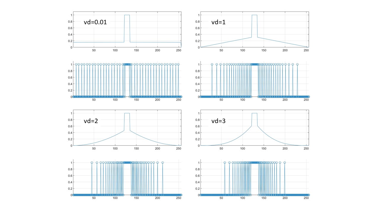
Fig.1: PDF (top panels),
and sampled ky lines (bottom panels) for various values of vd and
acceleration=4. Note how the sampling becomes more spread out at lower values
of vd (a), and become more focused near the center for higher values of vd.
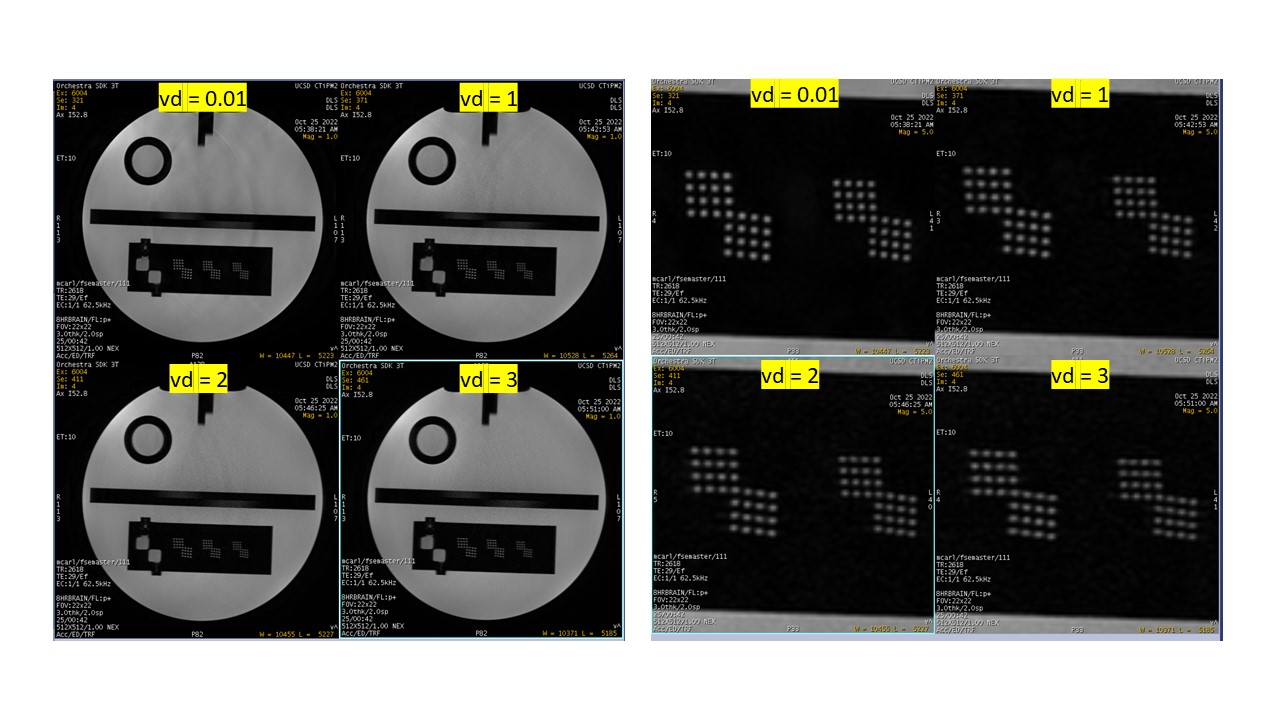
Fig.2:
ACR phantom experiments using the acceleration patterns shown in Fig.1. Images
are shown at the fully acquired FOV (to investigate undersampling aliasing
artifacts), and a zoomed-in portion (to investigate resolution performance).
Lower values of vd result in more aliasing artifacts (a), but result in sharper
images, while higher values of vd (d) show little-to-no aliasing but at the
expense of more image blurring.
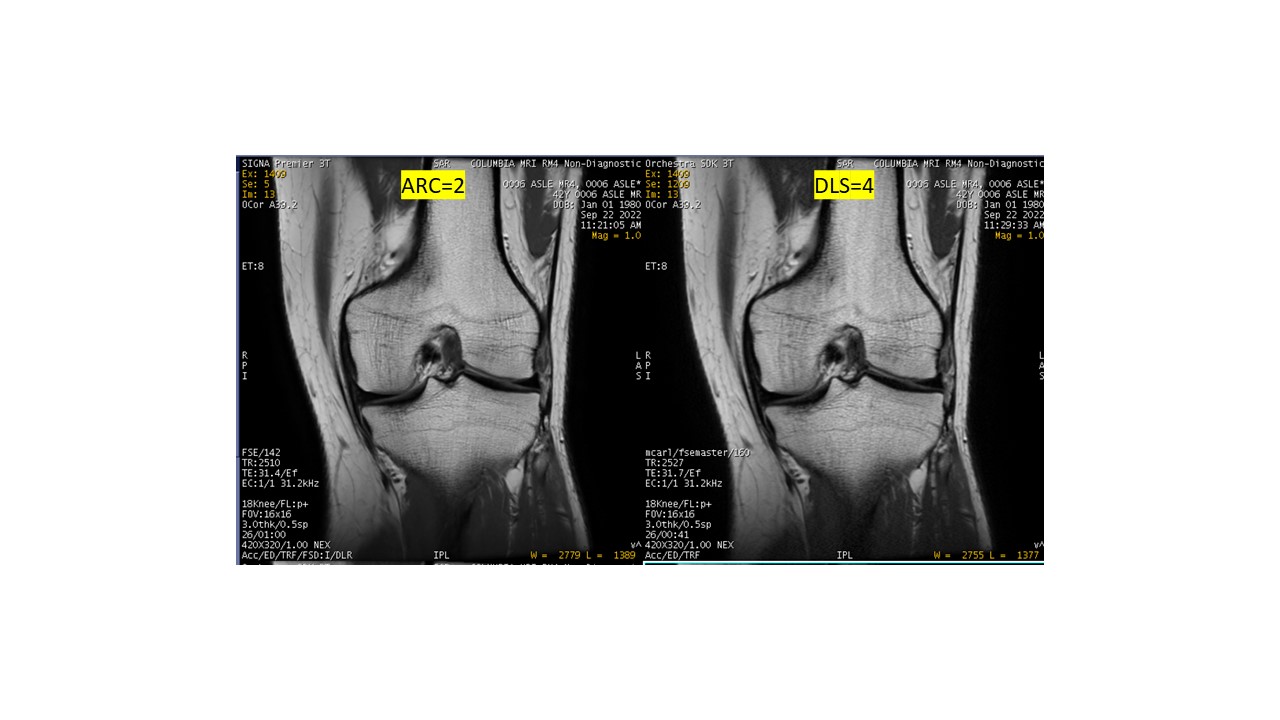
Fig.3:
In-vivo knee 2DFSE images. Shown are clinical ARC=2 images (left), and DL
variable undersampling=4 (right) with vd=2. Overall image quality is similar
between both sequences, with the image on the right only showing minor loss of
high-resolution features.
DOI: https://doi.org/10.58530/2023/4043