4042
Quantitative Evaluation of Deep Learning Reconstruction of Diffusion-weighted MRI using a DWI Phantom
Xiangchuang Kong1, Shengzhen Tao1, Eric H. Middlebrooks1, Thomas Benkert2, Xiangzhi Zhou1, and Chen Lin1
1Department of Radiology, Mayo Clinic, Jacksonville, FL, United States, 2Siemens Medical Solutions USA, Inc., Jacksonville, FL, United States, JACKSONVILLE, FL, United States
1Department of Radiology, Mayo Clinic, Jacksonville, FL, United States, 2Siemens Medical Solutions USA, Inc., Jacksonville, FL, United States, JACKSONVILLE, FL, United States
Synopsis
Keywords: Machine Learning/Artificial Intelligence, Diffusion/other diffusion imaging techniques, deep learning reconstruction; DWI phantom
In a quantitative phantom study, deep learning (DL) reconstruction is shown to improve the SNR ratio of DWI images while preserving measured ADC values. The average SNR gain is similar to that achieved with better gradient performance between Siemens Prisma and Vida. Such benefits of DL recon should allow better quality and/or shorter scanning of DWI in clinical applications.abstract
Introduction:Deep Learning (DL) based reconstruction has shown to be a promising technique to further improve image quality in various applications1. Currently, DL has been used for the interpretation and postprocessing of MRIs and for image acquisition and reconstruction In TSE and Haste sequence, deep learning reconstruction (DLR) trained by raw data on different vendors using supervised models within convolutional neural networks (CNN) and state of the art DL models showed comparable performance with conventional scan procedure in a remarkable reduction in acquisition time and denoising2-3.Nevertheless, there are few research on DWI using DL reconstruction (DWI-DLR) to figure out the impact of image quality and denoising. However, its performance and potential benefits in diffusion weighted imaging (DWI) has not been thoroughly evaluated.Purpose:The purpose of this study is to quantitatively measure the signal-to-noise (SNR) gain achieved with DL reconstruction and benchmark the gain against a higher performance gradient system; as well as validating the ADC value measured with DL reconstruction by scanning and comparing the results from a DWI phantom.
Methods:An early version of Quantitative Imaging Biomarkers Alliance (QIBA) DWI phantom was scanned on a Siemens 3T Vida scanner (60/200 gradient) using a prototype DWI sequence with both conventional recon and DL recon as well as on a Siemens 3T Prisma (80/200 gradient) with a conventional DWI sequence. A Siemens 20-channel head and neck coil was used in all acquisitions on both scanners. The same sequence parameters are used on both Vida and Prisma scanners, except the receiver bandwidth (rBW) optimized based on gradient performance to achieve minimal echo spacing and minimal TE, which, in turn, produced slightly different TR and scan time, as shown in Table 1. Two versions of DWI sequences both with through-plane parallel imaging acceleration (SMS) factor 2, but different in-plane parallel imaging acceleration (iPAT) factor 2 and 3 were tested. For each sequence, two identical acquisitions were performed consecutively to allow subtractions for measuring the noise in DWI images. The circular ROIs were then placed in each compartment of the DWI phantom on both the diffusion weight source image to measure the mean signal and the subtracted image to measure the standard deviation of noise in order to calculate the signal to noise ratio (SNR). The ratio of measured SNR for each compartment at different b-values was calculate between sequences. Only the average SNR ratios are presented in Table 2. Linear regressions were performed on mean ADC measurements of each vial between sequences with and without DL recon to evaluate the potential impact.
Results
Typical DWI images and the calculated ADC map are shown in figure 1 with circular ROIs placed in each compartment. The average SNR for sequences with DL recon is consistently higher than the average SNR with conventional recon, as shown in table 2, especially for higher iPAT factor and at lower b-values. The SNR gain is similar to that with better gradient performance and therefore shorter TE on Prisma for iPAT of 3 and greater for iPAT of 2. The ADC values measured with DL recon is consistent with ADC values measured with conventional recon as demonstrated by good correlations (R2 = 0.9959 – 0.9994) in figure 2.
Discussions:Reduced Distortion due to shorter ETL with higher iPAT while the loss of SNR can be recovered by DL Some Blurring and ghosting artifacts are observed with DL, addition investigation will be carried out. Volunteer and patient studies are needed to confirm and validate phantom study results.
Conclusion:DL recon offer improved SNR in DWI images without compromising the accuracy of ADC measurement. The SNR gain with DL recon is similar to that with gradient performance difference between Vida and Prisma.
Acknowledgements
No acknowledgement foundReferences
- Bae SH, Hwang J, Hong SS, et al. Clinical feasibility of accelerated diffusion weighted imaging of the abdomen with deep learning reconstruction: Comparison with conventional diffusion weighted imaging. Eur J Radiol. 2022;154:110428.
- Kidoh M, Shinoda K, Kitajima M, et al. Deep Learning Based Noise Reduction for Brain MR Imaging: Tests on Phantoms and Healthy Volunteers. Magn Reson Med Sci. 2020;19(3):195-206.
- Kim M, Lee SM, Park C, et al. Deep Learning-Enhanced Parallel Imaging and Simultaneous Multislice Acceleration Reconstruction in Knee MRI. Invest Radiol. 2022.
Figures
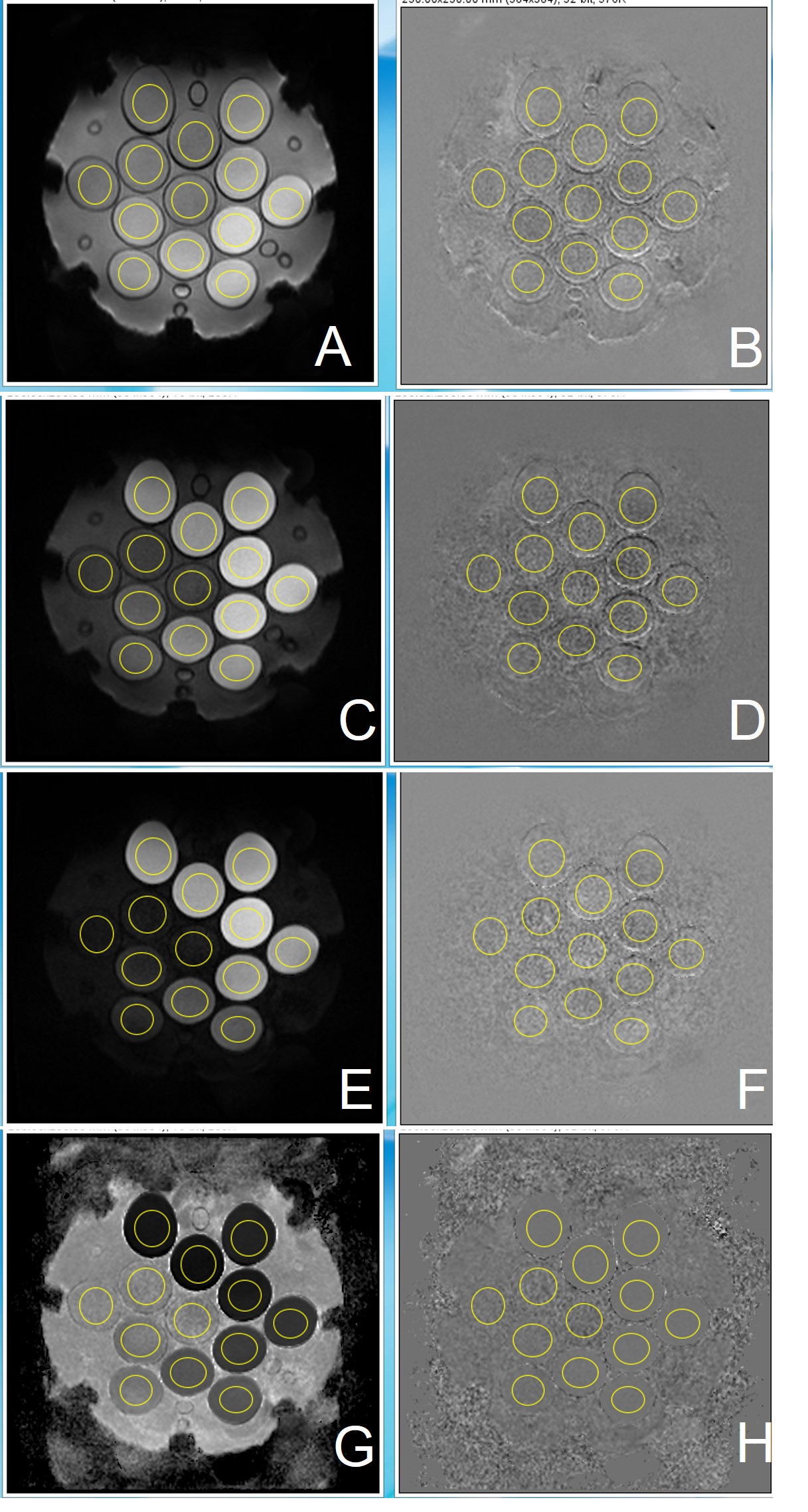
Figure1 DWI image used by DWI phantom
A: DL DWI b=1000; B: DL DWI b=1000 noise by 2 scan images;
C:DL DWI b=2000; D: DL DWI b=2000 noise by 2 scan images;
E: DL DWI b=3000; F: DL DWI b=3000 noise by 2 scan images;
G: DL DWI ADC; H: DL DWI ADC noise by 2 scan images;
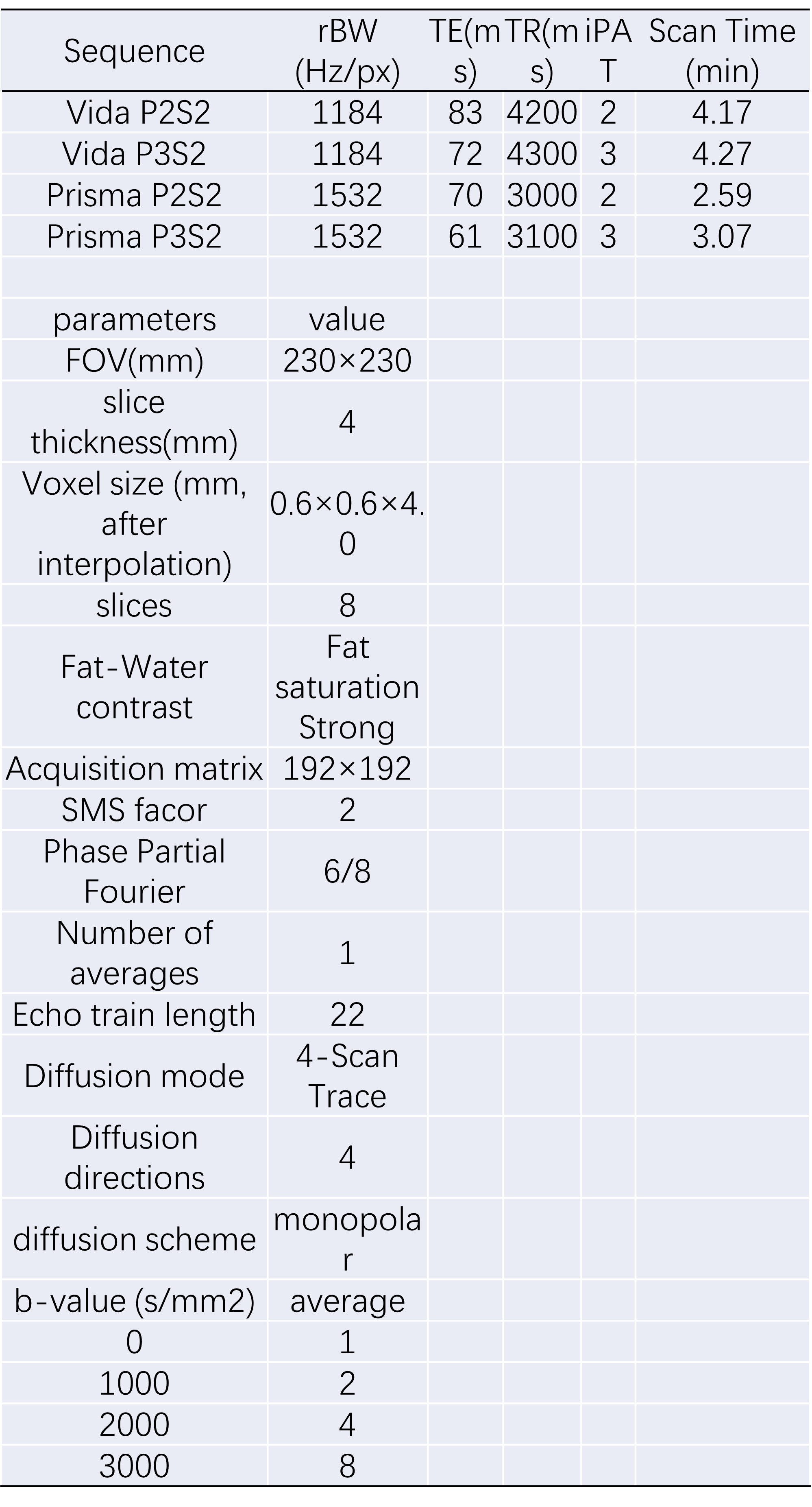
Table1 DLDWI sequence parameters
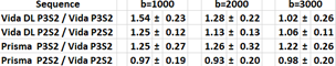
Table2:SNR ratio of different b values of DWI DL in Vida and Prisma.
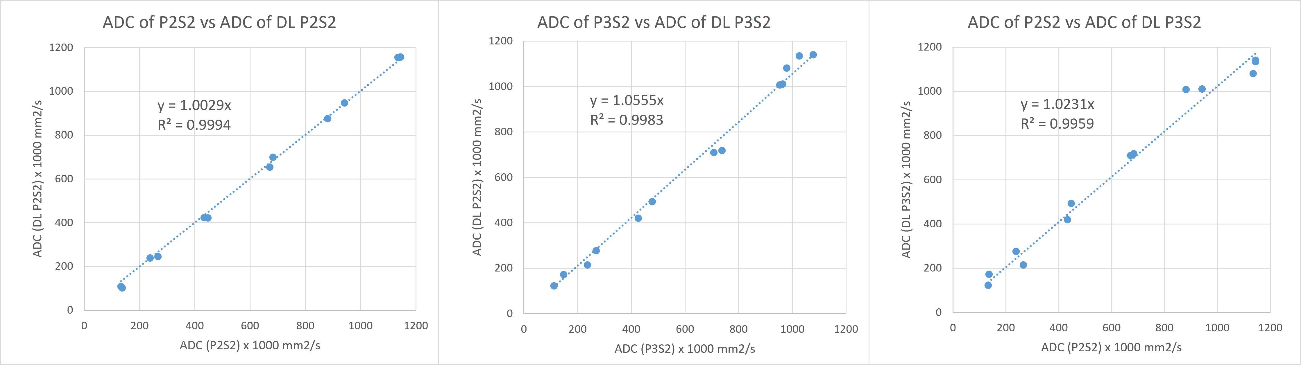
Figure2 Correlations of ADC measured with and without DL Recon with differet inplane parallel imaging accelerations
DOI: https://doi.org/10.58530/2023/4042