4040
Deep learning acceleration and reconstruction for single-channel signals1United Imaging, Houston, TX, United States, 2United Imaging Intelligence, Cambridge, MA, United States
Synopsis
Keywords: Machine Learning/Artificial Intelligence, Image Reconstruction
The capacity of the deep learning ReconNet3D model for single-channel image reconstruction with highly under-sampled k-spaces is demonstrated. Without the need for coil sensitivity information, the proposed method can achieve an acceleration factor of 4 on a dual-channel VTC coil. Supporting high-factor acceleration with limited coil channels can be very beneficial for imaging with surface coils or preclinical animal scans.Introduction
K-space under-sampling is the mainstream strategy for MRI acceleration. Generally speaking, multi-channel receiver coils are implicitly required so that the spatial information of each coil element can be utilized as another form of spatial decoding than the gradient pulses. Theoretically, the acceleration factor cannot exceed the number of coil channels, and thus for single-channel or dual-channel coils, such as surface coils or the volumetric transceiver coil, full k-space sampling is usually the only choice. However, recent developments in deep learning(DL) MR acceleration and reconstruction have suggested the possibility for highly under-sampled reconstruction with a single channel.In this work, we will demonstrate an initial attempt on directly reconstructing a highly under-sampled(R=4) k-space data from a dual-channel volumetric transceiver coil (VTC) on a channel-by-channel basis.
Methods
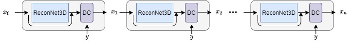
A recently developed deep learning model based on cascaded CNN , namely ReconNet3D, was employed for this study. The model design is shown in Figure.1, which is designed to perform k-space to image reconstruction with single-channel input and single-channel output. Therefore, each channel's data were independently reconstructed requiring no a priori information (such as coil sensitivity maps or k-space convolution kernel) from other channels, and the model was trained and tested as such. Inline image reconstruction pipeline with the ReconNet3D model was implemented on a 3T scanner (uMR890, United Imaging, Shanghai, China) for direct Dicom reconstruction.For noise and SNR evaluation, a sphere system phantom (CaliberMRI, Colorado) was scanned using the dual-channel VTC of the scanner. 3D GRE scans with 4x Poisson disc under-sampled k-space, as well as a fully sampled reference, were both performed 50 times. The images of each VTC channel were first reconstructed by the ReconNet3D model and then coil combined together. Voxel-wise mean and standard deviation (std) among repetitions was measured to generate the SNR maps.
Similar setups were also adopted for volunteer brain scans. Key scanning parameters included: TE/TE=3.7/20ms, FA=15°, voxel size = 1x1x3mm3.Scan time was 1:41 for 4x ReconNet3D acquisition, while it was 6:46 for full sampling. The option of parallel imaging was not available when using VTC, and thus was not tested.
Results
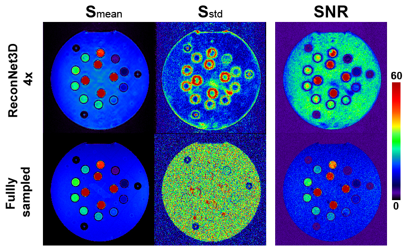
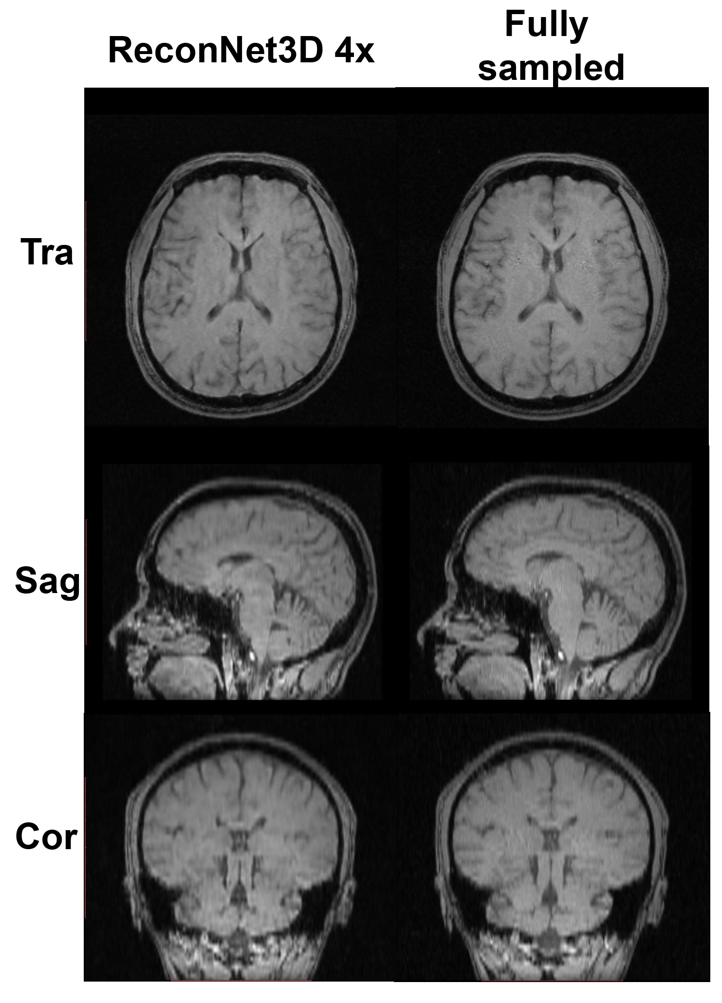
Figure.2 shows the mean images, signal std maps and SNR maps of the phantom. The ReconNet3D results show slight high spatial frequency artifacts in the backgrounds, but signal std of the phantom body was much lower than fully sampled results, thus yielding a much higher global SNR.Figure.3. compares brain images between 4x ReconNet3D and fully sampled results. The ReconNet3D images were slightly blurred compared to the fully sampled ones, but show no additional artifacts or elevated noise.
Discussion
The basic principle of substituting the spatial information from gradient encoding with those from the hardware4 (i.e. coil elements) has been well proven and implemented for parallel imaging (PI), compressed sensing (CS)5 and DL methods6.However, from the perspective on k-space's physical and mathematical properties, that each point in k-space is an overall integration of all excited spins, and the k-coordinates and spatial encodings are linearly connected. In principle, a single under-sampled k-space can be reconstructed, with a certain level of reliability, based merely on its own collected lines. This is difficult for routine model-based reconstruction methods such as PI and CS, as these methods can only see the information from the current data set. For DL methods, however, their unique training stage can be considered as the integration of the a prior information from numerous training data, which is generally on the order of thousands to millions. Therefore DL methods can 'see' information beyond the current data set, and hold the potential to reconstruct any single k-space. In this work, we have demonstrated the feasibility of the recently developed ReconNet3D for highly accelerated reconstruction with single-coil imaging.
Quantitative noise and SNR evaluation on the phantom clearly demonstrated that the ReconNet3D model, even with a 4x acceleration, yielded even higher SNR than the fully sampled results. On one hand, the Poisson Disc acquisition collected the outer parts of k-space in a more sparsely manner, and on the other, the ReconNet3D model has learned the integrated a prior information from all the training data. As a result, the ReconNet3D results enjoy much improved SNR, albeit with observable yet negligible blurriness.
Similarly for brain imaging, the ReconNet3D images showed highly similar image quality (Fig.3) to fully sampled images. However, by supporting much faster scan, pulsation artifacts caused by major cerebral arteries and visible in fully sampled images were eliminated in ReconNet3D image, demonstrating the benefit of acceleration even with VTC.
Conclusion
The capacity of a ReconNet3D deep learning model was demonstrated for highly accelerated image reconstruction using dual-channel VTC, suggesting the potential for imaging acceleration with surface coils or coils with limited channel counts, and thus potentially beneficial for preclinical imaging on live animals.Acknowledgements
No acknowledgement found.References
1. Dobbs NW, Budak MJ, White RD, Zealley IA. MR-Eye: High-Resolution Microscopy Coil MRI for the Assessment of the Orbit and Periorbital Structures,Part 1: Technique and Anatomy. Ajnr 2020;41(6):947-950.
2. Solis-Najera SE, Martin R, Vazquez F, Rodriguez AO. Surface coil with reduced specific absorption rate for rat MRI at 7 T. Magma (New York, NY2015;28(6):599-608.
3. Chen, E.Z., Ye, Y., Chen, X., Lyu, J., Zhang, Z., Hu, Y., Chen, T., Xu, J. and Sun, S., 2021. Accelerating 3D MULTIPLEX MRI Reconstruction with DeepLearning. arXiv preprint arXiv:2105.08163.
4. Ra JB, Rim CY. Fast imaging using subencoding data sets from multiple detectors. Magn Reson Med 1993;30(1):142-145.
5. Lustig M, Donoho D, Pauly JM. Sparse MRI: The application of compressed sensing for rapid MR imaging. Magn Reson Med 2007;58(6):1182-1195.
6. Ramzi Z, Ciuciu P, Starck J-L. Benchmarking MRI Reconstruction Neural Networks on Large Public Datasets. Applied Sciences. 2020;10(5):1816.https://doi.org/10.3390/app10051816
Figures