4032
Performance Evaluation of Deep Learning-based Image Reconstruction for Head and Neck Imaging Protocol
Amaresha Shridhar Konar1, Jaemin Shin2, Ramesh Paudyal1, Abhay Dave3, Maggie Fung2, Suchandrima Banerjee4, Vaios Hatzoglou5, and Amita Shukla-Dave1,5
1Medical Physics, Memorial Sloan Kettering Cancer Center, New York City, NY, United States, 2GE Healthcare, New York City, NY, United States, 3Touro College of Osteopathic Medicine, New York, NY, United States, 4GE Healthcare, Menlo Park, CA, United States, 5Radiology, Memorial Sloan Kettering Cancer Center, New York City, NY, United States
1Medical Physics, Memorial Sloan Kettering Cancer Center, New York City, NY, United States, 2GE Healthcare, New York City, NY, United States, 3Touro College of Osteopathic Medicine, New York, NY, United States, 4GE Healthcare, Menlo Park, CA, United States, 5Radiology, Memorial Sloan Kettering Cancer Center, New York City, NY, United States
Synopsis
Keywords: Machine Learning/Artificial Intelligence, Quantitative Imaging
MRI has excellent extracranial soft-tissue contrast to detect tumors in the head and neck (HN) region. Technical challenges arise due to MRI related artifacts. In routine radiological practice, HN MR imaging protocols are optimized specifically to the subsites. We aimed to evaluate the performance of the HN imaging protocol that include qualitative T1w, T2w, and quantitative diffusion MRI powered by a novel deep learning (DL) based reconstruction (recon) using the ACR and QIBA diffusion phantoms. This phantom study showed that qualitative T1w and T2w images and multiple b-value DWI data powered with DL recon substantially improves the image quality.Purpose:
In routine radiological practice, HN MR imaging protocols are optimized specifically to the subsite, such as the oral cavity, oropharynx, nasopharynx, nasal cavity, larynx, neck, parotid, and thyroid. The qualitative high-resolution, multiplanar T1- and T2-weighted (w) images are used to assess the location and extent of the HN tumors. However, quantitative analysis of diffusion MRI, a technique to measure tumor cellularity, can provide added value to the structural and anatomical evaluation of HN tumors by prognostic prediction and assessment of treatment response1-4. A novel deep learning-based MRI reconstruction pipeline was designed to address fundamental image quality limitations of conventional reconstruction to provide high-resolution, low-noise MR images5. This pipeline’s unique aims were to convert truncation artifacts into improved image sharpness while jointly denoising images to improve image quality5. This new approach, now commercially available as AIR Recon DL (GE Healthcare, Waukesha, WI), includes a deep convolutional neural network (CNN) to aid in the reconstruction of raw data, ultimately producing clean, sharp images. In this study, we evaluate the HN imaging protocol's performance, including qualitative T1w, T2w, and quantitative diffusion MRI powered by DL reconstruction, using the ACR and QIBA diffusion phantoms.Methods:
MRI data acquisition: HN MRI protocol consisted of T1 and T2w imaging followed by multi-b-value DWI on a 3 T MRI scanner (SIGNA Premier, GE Healthcare) using a 21-channel HN unit and at 1.5 T (Signa, GE Healthcare) using 19 channel HN unit. The medium-size ACR phantom was used for T1w and T2w imaging, and the QIBA diffusion phantom was used for DW-MRI. The acquisition parameters for T1w imaging were: FOV=25 cm, slice thickness=5 mm, slice gap=5 mm, the number of slices=11, and TR/TE= 550/8.1 ms, and for T2w imaging: TR/TE=2228/102 ms. The multi b-value (b=0, 50, 500, 1000 s/mm2) DW images were acquired using a single shot spin echo planar imaging (SS-SE-EPI) sequence with TR/TE=4000/77 (minimum) ms, the field of view (FOV)=20 cm, slice thickness=5 mm, number of excitation (NEX)=1, 4 and 6 for b=50, 500,1000 s/mm2, respectively. Another set of multi-b-value DW-MRI data was acquired using similar acquisition parameters by changing the NEX=1 for all the b-value. The qualitative and quantitative images were reconstructed using the standard and AIR Recon DL methods. The DL reconstruction was tested for all three settings, i.e., low, medium, and high, available at the scanner.MR image assessment: The T1w and T2w images acquired using the medium-size ACR phantom were reconstructed with and without DL recon and analyzed for the following assessment: 1. High contrast spatial resolution, 2. Percent Integral Uniformity (PIU), 3. Low contrast object detectability, 4. Gibbs (ringing) artifact. Similarly, the images obtained from the QIBA (DWI) phantom were reconstructed for standard and reduced NEX using the option of DL to measure the ADC values for the combinations mentioned above. The coefficient of variation (CV in %) was calculated and reported for all the combination measured ADC values.
Results:
Based on the four assessments performed on the ACR phantom study, the DL-based T1w and T2w images showed superior overall image quality compared to the images reconstructed without the DL method. The images acquired and reconstructed on 1.5T, and 3T with and without DL passed the Percent Integral Uniformity (PIU) test and the low contrast object detectability test. The high contrast spatial resolution test and Gibbs or ringing artifacts test showed improved image quality for DL-based images, as shown in Figure 1. A minimal difference was observed between the three different settings (low, medium, and high) of DL-based image reconstruction for both 3T and 1.5T scanners. Reduced ringing artifacts and improved image quality were observed in DL-reconstructed images obtained from the QIBA phantom for selected b-values, as shown in Figure 2. Diffusion images reconstructed using DL for reduced NEX=1 showed comparable image quality to standard NEX. The ADC maps obtained using the multi-b-value diffusion images reconstructed with and without DL for both standard and reduced NEX are shown in Figure 3. Figure 4 exhibits a bar plot for all combinations' measured mean ADC values. We did not observe a significant difference in measured ADC values between the images reconstructed with and without DL. The mean ADC values obtained from reduced NEX were comparable to Standard NEX. CV calculated for the measured ADC values are tabulated in Table 1, showing lower CV for DL based method.Discussion and Conclusion:
The SNR improvement offered by the DL Recon also provides opportunities for increasing spatial resolution. The ACR phantom study showed significant improvement by reducing ringing artifacts and increasing image sharpness. The different settings (low, medium, and high) available on the scanner to set the DL reconstruction did not show a significant difference between them. We observed a better reduction in the noise for the high setting than low or medium. The ADC values obtained from the QIBA diffusion phantom study showed similarities between the methods. Reduced NEX=1 significantly minimizes the image acquisition time, and the images reconstructed using the DL method showed comparable image quality and similar ADC values to the standard NEX. In conclusion, this phantom study showed that applying DL reconstruction on qualitative T1w and T2w images and DWI data substantially improves image quality.Acknowledgements
Funding support from National Institutes of Health Grant: U01 CA211205 (ASD)References
- Paudyal R, Chen L, Oh JH, et al. Nongaussian Intravoxel Incoherent Motion Diffusion Weighted and Fast Exchange Regime Dynamic Contrast-Enhanced-MRI of Nasopharyngeal Carcinoma: Preliminary Study for Predicting Locoregional Failure. Cancers (Basel). Mar 6 2021;13(5)doi:10.3390/cancers13051128
- Paudyal R, Konar AS, Obuchowski NA, et al. Repeatability of Quantitative Diffusion-Weighted Imaging Metrics in Phantoms, Head-and-Neck and Thyroid Cancers: Preliminary Findings. Tomography. Mar 2019;5(1):15-25. doi:10.18383/j.tom.2018.00044
- Paudyal R, Oh JH, Riaz N, et al. Intravoxel incoherent motion diffusion-weighted MRI during chemoradiation therapy to characterize and monitor treatment response in human papillomavirus head and neck squamous cell carcinoma. J Magn Reson Imaging. Apr 2017;45(4):1013-1023. doi:10.1002/jmri.25523
- Riaz N, Sherman E, Pei X, et al. Precision Radiotherapy: Reduction in Radiation for Oropharyngeal Cancer in the 30 ROC Trial. JNCI: Journal of the National Cancer Institute. 2021;113(6):742-751. doi:10.1093/jnci/djaa184
- Lebel RM. Performance characterization of a novel deep learning-based MR image reconstruction pipeline. arXiv preprint arXiv:200806559. 2020;
Figures
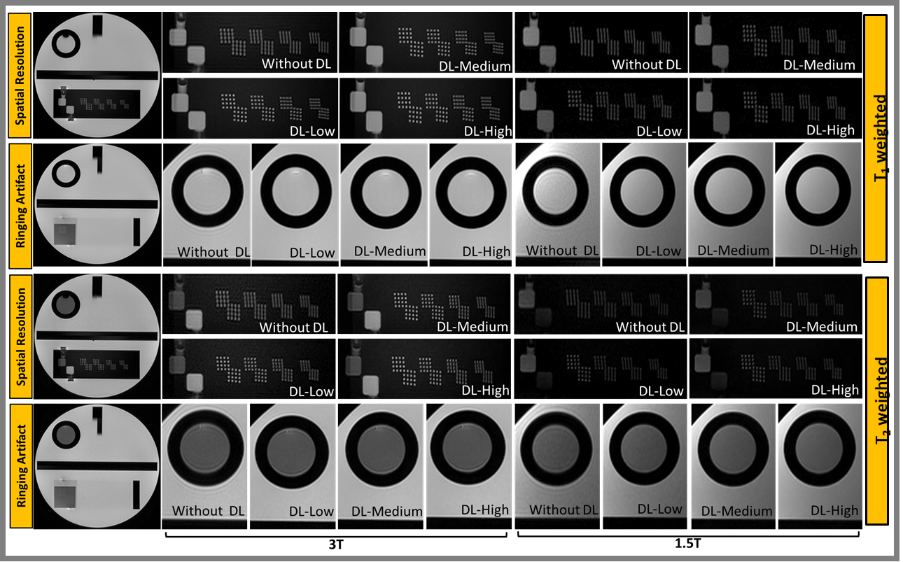
Figure 1:
ACR phantom
study performed on 3T and 1.5T to assess high contrast spatial resolution
and Gibbs artifact: The DL-based T1w and
T2w images showed superior overall image quality compared to those without DL.
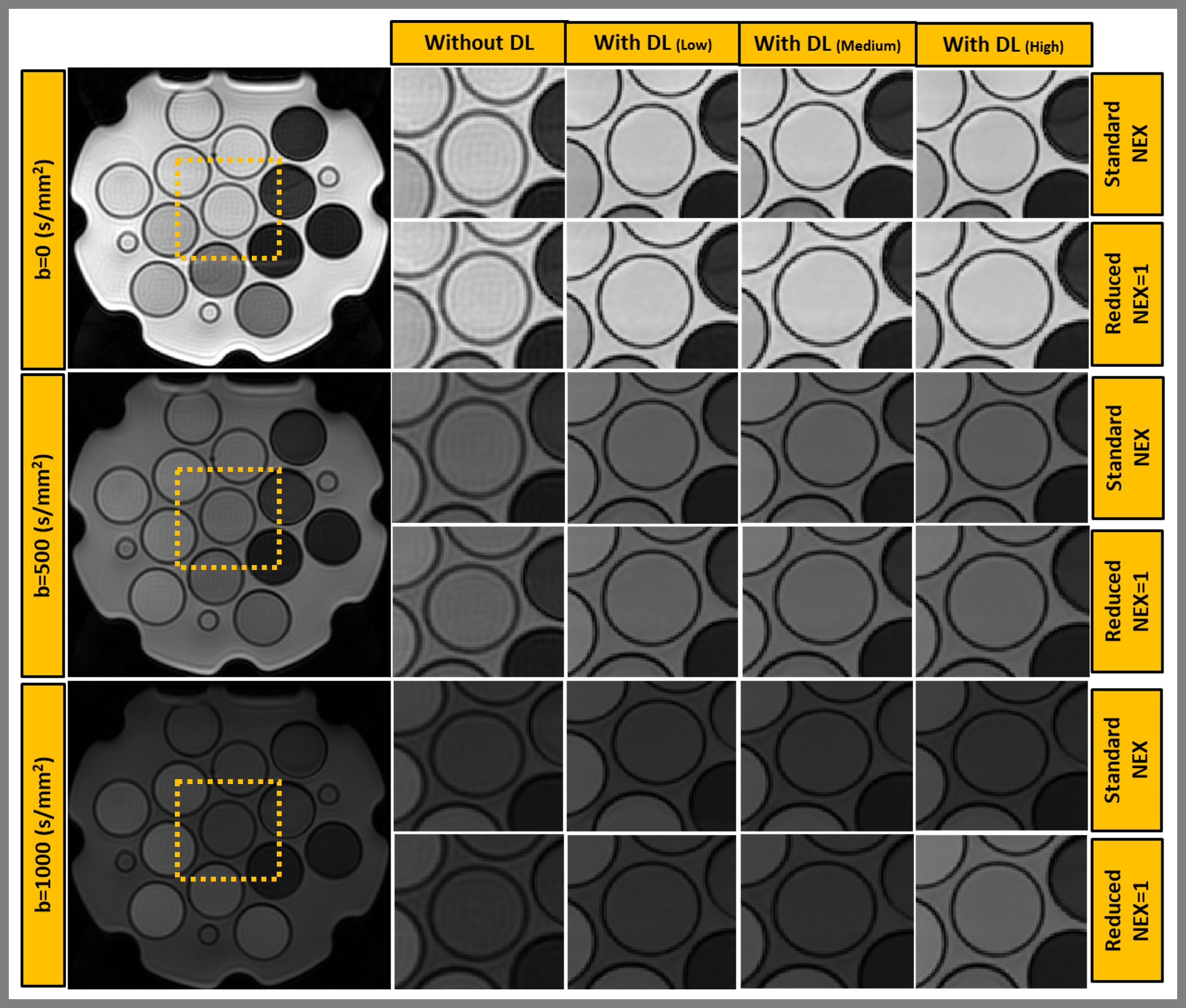
Figure 2: NIST/QIBA ice-water diffusion phantom imaged at 0°C showed reduced ringing artifacts, and improved image sharpness
for DL reconstructed images. Central vial magnified to show the reduction in
ringing artifacts and improved image quality. Reduced NEX=1 showed a comparable
result to standard NEX.
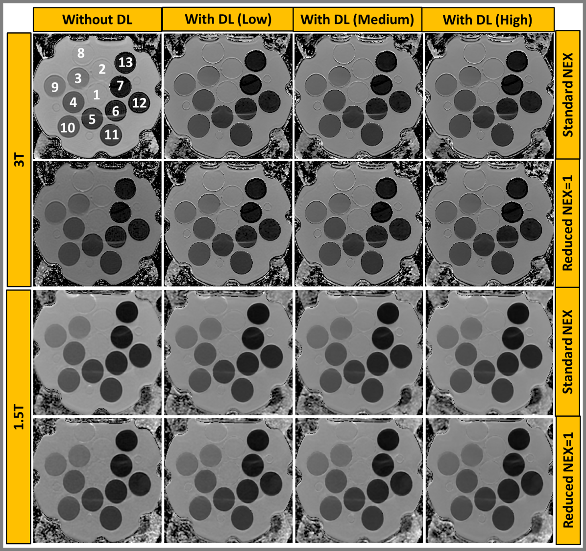
Figure 3: Apparent
Diffusion Coefficient (ADC ×10-3 mm2/s) maps were obtained from images reconstructed with and without DL on a 3T and a 1.5T.
The reduced NEX=1 showed comparable ADC maps to the standard NEX.
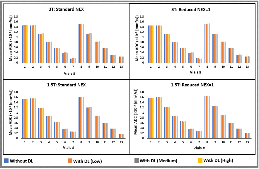
Figure 4:
Mean ADC values for the 13 vials: Values were obtained from DL-reconstructed
images for all three settings (low, medium, and high) and images without DL
reconstruction. Images were acquired with standard NEX and reduced NEX=1 on 3T and 1.5T scanners.

Table 1:
Coefficient of variation (in %) calculated for all the combinations of experiments
performed on a 3T and a 1.5T scanner
DOI: https://doi.org/10.58530/2023/4032