4018
Study of Oxidative Stress due to X-ray Irradiation in Mouse Brain using TOLD MRI Imaging
Raj Kumar Parajuli1, Megumi Ueno2, Saaya Suzuki1, Akira Sumiyoshi1, Takayuki Obata1, Ichio Aoki1, and Ken-ichiro Matsumoto2
1Department of Molecular Imaging and Theranostics, National Institutes for Quantum Science and Technology (QST), Chiba, Japan, 2Department of Radiation Regulatory Science Research, National Institutes for Quantum Science and Technology (QST), Chiba, Japan
1Department of Molecular Imaging and Theranostics, National Institutes for Quantum Science and Technology (QST), Chiba, Japan, 2Department of Radiation Regulatory Science Research, National Institutes for Quantum Science and Technology (QST), Chiba, Japan
Synopsis
Keywords: Oxygenation, Brain, Oxidative stress, TOLD, X-ray irradiation
This study aims to evaluate the metabolic changes caused on the brain tissues due to radiation exposure to healthy tissues while tumor treatment. The tissue oxidization and reduction mechanism represented by redox imaging could be a suitable index to evaluate in vivo oxidative stress in living animals that could evaluate metabolic damages due to irradiation. We adopted tissue oxygen level dependent (TOLD) MRI imaging to analyze TOLD signal for the control group mice and the irradiated mice. The result shows the distinct oxygen consumption levels in the control and the irradiated mice.Introduction
Radiotherapy is a sophisticated cancer/tumor treatment modality. However, it is likely to be affected from the late-onset side effects such as mucosal damages or radiation-induced reactions which could oxidize and/or reduce the biologically important molecules to chemically modify them. The tissue oxidization and reduction mechanism represented by redox imaging could be a suitable index to evaluate in vivo oxidative stress (Singh et al. 2019). Oxygen-sensitive MRI is considered as an attractive method to analyze the tumor progression since it is non-invasive and avoids the need for an exogenous reporter agent (O’Connor et al. 2019). Tissue oxygen level-dependent (TOLD) MRI signal is induced by the proton T1-shortening effect of molecular oxygen (Matsumoto et al. 2006). Since oxygen has two unpaired electrons on the molecule, oxygen can work as a T1-contrast agent like the gadolinium complex. Oxygen is a relatively lipophilic molecule. Lipid-rich brain tissue can show TOLD response sensitively. The tissue oxygen concentration is a result of oxygen supply and oxygen consumption. Tissue oxygenation level induced by an excess supply of oxygen probably be increased or suppressed when the tissue oxygen consumption level was varied after irradiation. Immunohistochemical studies show that the inhalation of carbogen (95% oxygen (O2) plus 5% carbon dioxide (CO2)) increases the oxygen diffusion (Kaanders et al. 2002). Therefore, the carbogen breathing probably makes a large excess oxygen supply and can push up resolved oxygen level in tissue very quickly to the maximum level.In this paper, the influence in the T1-maps of the mouse brain due to inhalation of normal air, carbogen, and 100% O2 were visualized and compared. Furthermore, TOLD MRI signal in the brain induced by carbogen breathing was compared between irradiated and non-irradiated mice. Effects of radiation on the brain tissue oxygen metabolism were analyzed.Materials and Methods
At first, T1-maps were acquired using rapid acquisition with relaxation enhancement with variable repetition time (RAREVTR) sequence for the control (unirradiated) C3H/He modal mice inhaling air, carbogen, and 100% O2 separately. Next, X-ray-irradiated, and control mice were used for TOLD analysis. 10 Gy x-ray irradiation on mouse brain through 1.0 cm silt of 5 cm thick lead blocks was performed at TITAN-E320 x-ray machine. Bruker’s 7T BioSpec MRI system with 1H receive-only four-channel phased array MRI cryogenic coil was used to acquire T1-weighted images by repeated scans of multiple-slice fast low angle shot (FLASH) sequence (repetition time (TR) = 164.0 ms, echo time (TE) = 2.3 ms, flip angle (FA) = 45°). The T1-weighted images with slice thickness of 1 mm without slice gap were acquired repeatedly for 24 times every 52 sec 480 ms. During the MRI scan, 1.5-2% isoflurane anesthetized mouse was simultaneously inhaled with air for the first initial 7th baseline image (7 min), and the gas was changed to carbogen for 8th to 16th image (7 min) and finally switched back to air again for 17th to 24th image (7 min).Results
Figure 1 shows the T1-maps for the controlled mouse when inhaled with normal air (Fig. 1a), carbogen (Fig. 1b), and 100% O2 (Fig. 1c) individually for 13 minutes each scan. Signal enhancement during carbogen inhalation was distinctly observed with respect to normal air and 100% O2 inhalation. Figure 2 shows the T1-weighted images of mouse brain induced by carbogen inhalation with respect to normal air inhalation for the control group mouse (Fig. 2a-c) and 10 Gy x-ray irradiated mouse on the same day after a few hours of irradiation (Fig. 2d-f) and 8 days after of x-ray irradiation (Fig. 2g-i) at 4th min (air inhalation), 12th min (carbogen inhalation) and 20th min (air inhalation) instant of MRI scan respectively. T1-weighted images were processed further using ImageJ software package to evaluate the normalized signal intensity of TOLD. Figure 3 shows the statistical abrupt increase in TOLD signal at 7th min as soon as the carbogen is inhaled by the mouse and diminish of the TOLD signal at 14th min when the carbogen supply was switched to normal air supply.Discussions and Conclusion
Comparing the T1-maps of the mice brain acquired for the inhalation of air, carbogen, and 100% O2, the TOLD signal were enhanced relatively high during carbogen inhalation than that of normal air and 100% O2 inhalation. This illustrates the advantage of using carbogen over 100% O2 for the TOLD signal analysis in this study. When the breathing gas was switched to air, the decay rate of the T1-weighted TOLD signal could reflect the oxygen consumption rate in the tissue. Oxygen consumption may have been decreased in the irradiated mouse brain with respect to the control group mouse brain and it was because of the damages in the mouse brain tissues due to x-ray irradiation. The results showed the possibility of the study of mouse brain tissue damages using the MRI TOLD analysis method. Detailed analysis of the TOLD imaging in mouse brain such as by varying the x-ray doses, days elapsed after x-ray irradiation, responses due to nitroxyl contrast agents are planned for future study.Acknowledgements
No acknowledgement found.References
- Singh A, Kukreti R, Saso L, Kukreti S. Oxidative stress: A key modulator in neurodegenerative diseases. Molecules 2019; 24:1583.
- O’Connor JPB, Probinson SP, Waterton JC. Imaging tumor hypoxia with oxygen-enhanced MRI and BOLD MRI. Br J Radiol 2019; 92:20180642.
- Matsumoto K, Bernardo M, Subramanian S, Choyke P, Mitchell JB, Krishna MC, Lizak MJ. MR assessment of changes of tumor in response to hyperbaric oxygen treatment. Magn Reson Med 2006; 56: 240–246.
- Kaanders JHAM, Bussink J, Kogel AJVD. ARCON: a novel biology-based approach in radiotherapy. Oncology 2002; 3:728-737.
Figures
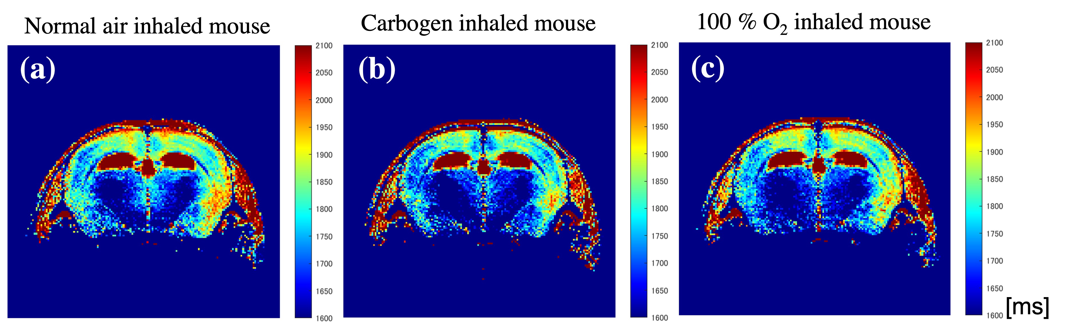
Fig. 1. Influence in the T1-maps of the control group mouse brain when inhaled with (a) normal air, (b) carbogen and (c) 100% oxygen.
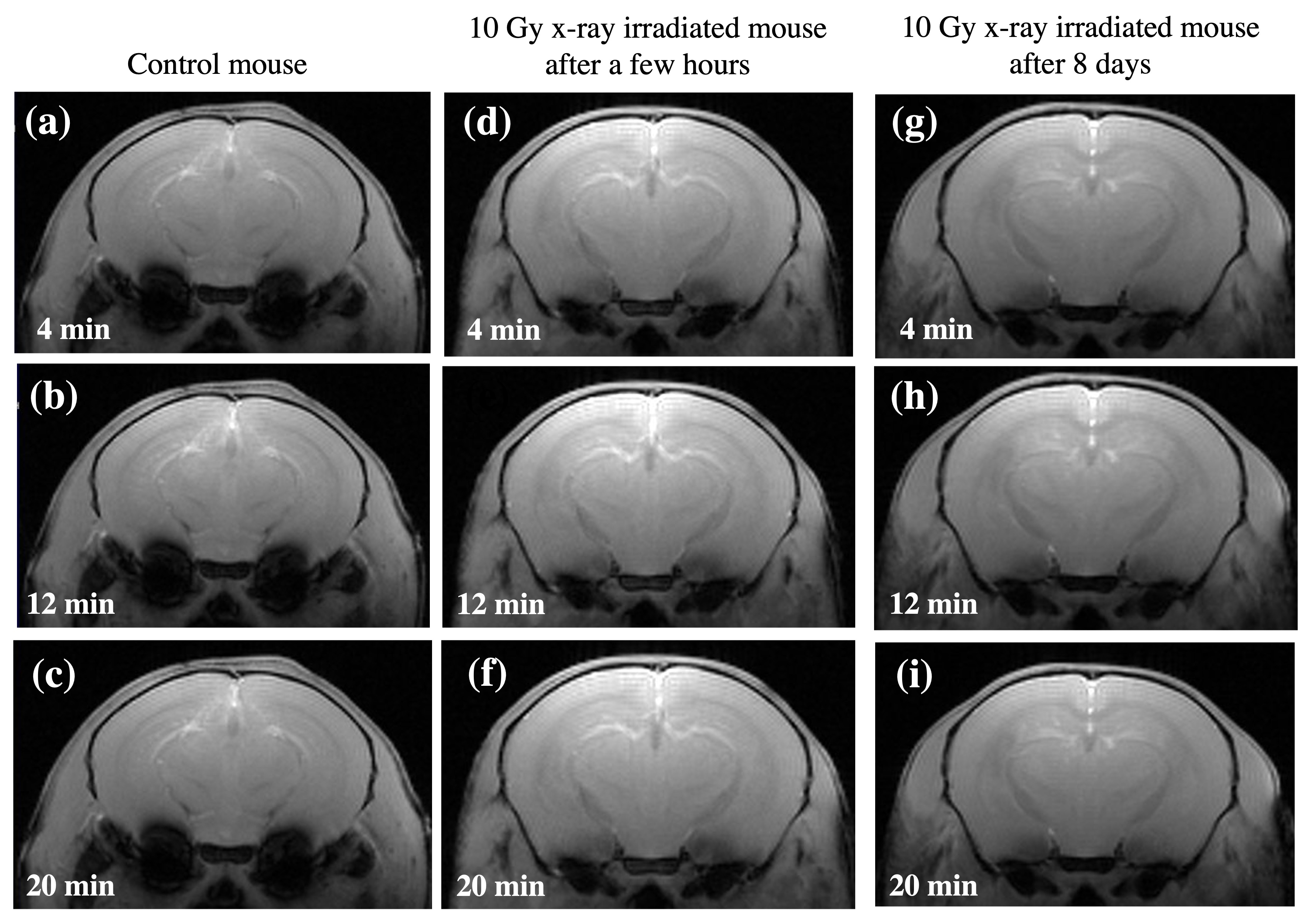
Fig. 2. T1-weighted images of control group mouse brain (Fig. 1a, b, c), 10 Gy x-ray irradiated mouse on same day after a few hours (Fig. 1d, e, f), and 8 days after irradiation (Fig. 1d, e, f) acquired at 4 min (air inhalation), 12 min (carbogen inhalation) and 20 min (air inhalation) respectively.
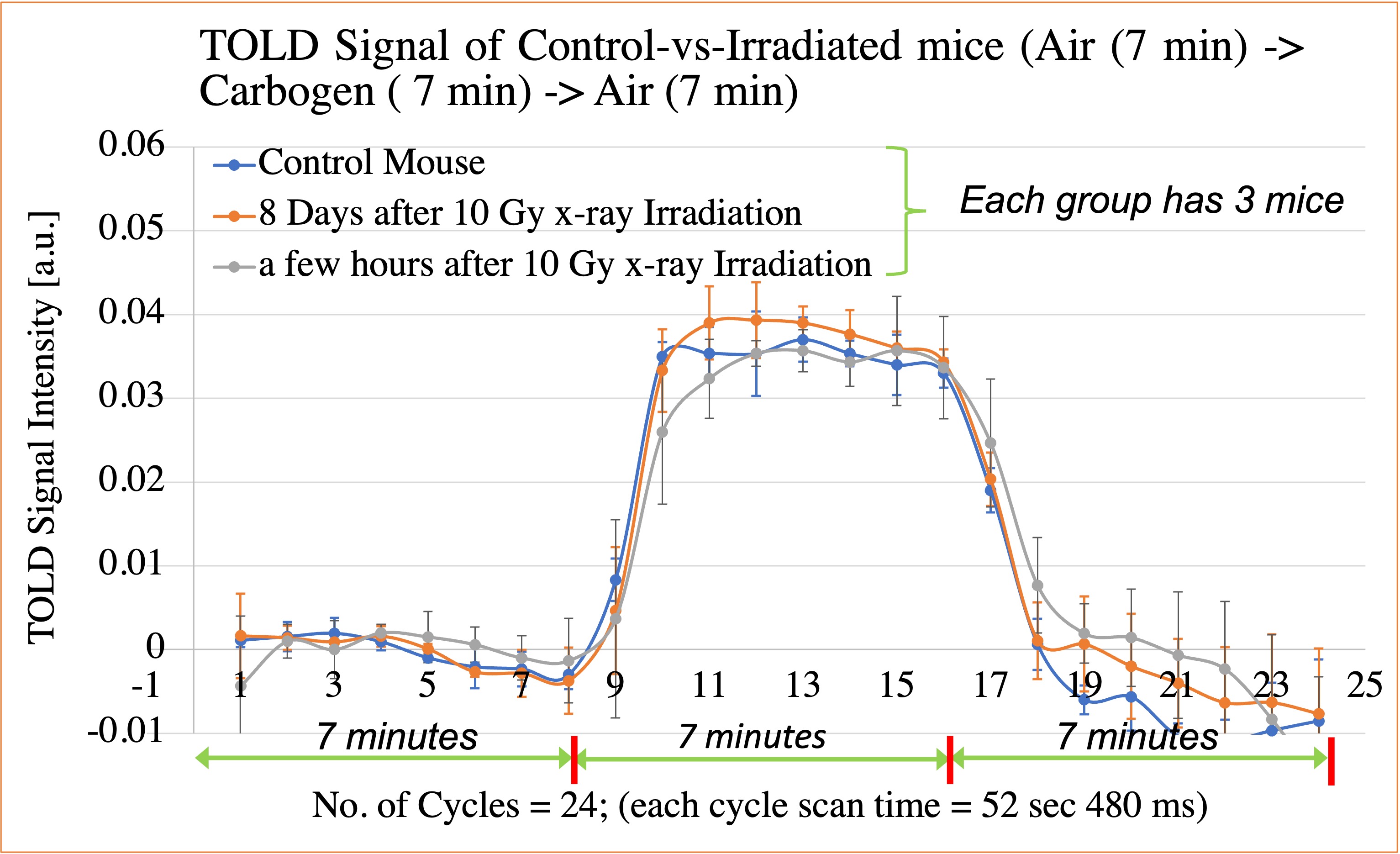
Fig. 3. Statistical analysis of the TOLD signal from T1-weighted images of the control group mouse brain, 10 Gy x-ray irradiated mouse on the same day after a few hours, and 8 days after irradiation respectively.
DOI: https://doi.org/10.58530/2023/4018