3990
Diffusion-weighted MRI of rectal cancers: Utility of reduced FOV single-shot EPI with tilted 2D RF excitation pulses
Atsuo Inoue1, Masahiro Tanabe1, Kenichiro Ihara1, Keiko Hideura1, Thomas Benkert2, Hiroshi Imai3, Masatoshi Yamane4, Takahiro Yamaguchi4, Mayumi Higashi1, and Katsuyoshi Ito1
1Department of Radiology, Yamaguchi University Graduate School of Medicine, Yamaguchi, Japan, 2MR Application Predevelopment, Siemens Healthcare GmbH, Erlangen, Germany, 3MR Research and Collaboration, Siemens Healthcare K.K., Tokyo, Japan, 4Department of Radiological Technology, Yamaguchi University Hospital, Yamaguchi, Japan
1Department of Radiology, Yamaguchi University Graduate School of Medicine, Yamaguchi, Japan, 2MR Application Predevelopment, Siemens Healthcare GmbH, Erlangen, Germany, 3MR Research and Collaboration, Siemens Healthcare K.K., Tokyo, Japan, 4Department of Radiological Technology, Yamaguchi University Hospital, Yamaguchi, Japan
Synopsis
Keywords: Pelvis, Diffusion/other diffusion imaging techniques, Rectum
This study evaluated the image quality of reduced FOV DWI using 2D spatially-selective RF pulses with tilted excitation plane (tilted r-DWI) for rectal cancer compared with full-FOV DWI (f-DWI) using readout segmented echo-planar imaging (RS-EPI). Two radiologists evaluated the MR images, and SNR, CNR, and ADC values of the rectal lesions were quantitatively evaluated. All image quality scores and CNR were significantly higher in tilted r-DWI than in f-DWI with RS-EPI. SNR and ADC values showed no significant deference between them. Tilted r-DWI provides better image quality with less artifacts and higher rectal lesion conspicuity than f-DWI with RS-EPI.Introduction
Several studies have shown that diffusion-weighted MR imaging (DWI) can help to detect, localize, and further characterize colorectal cancers in clinical practice 1. However, conventional DWI using single-shot echo-planar imaging (SS-EPI) sequence has limited spatial resolution and is sensitive to motion, magnetic field inhomogeneity, and susceptibility artifacts, leading to geometrical distortion and image blurring. Readout segmented (RS)-EPI using readout segmentation of long variable echo-trains (RESOLVE) technique has been an alternative sequence for obtaining DWI. DWI with RS-EPI exhibits reduced spatial distortion, improved resolution and improved susceptibility sensitivity compared with DWI using SS-EPI sequence. Several previous studies demonstrated that DWI based on RS-EPI is a clinically promising technique to improve the image quality for the purpose of evaluating lesions in patients with rectal cancers compared with DWI using SS-EPI sequence. Meanwhile one of the disadvantages of DWI with RS-EPI was the increased acquisition time. Recently, DWI with SS-EPI sequences performed with a reduced phase direction field-of-view (FOV) technique by using two-dimensional (2D) spatially-selective radiofrequency (RF) pulses with tilted excitation plane has been developed and applied to MR examinations 2. This reduced FOV DWI (tilted r-DWI) technique allows direct excitation of small rectangular areas and provides high quality images with improved spatial resolution and less ghosting and susceptibility artifacts related to motion and intestinal gas compared to conventional full-size FOV DWI (f-DWI) 3-5. However, there has been no reports assessing the utility of tilted r-DWI for the imaging of rectal cancers. The purpose of this study was to evaluate the image quality qualitatively and quantitatively as well as apparent diffusion coefficient (ADC) values of tilted r-DWI for the rectal cancers compared with f-DWI using RS-EPI.Materials and methods
The study group included 22 patients with rectal cancer who underwent pelvic MR examinations including both f-DWI (RESOLVE) and tilted r-DWI sequence on a 3T scanner (MAGNETOM Prisma or Skyra, Siemens Healthcare, Erlangen, Germany) (Table 1). Two experienced radiologists independently participated in the qualitative image analysis. The datasets of f-DWI and tilted r-DWI were analyzed in random order for the following parameters such as artifacts and image quality using a 4-point scale: presence of ghost/motion artifacts from physiological movement, susceptibility artifacts at tissue-air interface; anatomical visualization of the rectum; interslice signal homogeneity; overall image quality; and rectal lesion conspicuity. For quantitative analysis, signal-to-noise ratio (SNR), contrast-to-noise ratio (CNR) and ADC values were measured using regions of interest (ROIs). Wilcoxon signed-rank test was performed to compare the qualitative visual assessment and the quantitative assessment in SNR and CNR. For comparison of the ADC values, paired-sample t-test was performed.Results
In the qualitative visual assessment, all image quality scores were significant higher (P < 0.01 for all) in tilted r-DWI than in f-DWI with RS-EPI: anatomical visualization of the rectum, 3 (1) vs. 2 (1); signal homogeneity, 3 (0) vs. 2 (1); presence of ghosting artifacts, 3 (0) vs. 2 (1); presence of susceptibility artifacts, 3 (1) vs. 2 (0); rectal lesion conspicuity, 4 (1) vs. 2 (0); and overall image quality, 3 (1) vs. 2 (0), respectively (Table 2). In the quantitative evaluation, CNR in tilted r-DWI was significantly higher than in f-DWI with RS-EPI (4.92 ± 2.73 vs. 3.58 ± 2.04, P<0.01). On the other hand, SNR showed no significant difference between tilted r-DWI and f-DWI with RS-EPI (130 ± 107 vs. 123 ± 80, P=0.86). Regarding the ADC values, there was no significant difference between median ADC values of the rectal cancers calculated from tilted r-DWI and those from f-DWI with RS-EPI (921 ± 162 vs. 898 ± 134, P=0.27) (Table 3).Discussion
This study showed that tilted r-DWI can provide significantly better image quality with less artifacts and higher conspicuity of anatomical structure and rectal cancers than f-DWI with RS-EPI. Tilted r-DWI using 2D selective RF pulses and reduced FOV in the phase-encoding direction improved aliasing artifacts from tissue and fat outside the reduced FOV, minimized susceptibility artifacts by reducing the air-tissue interface, and improved conspicuity provided by higher in-plane resolution, compared to f-DWI with RS-EPI. In the objective evaluation, CNR was significantly higher in tilted r-DWI compared to f-DWI with RS-EPI consistent with previous studies. Conversely, there was no significant difference in SNR. Several previous reports have shown that SNR decreases with decreasing FOV. Theoretically, SNR decreases with smaller voxel size. However, in this study, the number of excitations was increased, presumably maintaining an SNR comparable to that of f-DWI. With respect to ADC assessment, no significant difference was found between the median ADC values of f-DWI with RS-EPI and tilted r-DWI. This fact suggested that ADC values measured on tilted r-DWI may be used to predict response to neoadjuvant chemoradiation therapy or as a noninvasive marker of tumor aggressiveness as the results of previous studies.Conclusion
Tilted r-DWI using 2D selective RF pulses and reduced FOV in the phase-encoding direction provides better image quality with less artifacts and higher rectal lesion conspicuity than f-DWI with RS-EPI, indicating the feasibility of this MR sequence in evaluating rectal cancer in clinical practice.Acknowledgements
No acknowledgement found.References
1. Soyer P, Lagadec M, Sirol M, et al. Free-breathing diffusion-weighted single-shot echo-planar MR imaging using parallel imaging (GRAPPA 2) and high b value for the detection of primary rectal adenocarcinoma. Cancer imaging : the official publication of the International Cancer Imaging Society 2010;10:32-39.2. Finsterbusch J. Fast-spin-echo imaging of inner fields-of-view with 2D-selective RF excitations. Journal of magnetic resonance imaging : JMRI 2010;31(6):1530-1537.
3. Finsterbusch J. Improving the performance of diffusion-weighted inner field-of-view echo-planar imaging based on 2D-selective radiofrequency excitations by tilting the excitation plane. Journal of magnetic resonance imaging : JMRI 2012;35(4):984-992.
4. He YL, Hausmann D, Morelli JN, Attenberger UI, Schoenberg SO, Riffel P. Renal zoomed EPI-DWI with spatially-selective radiofrequency excitation pulses in two dimensions. European journal of radiology 2016;85(10):1773-1777.
5. Hwang J, Hong SS, Kim HJ, et al. Reduced field-of-view diffusion-weighted MRI in patients with cervical cancer. The British journal of radiology 2018;91(1087):20170864.
Figures
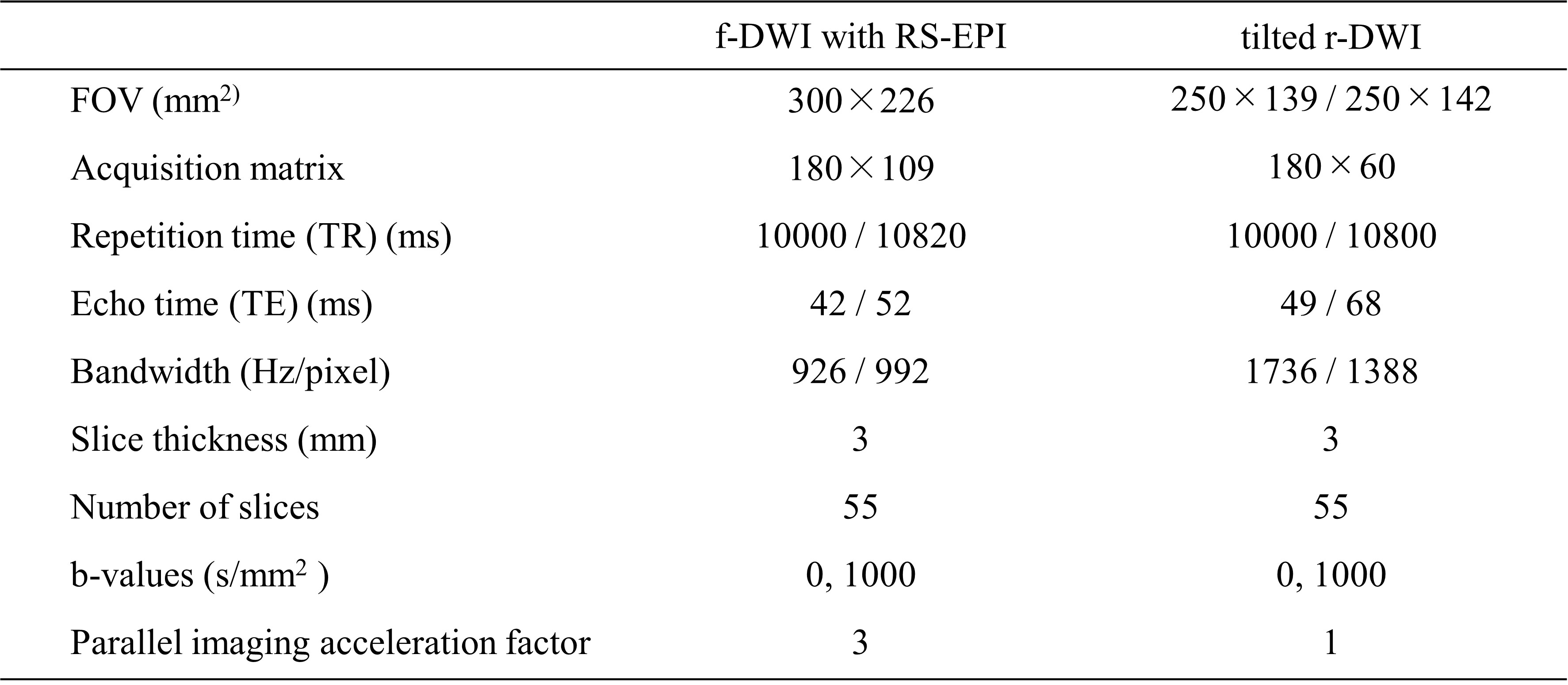
Table 1. Imaging parameters for f-DWI with RS-EPI and tilted r-DWI.
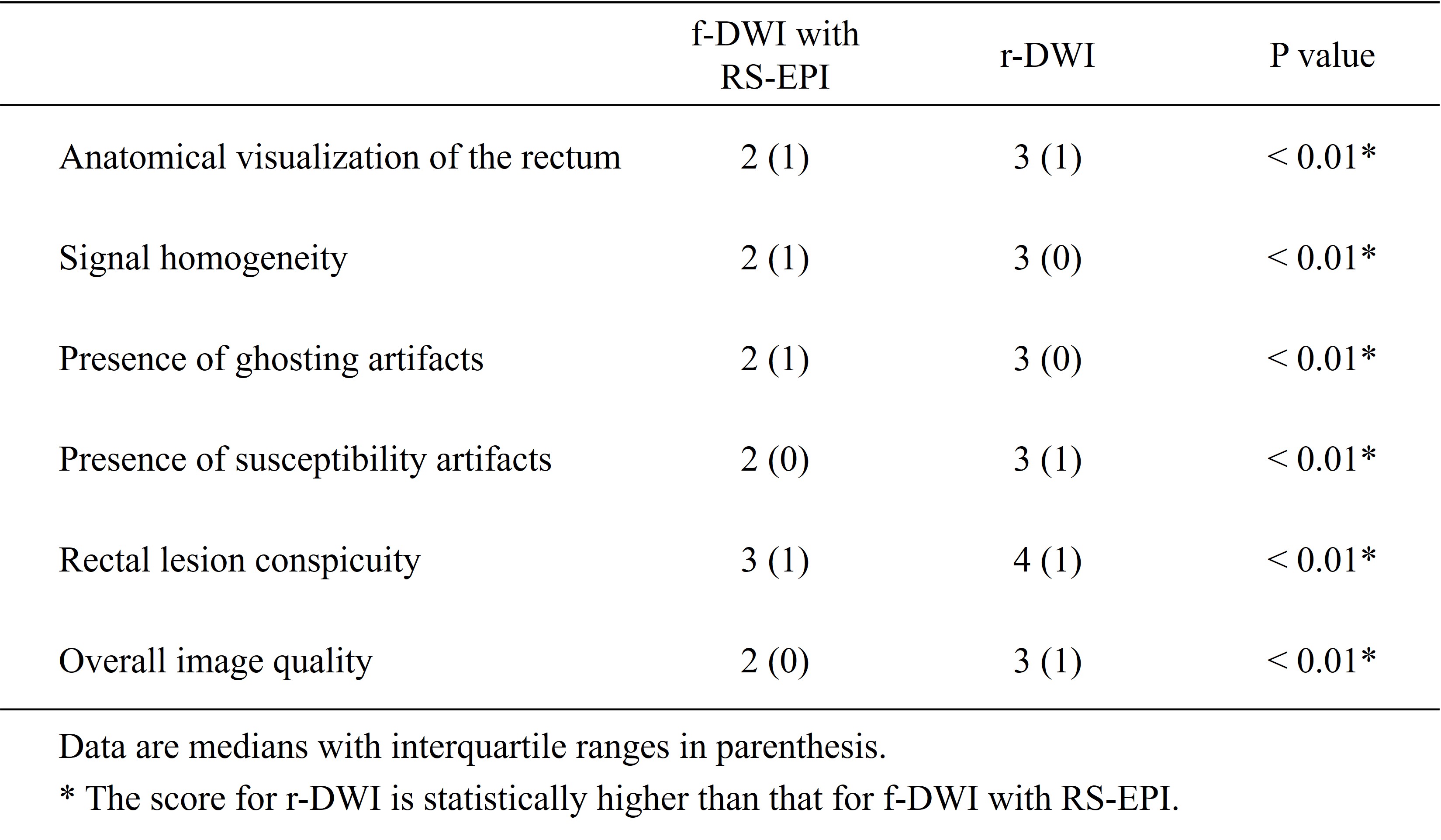
Table 2. Comparison of image quality and conspicuity between f-DWI with RS-EPI and tilted r-DWI.
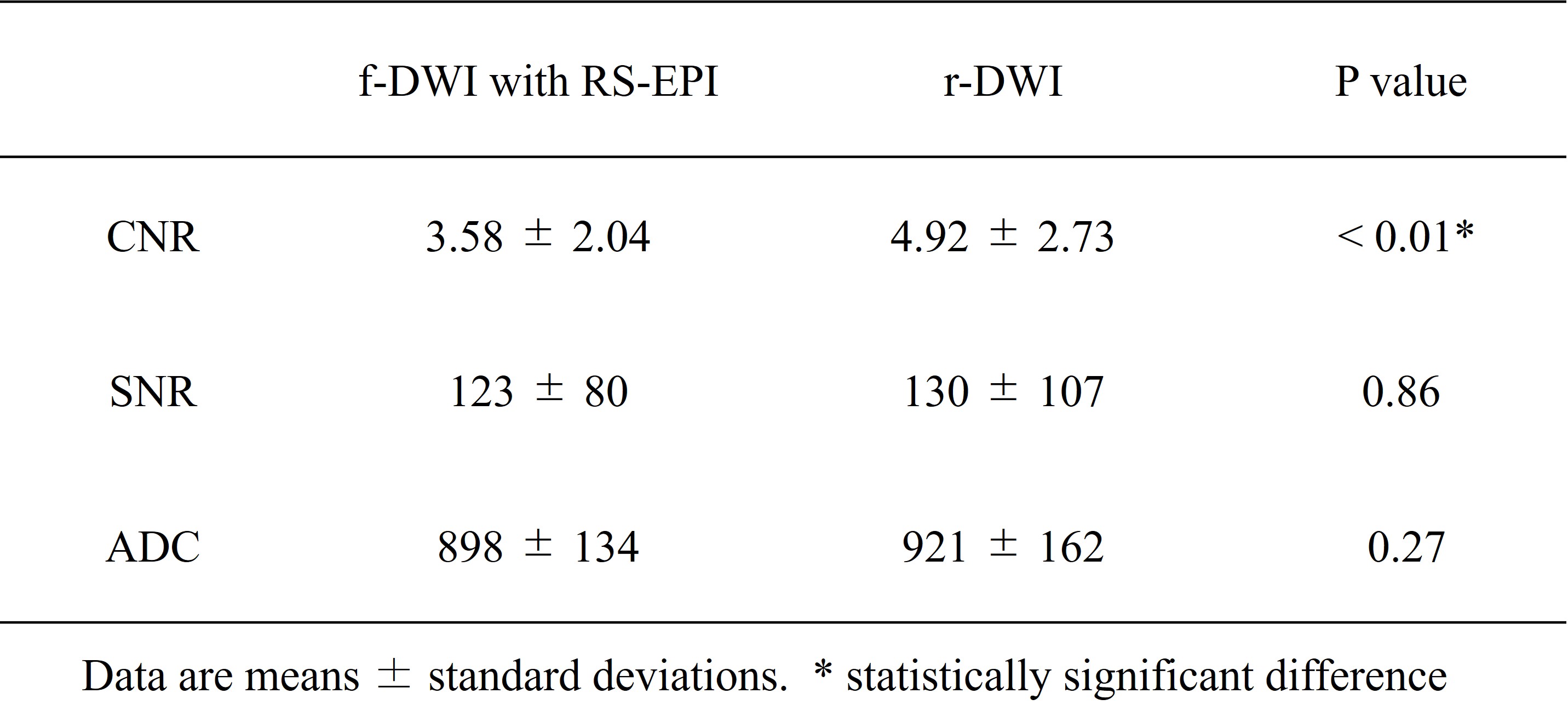
Table 3. Comparison of CNR, SNR and ADC values between f-DWI with RS-EPI and tilted r-DWI.
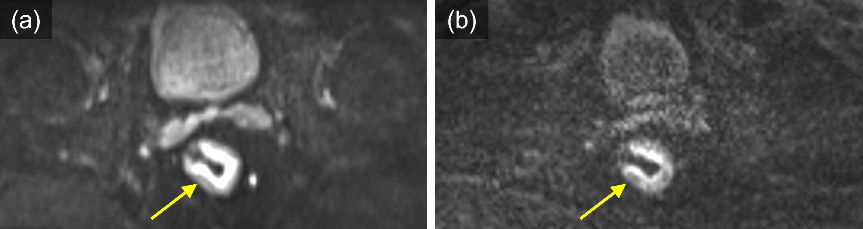
Figure 1. MR images from a patient with rectal cancer. Lesion conspicuity in tilted r-DWI (a) is significantly better than that in f-DWI with RS-EPI (b).
DOI: https://doi.org/10.58530/2023/3990