3982
Modified reduced FOV diffusion-weighted MRI using tilted 2D RF excitation pulses in patients with pancreatic ductal adenocarcinoma1Department of Radiology, Yamaguchi University Graduate School of Medicine, Ube, Japan, 2Radiology, Yamaguchi University Graduate School of Medicine, Ube, Japan, 3MR Application Predevelopment, Siemens Healthcare GmbH, Erlangen, Germany, 4MR Research and Collaboration, Siemens Healthcare K.K., Tokyo, Japan, 5Department of Radiological Technology, Yamaguchi University Hospital, Ube, Japan, 6Yamaguchi University Hospital, Ube, Japan
Synopsis
Keywords: Pancreas, Diffusion/other diffusion imaging techniques
This study evaluated the image quality of modified reduced field-of-view (FOV) DWI using 2D spatially-selective RF pulses with tilted excitation plane (tilted r-DWI) for pancreatic ductal adenocarcinomas (PDACs) compared with conventional full-FOV DWI (f-DWI). Two radiologists evaluated the MR image quality, and SNR, CNR, and ADC values of the pancreatic lesions were quantitatively assessed. Image quality scores and CNR of tilted r-DWI were significantly higher than that of f-DWI. The ADC values of PDACs showed no significant deference between them. Tilted r-DWI provides better image quality with less artifacts and higher pancreatic lesion conspicuity than f-DWI.Introduction
The value of diffusion-weighted (DW) imaging in the diagnosis of pancreatic ductal adenocarcinomas (PDAC) has been recognized because of its excellent contrast resolution showing hyperintensity of PDAC compared with the rest of the pancreatic parenchyma. Meanwhile, PDACs are often not depicted on DWI as hyperintense with clearly defined borders with the surrounding pancreatic parenchyma, probably due to its intrinsic drawbacks of DWI using single-shot echo-planar imaging (EPI) technique. These included blurring in the phase-encoding direction, low in-plane spatial resolution, susceptibility artifacts from adjacent intestinal gas, and chemical shift, which result in ghosting and geometric distortion.Single-shot EPI performed with a reduced field-of-view (FOV) in the phase-encoding direction combined with two-dimensional (2D) spatially-tailored radiofrequency (RF) excitation pulses has been developed to overcome these limitations. This reduced FOV DWI (r-DWI) technique allows direct excitation of a small rectangular volume of interest, leading to decreased ghosting and susceptibility artifacts, increased spatial resolution, and improved image quality 1-3. However, given the limited sampling of excitation k-space of the 2D RF pulses in r-DWI, aliasing artifacts in the phase-encoding direction which appear within the image plane cannot be completely eliminated due to side excitation caused by the periodicity of the excited profile 4.
Recently, modified r-DWI using the spatially-tailored 2D RF pulses with tilted excitation plane (tilted r-DWI) has been developed to resolve this issue. Tilted r-DWI can eliminate aliasing artifacts in the phase-encoding direction since the unwanted side excitations can be positioned in the blind spot between the image plane and the slices to be acquired 1. Improved image quality of the tilted r-DWI using the 2D RF excitation plane has been demonstrated for the normal pancreas 5. However, tilted r-DWI has not yet been used for the imaging of the pancreatic adenocarcinomas. Therefore, the purpose of this study was to evaluate the impact on image quality and quantitative apparent diffusion coefficient (ADC) values of tilted r-DWI for the pancreatic adenocarcinomas in comparison to the conventional full-FOV DWI (f-DWI).
Materials and methods
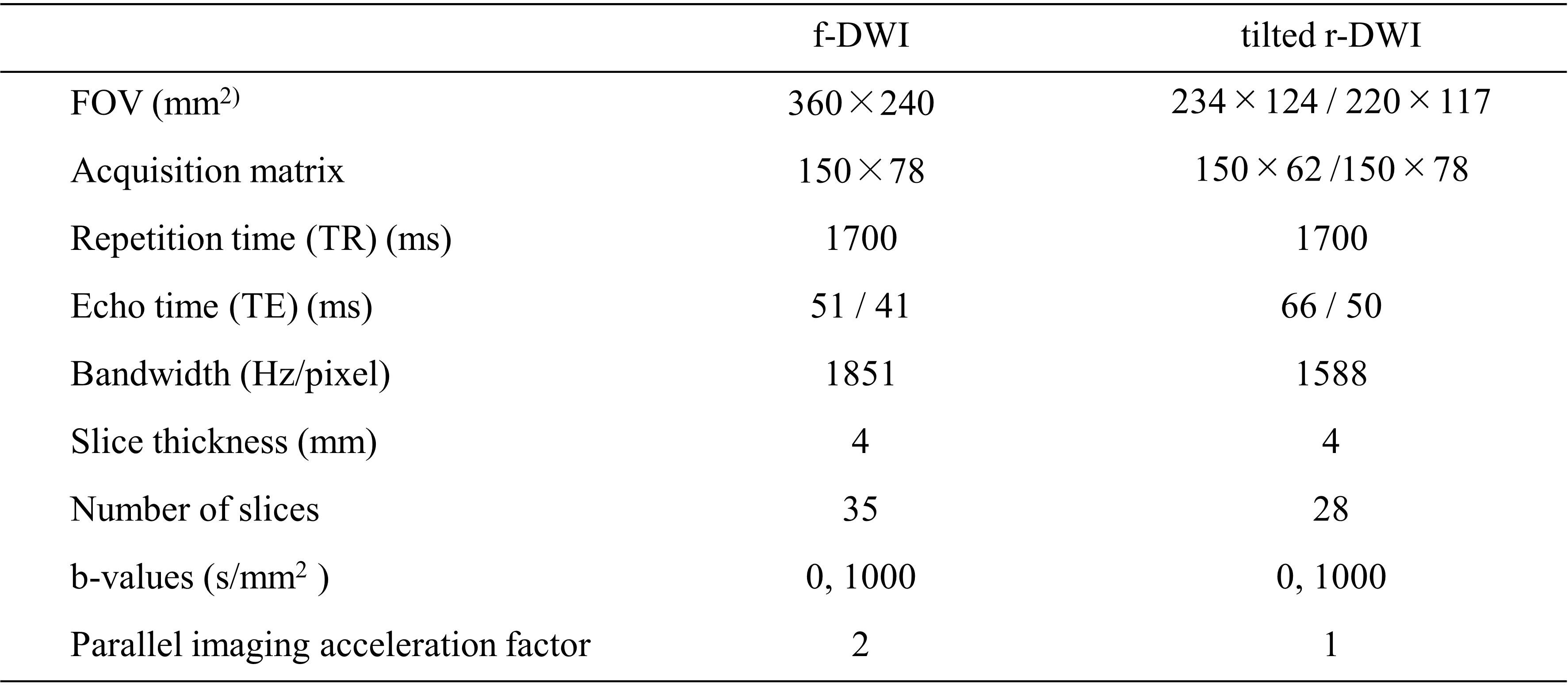
This study included 18 patients with pathologically confirmed PDAC. Patients were scanned on a 3T MR system (MAGNETOM Prisma or Skyra, Siemens Healthcare, Erlangen, Germany). In all patients, the pancreatic MR protocol included both f-DWI with a full-size FOV and tilted r-DWI with a reduced-size FOV using the spatially-tailored 2D RF pulses with tilted excitation plane (Table 1). Two experienced radiologists independently performed the qualitative image analysis. Each data set of f-DWI and tilted r-DWI was randomly analyzed using a 4-point scale to evaluate the following parameters of artifacts and image quality: presence of blurring or ghost artifacts, susceptibility artifacts, and aliasing artifacts; anatomic visualization of the pancreas; interslice signal homogeneity; overall image quality; and conspicuity of the PDAC. As the quantitative analysis, signal-to-noise ratio (SNR), contrast-to-noise ratio (CNR) and ADC values were measured using regions of interest (ROIs). Wilcoxon signed-rank test was performed to compare the qualitative scores and the quantitative values.Results
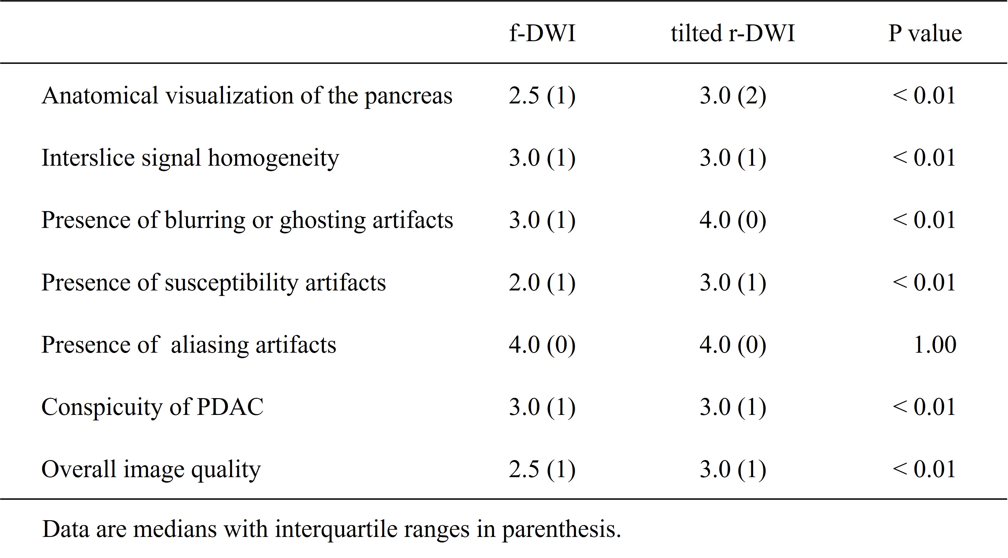
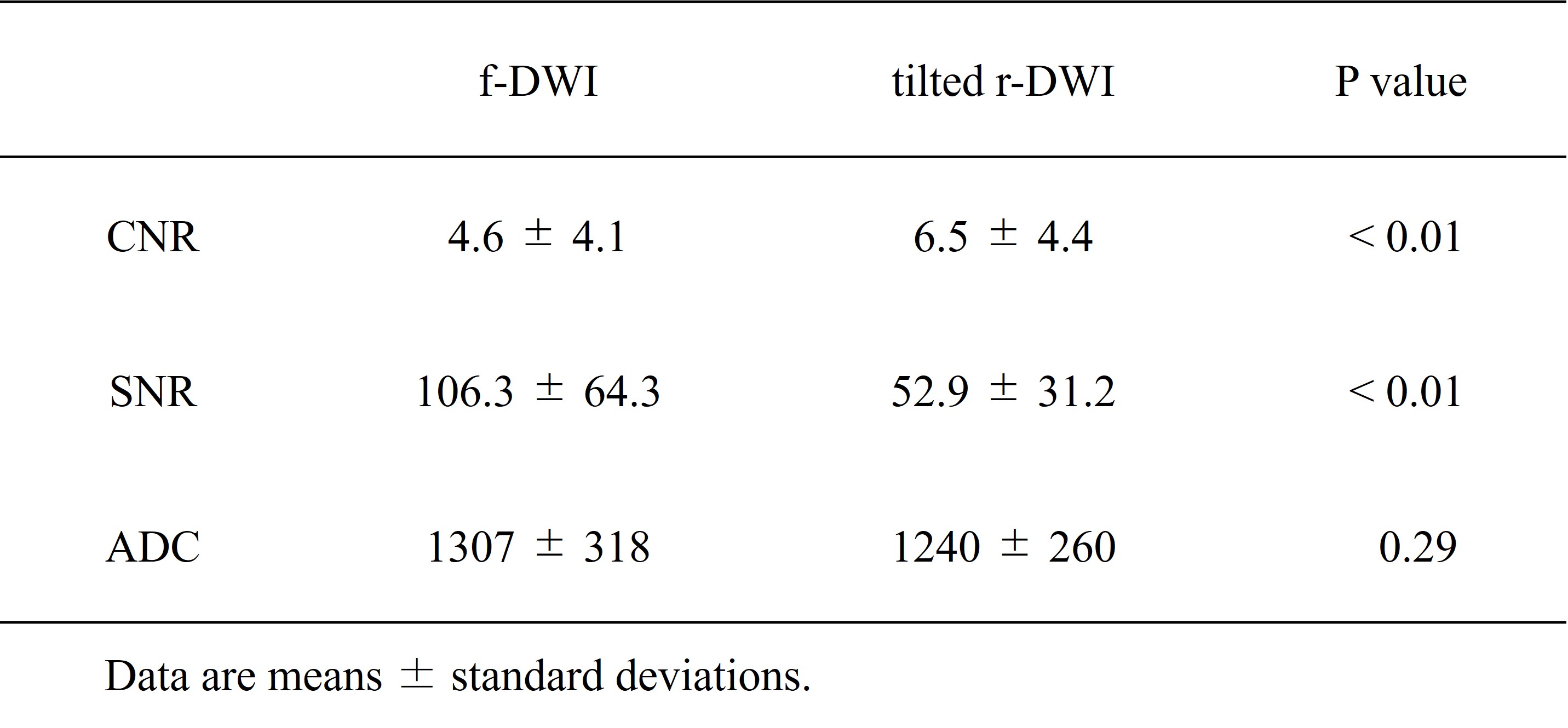
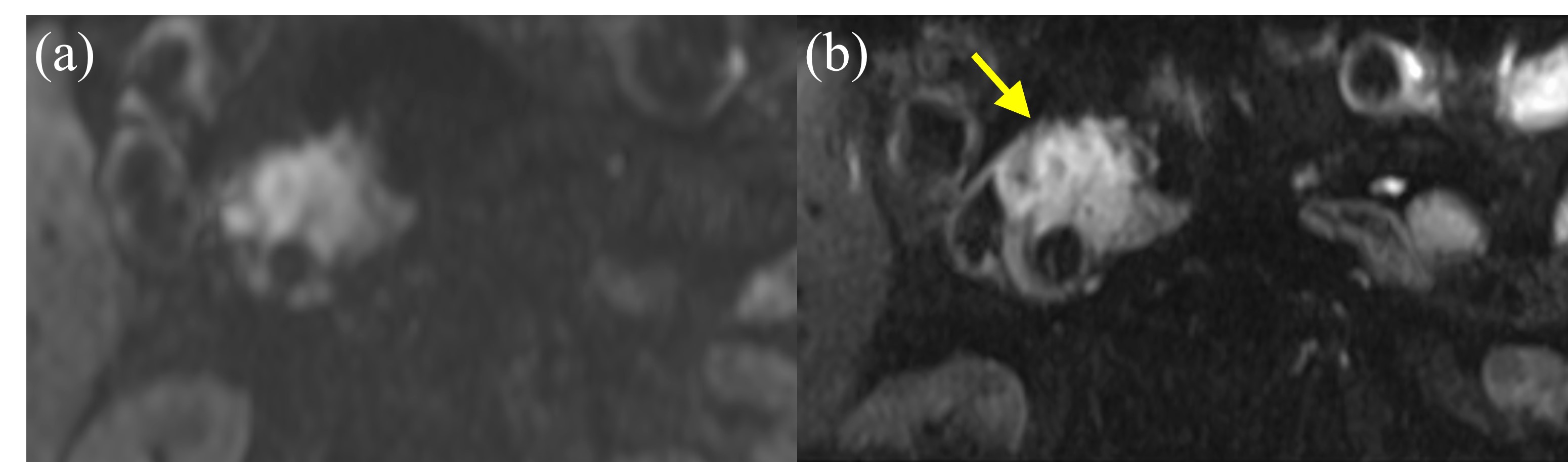
In the qualitative visual assessment, all image quality scores in tilted r-DWI were significantly higher than those in f-DWI (anatomic visualization of the pancreas, 3.0 [2] vs. 2.5 [1], p<0.01; interslice signal homogeneity, 3.0 [1] vs. 3.0 [0], p<0.01; presence of blurring or ghosting artifacts, 4.0 [0] vs. 3.0 [0], p<0.01; presence of susceptibility artifacts, 3.0 [1] vs. 2.0 [1], p<0.01; presence of aliasing artifacts, 4.0 [0] vs. 4.0 [0], p=1.00; conspicuity of PDAC, 3.0 [1] vs. 3.0 [1], p<0.01; and overall image quality, 3.0 [1] vs. 2.5 [1], p<0.01) (Table 2).The CNR of PDAC was significantly higher in tilted r-DWI than in f-DWI (6.5 ± 4.4 vs. 4.6 ± 4.1, p<0.01). Conversely, SNR of tilted r-DWI was significantly lower than that of f-DWI (52.9 ± 31.2 vs. 106.3 ± 64.3, p<0.01). No significant difference was observed between mean ADC values of the PDAC calculated from tilted r-DWI (tilted r-ADC) and those from f-DWI (f-ADC) (1240 ± 260 vs. 1307 ± 318, p=0.29) (Table 3).
Discussion
In this study, tilted r-DWI provided higher subjective image quality scores and better CNR compared with f-DWI without affecting ADC quantification, while SNR decreases. The higher in-plane resolution for tilted r-DWI using the reduced FOV in the phase-encoding direction than that for f-DWI contributed to the improved conspicuity of PDAC as well as pancreatic parenchyma. Regarding aliasing artifacts, there was no statistical difference between tilted r-DWI and f-DWI. However, tilted r-DWI achieved the highest score of 4 in all patients, indicating that no aliasing artifacts were observed, and showing the benefit of the use of tilted excitation plane in r-DWI using the spatially-tailored 2D RF pulses. Several studies reported that the r-DWI sequence with a smaller FOV and higher matrix size could result in a lower SNR compared with the f-DWI. However, CNR of tilted r-DWI, which is important for assessment of lesions, was significantly higher than that of f-DWI, which potentially can be attributed to fewer artifacts.Conclusion
The r-DWI using 2D RF techniques with a tilted excitation plane was shown to significantly improve image quality and CNR and reduce image artifacts compared to f-DWI techniques in the MRI of PDAC without significantly affecting the ADC quantification.Acknowledgements
No acknowledgement found.References
1. Finsterbusch J. Improving the performance of diffusion-weighted inner field-of-view echo-planar imaging based on 2D-selective radiofrequency excitations by tilting the excitation plane. Journal of magnetic resonance imaging : JMRI 2012;35(4):984-992.
2. He YL, Hausmann D, Morelli JN, Attenberger UI, Schoenberg SO, Riffel P. Renal zoomed EPI-DWI with spatially-selective radiofrequency excitation pulses in two dimensions. European journal of radiology 2016;85(10):1773-1777.
3. Hwang J, Hong SS, Kim HJ, et al. Reduced field-of-view diffusion-weighted MRI in patients with cervical cancer. The British journal of radiology 2018;91(1087):20170864.
4. Zhao L, Madore B, Panych LP. Reduced field-of-view MRI with two-dimensional spatially-selective RF excitation and UNFOLD. Magnetic resonance in medicine 2005;53(5):1118-1125.
5. Tanabe M, Higashi M, Benkert T, et al. Reduced Field-of-View Diffusion-Weighted Magnetic Resonance Imaging of the Pancreas With Tilted Excitation Plane: A Preliminary Study. Journal of magnetic resonance imaging : JMRI 2021;54(3):715-720.
Figures