3970
Volume isotropic thin-slice high-quality brain DWI with no fat-suppression pre-pulse1Radiology, Eastern Chiba Medical Center, Chiba, Japan, 2Philips Japan, Tokyo, Japan, 3Philips Australia & New Zealand, North Ryde, Australia, 4Faculty of Health Sciences, Institute of Medical, Pharmaceutical and Health Sciences, Kanazawa University, Kanazawa, Japan, 5General Medical Services, Chiba University Graduate School of Medicine, Chiba, Japan, 6Orthopaedic Surgery, Eastern Chiba Medical Center, Chiba, Japan
Synopsis
Keywords: Data Acquisition, Diffusion/other diffusion imaging techniques
We hypothesized that LIPO-only (LION) DWI might be one of the best solutions to improve the image quality of volumetric thin-slice DWI if it is further optimized to increase the robustness of fat suppression. We investigated the clinical usefulness of proposed LION fat suppression combined with thin-slice DWI for brain volumetric DWI in patients with acute stroke. LION-DWI has improved SNR compared to conventional DWI and has superior visual lesion detectability in patients with acute stroke. Therefore, it is possible to improve the image quality and shorten the imaging time of thin-slice DWI by changing the fat suppression to LION.Introduction
Diffusion-weighted imaging (DWI) has a high sensitivity exceeding 90% for detecting acute ischemia1,2 and is routinely used for this purpose in clinical practice. On the other hand, false-negative rates are high in the brainstem and cerebellum, and infarcts are often small and inconspicuous, complicates the diagnosis of stroke using EPI-based DWI sequences3. To solve this problem, a study using thin-slice DWI with a slice thickness of 2.5 mm reported better detection of ischemic lesions than the common 5 mm slice thickness4. In addition, thin-slice DWI enables multi-planar reconstruction, which allows observation from multiple directions in addition to axial cross section. However, thin-slice DWI has a potentially lower signal-to-noise ratio (SNR) and requires longer imaging time to ensure lesion detection.Slice selection gradient reversal (SSGR, LIPO)5 technique is commonly used in combination with fat-suppression pre-pulse (SPIR/SPAIR) to increase the robustness of fat suppression at high fields. We hypothesized that LIPO-only (LION) DWI might be one of the best solutions to improve the image quality of volumetric thin-slice DWI if it is further optimized to increase the robustness of fat suppression. LION-DWI is expected to improve SNR because it does not require pre-pulse type fat suppression. In this study, we investigated the clinical usefulness of the proposed LION fat suppression combined with thin-slice DWI for brain volumetric DWI in patients with acute stroke.
Methods
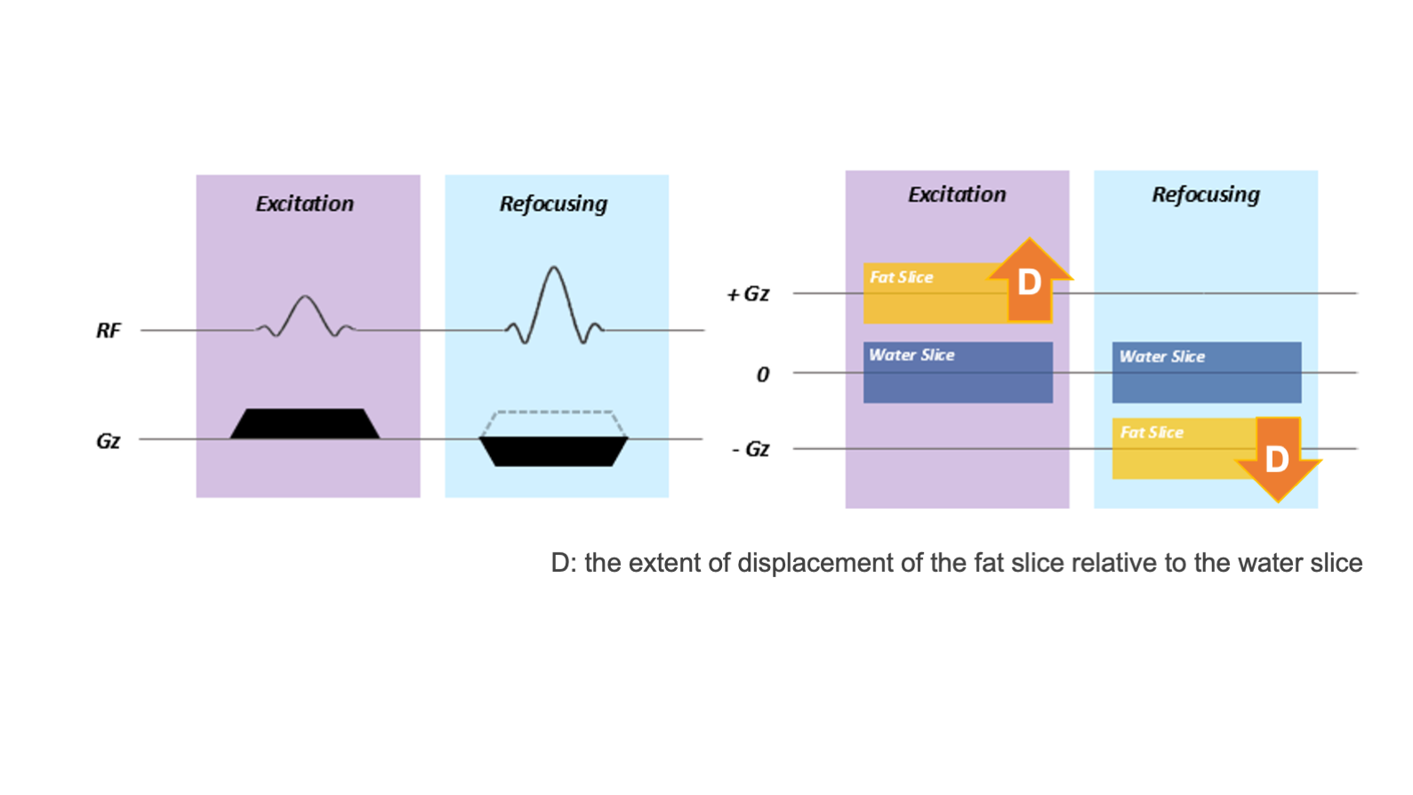
The extent of displacement of the fat slice relative to the water slice (D) is defined by D=∂*B0/Gz, where ∂ is relative chemical Shift (ppm), B0 is field strength (T), and Gz is slice selection gradient amplitude (mT/m)5 (Fig. 1). LIPO performance is better with low gradient strength and low RF transmit bandwidth (BW) of excitation and refocusing pulses. This can be understood as the thicknesses of the fat slices being reduced as low BW means fat can partly or completely fall out of the transmission BW. The displaced fat slice should be >= slice thickness/25. LIPO performance ls also improved by applying thinner slice thickness since the slice thickness/2 condition is more easily met, it is suitable for volume DWI. In fact, B0 inhomogeneity can also cause water and fat boxes to be shifted further and usually closer to each other which is not a desirable condition for LIPO. To prevent the B0 inhomogeneity effects for robust LIPO performance, the ratio between excitation and refocusing selection gradient strengths has been optimized to increase the fat slice shifts from the excitation and refocusing pulses with respect to water in this study.Sixteen patients with suspected acute stroke in the brainstem or cerebellum (14 males, 2 females, mean age 69.7±12.1) were examined on 3.0T whole-body clinical systems (Ingenia CX, Philips Healthcare, Best, the Netherland) with a 20ch head-neck coil.
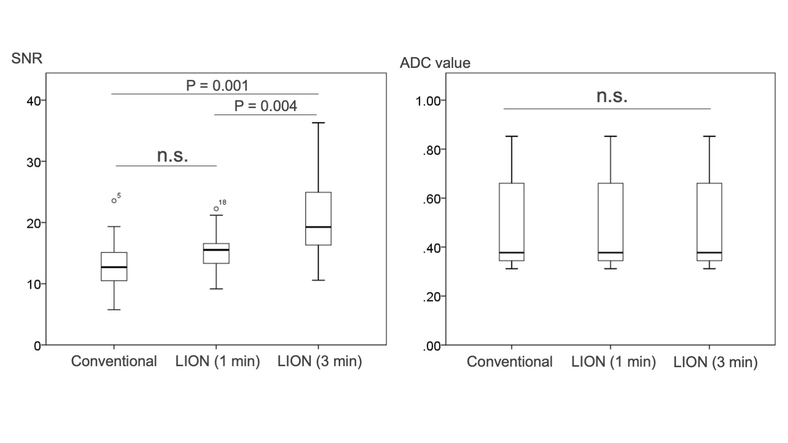
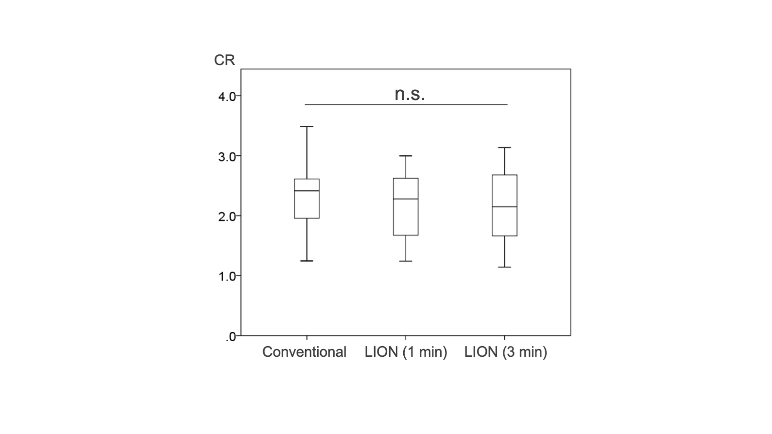
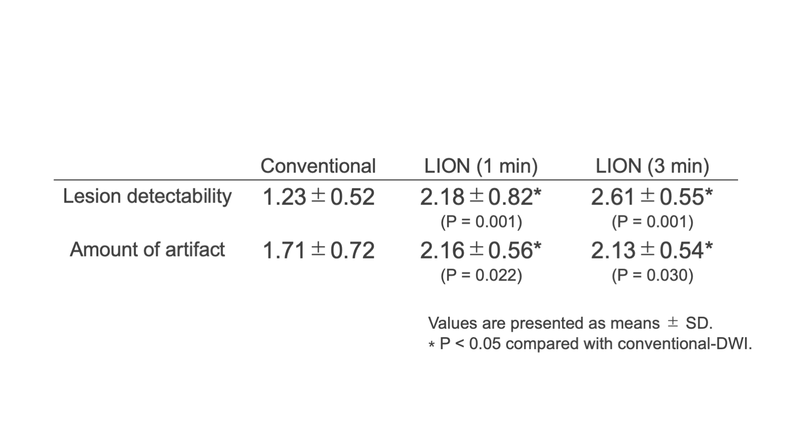
We acquired the following three patterns of thin-slice DWI. 1- conventional DWI using SPAIR for fat suppression (3 minutes), 2- LION-DWI (1 minute), and 3- LION-DWI (3 minutes). SNR and the ADC value of the lesion were measured. Contrast ratio (CR) were also measured in the lesion and the normal cerebellum. In addition, a subjective visual evaluation of lesion detectability and amount of artifact were performed on axial, coronal, and sagittal sections created by MPR. A 3-point scale was performed for the subjective visual evaluation scores as shown below (Excellent 3, Good 2, Poor 1).
Imaging parameters for thin-slice DWI were; Axial, voxel size = 2.0 mm3, 75 slices, gap 0 mm, FOV = 230 × 230 mm2, b-value = 1000 s/mm2, SENSE factor = 2, TR = 8000 ms, TE = 82 ms, conventional DWI--> fat suppression = SPAIR, NSA = 1 and total acquisition time = 3 min 14 sec. LION-DWI--> fat suppression = LION, and total acquisition time = 1 min 12 sec (NSA = 1), 3 min 05 sec (NSA = 3).
Results and Discussions
SNR of lesions were improved by LION-DWI compared with conventional DWI, on the other hand, the ADC values of the lesion did not differ significantly between imaging methods (Fig. 2). The CR between the lesion and the normal cerebellum tended to be slightly lower with LION-DWI compared to conventional DWI, but the difference was not significant (Fig. 3). Subjective visual evaluation showed that LION-DWI tended to have higher lesion detectability and lower amount of artifact (Fig. 4). Because LION-DWI (1 minute) was quantitatively and visually superior to conventional DWI (3 minutes), it is possible to shorten the examination time for thin-slice DWI for small lesions such as cerebellar and brainstem infarcts. Clinical cases imaged by each method are shown in Fig. 5. In conventional DWI, the SPAIR pulse induces a MT effect6, which reduces the signal in the white matter. Therefore, the contrast between white and gray matter is enhanced, and there is a risk of misdiagnosis as encephalitis or Jacob's disease. On the other hand, LION-DWI does not apply a pre-pulse, so the effect on white matter is minimal.Conclusion
LION-DWI has improved SNR compared to conventional DWI and has superior visual lesion detectability in patients with cerebellar or brainstem infarction. Therefore, it is possible to improve the image quality and shorten the imaging time of thin-slice DWI by changing the fat suppression to LION.Acknowledgements
No acknowledgement found.References
1. González RG, Schaefer PW, Buonanno FS, Schwamm LH, Budzik RF, Rordorf G, et al.Diffusion-weighted MR imaging: diagnostic accuracy in patients imaged within 6 hours of stroke symptom onset. Radiology. Radiological Society of North America; 1999;210: 155–62.
2. Warach S, Dashe JF, Edelman RR. Clinical outcome in ischemic stroke predicted by early diffusion-weighted and perfusion magnetic resonance imaging: a preliminary analysis. J Cereb Blood Flow Metab. 1996;16: 53–59.
3. Oppenheim C, Stanescu R, Dormont D, Crozier S, Marro B, Samson Y, et al. False-negative Diffusion-weighted MR Findings in Acute Ischemic Stroke. AJNR Am J Neuroradiol. 2000;21: 1434–1440.
4. Benameur K, Bykowski JL, Luby M, Warach S, Latour LL. Higher Prevalence of Cortical Lesions Observed in Patients with Acute Stroke Using High-Resolution Diffusion-Weighted Imaging. AJNR Am J Neuroradiol. 2006;27: 1987–1989.
5. Nagy Z, Weiskopf N. Efficient fat suppression by slice-selection gradient reversal in twice-refocused diffusion encoding. Magn Reson Med. 2008 Nov;60(5):1256-60. doi: 10.1002/mrm.21746.
6. Matsushita T, Fujii S, Kurozumi A, Nagano A, Ichiba Y, Ohno S, Tahara S. Improvement of Brain Contrast Using Spin Echo T1-weighted Image at 3 T MRI. Nihon Hoshasen Gijutsu Gakkai Zasshi. 2019;75(1):46-53.
Figures