3962
Integrated shimming technique can improve ultrahigh-b-value DWI in laryngeal and hypopharyngeal squamous cell carcinoma: comparison with conventional volume shimming1Department of Radiology, The First Affiliated Hospital of Guangxi Medical University, Nanning, China, 2MR Scientific Marketing, Siemens Healthineers Ltd., Wuhan, China, 3MR Application Predevelopment, Siemens Healthcare GmbH, Erlangen, Germany
Synopsis
Keywords: Data Analysis, Diffusion/other diffusion imaging techniques
This study aimed to investigate the value of integrated shimming (iShim) technique in the ultrahigh-b-value DWI images in laryngeal and hypopharyngeal squamous cell carcinoma (SCC) by comparing with the conventional volume shimming technique. Our results showed that the ultrahigh-b-value DWI images with iShim technique had significantly higher image quality based on subjective (edge, artifact, and confidence) and objective (signal-to-noise ratio, contrast, and contrast-to-noise ratio) assessments compared with conventional single-shot-EPI DWI images with volume shimming. Therefore, ultrahigh-b-value DWI images with iShim technique can be well applied in laryngeal and hypopharyngeal SCC examination.
Introduction
DWI is an import technique in the detection and characterization of laryngeal and hypopharyngeal squamous cell carcinoma (SCC). High-b-value (>1000 s/mm2) even ultrahigh-b-value (>2000 s/mm2) has been confirmed the b-value of choice for the detection of small changes in diffusion in tumors because of better contrast between malignant and background tissue [1-2]. However, high-b-value DWI is technically challenging due to poor in-plane spatial resolution, distortion, and artifacts using conventional singleshot echo planar imaging (ss-EPI) DWI with volume shimming, especially in complex anatomical structure in the laryngeal and hypopharyngeal. A novel technique called integrated shimming (iShim) has been proposed to reduce distortions and signal voids caused by local B0 inhomogeneity for EPI, and has demonstrated superior performances on diffusion imaging through distortion reduction and lesion detection in the neck region [3-4]. In this study, we assessed the value of ultrahigh-b-value (b=2000 s/mm2) DWI with iShim technique (iShim-DWI) in laryngeal and hypopharyngeal SCC by comparisons with conventional ss-EPI with volume shimming (c-DWI).Method
This prospective study was approved by the Institutional Review Board of our hospital. Twenty-three patients with laryngeal and hypopharyngeal SCC confirmed by pathology and without any treatment, were recruited between October 2021 and June 2022. All patients underwent MRI on a 3T MRI system (MAGNETOM Prisma; Siemens Healthcare, Erlangen, Germany) equipped with 18-element body matrix coil and an inbuilt 32- element spine matrix coil. Besides conventional T1WI and T2WI, a conventional c-DWI and a research application iShim-DWI were performed. The same parameters for the two DWI sequences were as follows: TR 5300ms, TE 65ms, FOV 140×200mm²,in-plane resolution 1.5×1.5mm²,Slice thickness 3mm, bandwidth 1750 Hz/pixel, b-values 0,50,100,150,200,800,1000,1500 and 2000 s/mm2; other different parameters were as follows: c-DWI /iShim-DWI, fat suppression, SPAIR/water excitation, total time 3min:59s/4min:19s.The image quality of the c-DWI and iShim-DWI with b = 2000 s/mm2 were compared by subjective and objective assessments. The subjective image quality was assessed according to the edge and detail of lesion, magnetic susceptibility artifact, and diagnostic confidence based on a 5-Likert scale by two radiologists with more than 5 years of experience. Objective image quality was measured by signal-to-noise ratio (SNR), contrast (C) and contrast-noise ratio (CNR). The formulas were as follows: SNR = SId/SD, CNR = (SId − SIm)/ SD, and C = (SId- SIm)/ (SId + SIm). Where, SId and SIm were the signal intensity of the lesion and muscle at the maximum level of the lesion, respectively; SD was the standard deviation (SD) of the background signal. Statistical analysis was performed using MedCalc 15.2.2 (MedCalc Software Ltd, Belgium). The differences in subjective and objective image quality between the two DWI groups was assessed by Wilcoxon signed rank test. A two-tailed p < 0.05 was considered significant. The agreement of the SNR, CNR, and C were also analyzed by the Bland-Altman method.
Results
The image quality of iShim-DWI was significantly better than that of c-DWI based on edge of lesion, susceptibility artifact, and confidence (Table 1), and the representational images are shown in Figure 1 and Figure 2.IShim-DWI had significantly higher SNR, CNR and C than those of c-DWI (p < 0.001, respectively), as shown in Table 2. Except one iShim-DWI image that showed lower SNR and CNR than c-DWI, all the other iShim-DWI images had higher SNR, CNR and C than c-DWI, and the corresponding mean differences were 45.1, 47.0, and 0.28, respectively (Figure 3).
Discussion
As we know, for c-DWI, the intrinsic complex mixtures of air- and bone-tissues interfaces in larynx and laryngopharynx, including the trachea, cartilage and vocal cords, and the piriform fossa, make this area more prone to susceptibility artifacts because of B0 inhomogeneity areas at air-bone interfaces. The iShim technique acquires field map for each slice first, and then dynamically updates the calculated center frequency according to field map prior to the acquisition of each slice. Therefore, iShim can overcome the effect of B0 inhomogeneity, which was verified in our study. Besides, in the iShim-DWI image, the edge-enhanced and motion correction techniques were added, further improving the sharpness of the edge of lesions. Therefore, In b=2000 s/mm2 DWI image, iShim-DWI exhibited lesions with clear edge, no geometric distortion or susceptibility artifacts, and presented superior image quality for better visualization of anatomical structures, improving diagnosis confidence of doctors.In addition, our study showed iShim-DWI had significant higher SNR, CNR and C of ultrahigh-b-value DWI image than those in c-DWI. One reason is that the iShim technique increased the signal strength of tissue through dynamically updating the center frequency to reduce the off-resonance excitation. The other reason is that the denoising method were added to iShim-DWI image in our study.
Conclusion
The iShim technique can improve the image quality of ultrahigh-b-value DWI compared with the conventional volume shimming, and is a clinically promising method in evaluation SCCs.Acknowledgements
No acknowledgement found.References
1. Koh DM, Blackledge M, Padhani AR, et al. Whole-body diffusion-weighted MRI: tips, tricks, and pitfalls. AJR Am J Roentgenol 2012;199(2):252–262.
2. Hu L, Zhou D, Zha Y, et al. Synthesizing High-b-Value Diffusion–weighted Imaging of the Prostate Using Generative Adversarial Networks, Radiology: Artificial Intelligence 2021; 3(5):e200237.
3. Taviani V, Nagala S, Priest AN, McLean MA, Jani P, Graves MJ. 3T diffusionweighted MRI of the thyroid gland with reduced distortion: preliminary results. Br J Radiol. 2013; 86:20130022.
4. Stemmer A, Kiefer B. Combination of Integrated SliceSpecific Dynamic Shimming and PixelWise Unwarpping of Residual EPI Distortions. ISMRM. 2015; 3729.
Figures
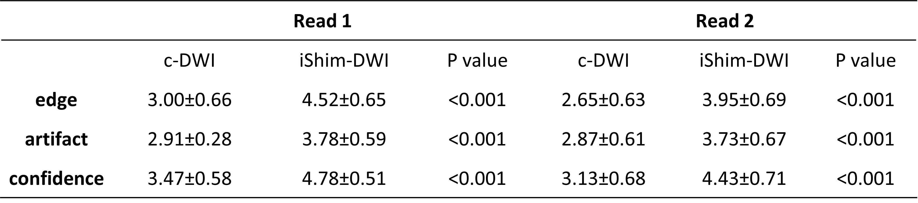
Table 1 Comparisons of edge of lesion, motion artifact, and diagnosis confidence between c-DWI and iShim-DWI images.
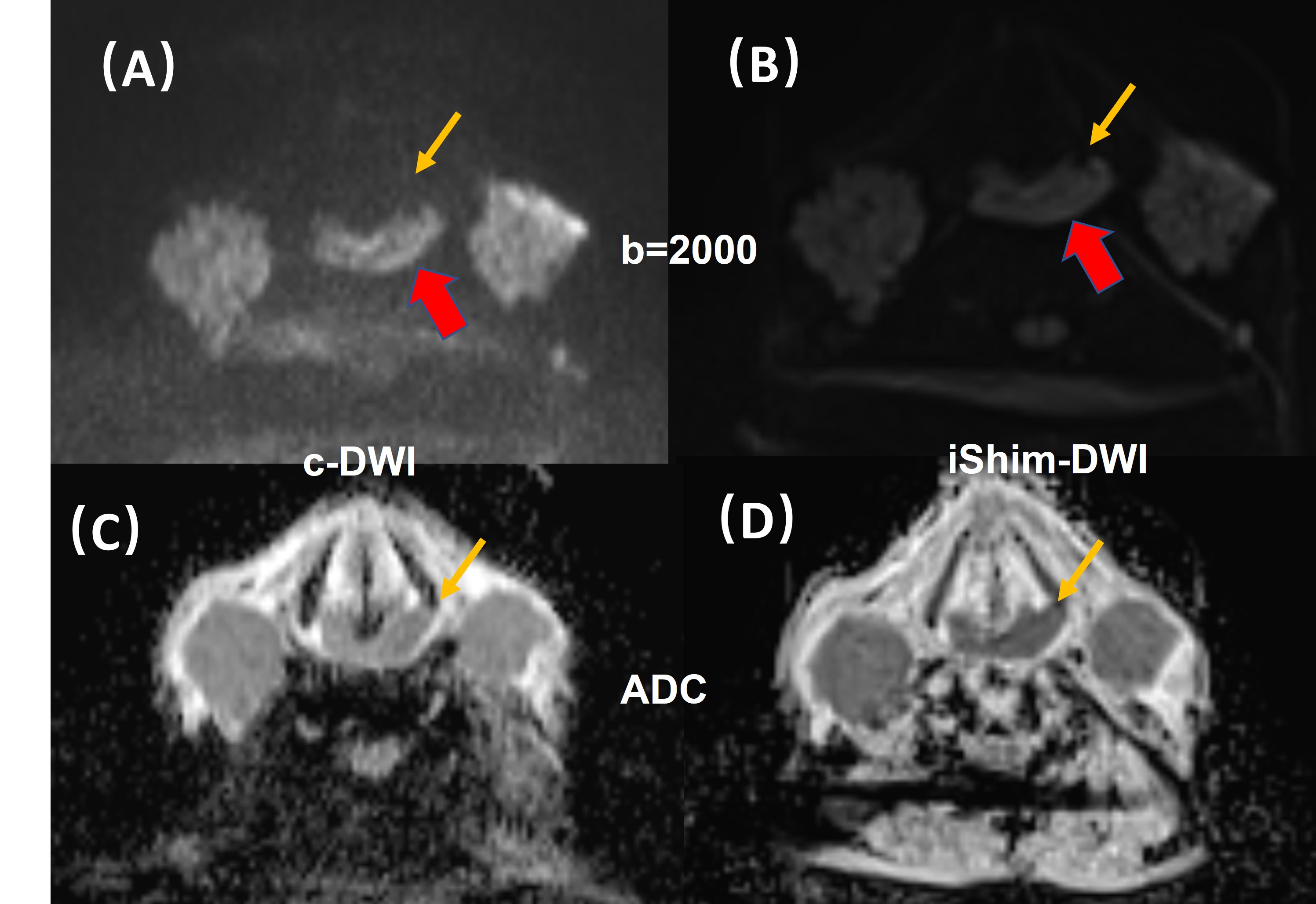
Figure 1. Comparison of (A) c-DWI and (B) iShim-DWI images with b=2000 s/mm2 and the corresponding ADC maps (C and D) for a 52-year-old male patient with hypopharyngeal carcinoma located in postcricoid region (red arrow). The edge of larger hypopharyngeal lesion, normal thyroid cartilage, and the invasion of the thyroid cartilage (yellow arrow) can be seen more clearly on the iShim-DWI images.
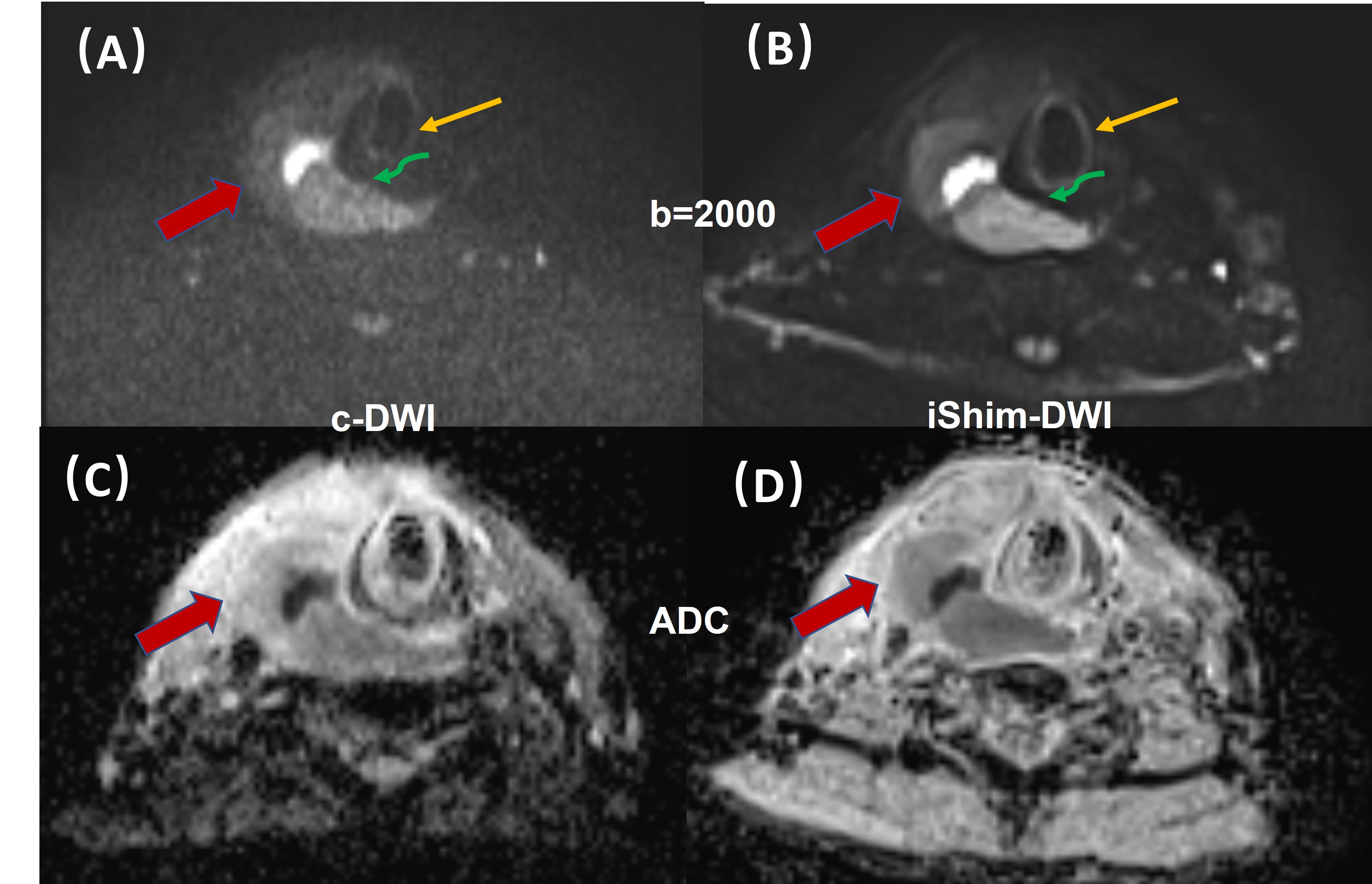
Figure 2. Comparison of (A) c-DWI and (B) iShim-DWI images with b=2000 s/mm2) and the corresponding ADC maps (C and D) for a 55-year-old male patient with hypopharyngeal carcinoma located in in right piriform fossa (red arrow). The edge of larger hypopharyngeal lesion, normal tracheal cartilage (yellow arrow), and the invasion of the cricoid cartilage(green arrow) can be seen more clearly on the iShim-DWI image.
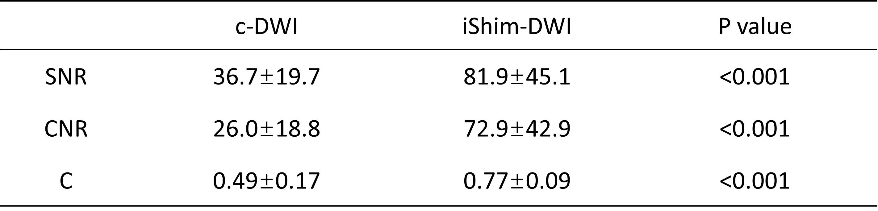
Table 2 Comparisons of SNR, C, and CNR between c-DWI and iShim-DWI using Wilcoxon signed rank test. Data are expressed as median(95%CI).
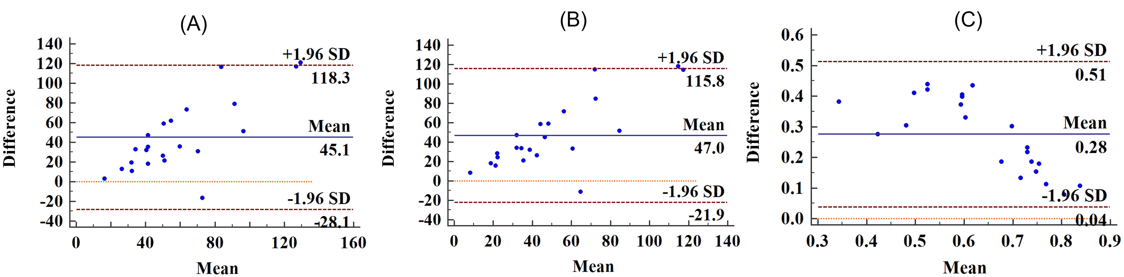
Figure 3. The Bland-Altman plots for SNR (A), CNR (B) and C (C) between c-DWI and iShim-DWI images.