3958
AcceleraTed deep-LeArning for model-free and multi-Shell (ATLAS) DWI1Department of Electrical and Computer Engineering, University of Arizona, Tucson, AZ, United States, 2Department of Biomedical Engineering, University of Arizona, Tucson, AZ, United States, 3Department of Medical Imaging, University of Arizona, Tucson, AZ, United States, 4Department of Applied Mathematics, University of Arizona, Tucson, AZ, United States
Synopsis
Keywords: Machine Learning/Artificial Intelligence, Diffusion/other diffusion imaging techniques
In this work, we aim to accelerate diffusion weighted MRI (dMRI) by predicting diffusion -weighted images (DWIs) across different shells using deep learning (DL), while remaining independent of a diffusion-model constraint. The proposed approach enables the predictions of unacquired DWIs in multiple shells from a small set of acquired DWIs from a given shell. This relaxes the need for applying multiple diffusion gradient weightings for obtaining a fully-acquired dataset over multiple shells. Without the constraint of a diffusion model, accurate diffusion metrics over multiple diffusion models can potentially be obtained by acquiring a small number of DWIs.
Introduction
Diffusion-weighted magnetic resonance imaging (dMRI) is a prominent technique, and a unique attribute of MRI, for qualitatively and quantitatively assessing microstructural characteristics of tissues. dMRI techniques such as Diffusion Tensor Imaging (DTI)1 have been utilized to study a wide range of structural and pathological processes such as neuronal connectivity2, ischemia3, tumor growth and response to therapy4. One of the major challenges of dMRI is the long data acquisition times required to obtain and leverage a large number of diffusion-weighted images (DWIs) for accurate estimation of the various diffusion metrics obtained from different models across multiple shells. Recently, deep learning (DL) techniques have been proposed to accelerate dMRI5,6,7,8,9. In this work, we present a novel DL technique, AcceleraTed deep-LeArning for model-free and multi-Shell (ATLAS), in predicting unacquired DWIs over multiple shells, given an accelerated acquisition in one shell.Methods
DWI datasets were acquired from the Human Connectome Project (HCP) public database10. 50 randomly selected datasets were used for training, 10 for validation, and 10 for testing. HCP data consists of N = 90 DWIs each at three shells (b = 1000, 2000, 3000 s/mm2) together with 18 b =0 images. The proposed ATLAS pipeline is illustrated in Figure 1. Two experiments were conducted. In the first experiment, k acquired DWIs at b = 1000 s/mm2 were used to predict N DWIs at b = 3000 s/mm2. One acquired b=0 DWI was also used in each case. The predicted DWIs were then used to estimate the diffusion tensor. Tensor-derived metrics, such as Fractional Anisotropy (FA) and Mean Diffusivity (MD), were calculated. The metrics derived from N=90 DWIs These DWIs were then used to estimate the diffusion tensor. Tensor-derived metrics, such as Fractional Anisotropy (FA) and Mean Diffusivity (MD), were calculated as illustrated in Figure 1. The metrics derived from N=90 DWIs were used as reference values in subsequent analysis and metrics derived from using only K DWIs at each shell were also calculated for comparison.
ATLAS DL process was carried out using a Unet11, comprising contracting and expansive paths and channel attention. The input tensor dimensions were 64 X 64 X 5 X (K+1) and the output tensor dimensions were 64 X 64 X 5 X (N-K or N). The b=0 s/mm2 image was incorporated as input to the CNN. The network was implemented in Python using Keras with Tensorflow backend and executed on an NVIDIA RTX 3090 GPU. Overlapping patches were utilized for improved estimation. Experiments were conducted with K=6,9, and 12 to represent different acceleration ratios. An Adam optimizer with batch size = 16 was used to train each network for up to 50 epochs with initial learning rate = 10-4, which was reduced by half when the metric stopped improving. The network was optimized over an L1-norm loss for training. Early stopping was adopted when the mean absolute error loss improvement fell below 10-5. MRtrix software12 was used for tensor and DTI metric calculations. DTI metrics from accelerated acquisitions were calculated and compared to the DTI metrics obtained from the reference datasets.
Results and Discussion
Figure 2(b) depicts DWI reconstructions for qualitative assessment of ATLAS’s performance in predicting N DWIs in b = 3000 s/mm2 shell, for a given input K = 6 for b = 1000 s/mm2 in Figure 2(a). Upon direct comparison of predictions made by ATLAS with conventionally acquired DWIs in b = 3000 s/mm2 in Figure 2(c), it is demonstrated that ATLAS can delineate structural features across multiple shells. The predicted images yield a much higher SNR than conventionally acquired b = 3000 s/mm2 images. Figure 3 shows DTI metrics calculated utilizing conventional DTI modeling, compared to the ATLAS DL pipeline, for different acceleration ratios. For increased accelerated ratios, ATLAS retains high-quality tensor maps that appear to be comparable to DTI metrics acquired from 90 DWIs. In contrast, conventional DTI experiences considerable image deprecation for increased acceleration factors. Figure 4(a-b) shows plots of two-dimensional correlation histograms comparing the predicted FA values for each computational framework to the reference FA values over a test cohort of n=10 subjects for b = 1000 s/mm2 and b = 3000 s/mm2. The plots indicate increased overestimation of FA values with an increase in the acceleration factor for conventional DTI, while the ATLAS DL framework remains tightly distributed with respect to the reference, although some underestimation is apparent.Overall, the results show that ATLAS is able to effectively predict fully-acquired DWIs and obtain high-quality tensor metrics over multiple shells, when given an accelerated input of DWIs for b = 1000 s/mm2. Although ATLAS is in early development, it demonstrates decent performance and the benefits from relaxing the need for full acquisitions of DWIs acquired across multiple shells. Moreover, without being constrained to a diffusion model, it can be potentially adapted to diffusion models other than DTI.
Conclusions
We developed a novel DL technique, ATLAS, which can predict DWIs over multiple shells from accelerated single-shell data, enabling high-accuracy predictions of DTI metrics, and significantly reducing the requirements of acquiring large DWI datasets over multiple shells. ATLAS also has the potential to be used with various diffusion models.Acknowledgements
We would like to acknowledge grant support from the Arizona Biomedical Research Centre (CTR056039), Arizona Alzheimer’s Consortium, and the Technology and Research Initiative Fund (TRIF).
References
1. Basser PJ, Mattiello J, LeBihan D. Estimation of the effective self-diffusion tensor from the NMR spin echo. J Magn Reson B 1994;103:247–254.
2. Xue, R., van Zijl, P.C., Crain, B.J., Solaiyappan, M. and Mori, S. (1999), In vivo three‐dimensional reconstruction of rat brain axonal projections by diffusion tensor imaging. Magn. Reson. Med., 42: 1123-1127. https://doi.org/10.1002/(SICI)1522-2594(199912)42:6%3c1123::AID-MRM17%3e3.0.CO;2-H
3. Bockhorst KH, Narayana PA, Liu R, Ahobila-Vijjula P, Ramu J, Kamel M, Wosik J, Bockhorst T, Hahn K, Hasan KM, Perez-Polo JR. Early postnatal development of rat brain: in vivo diffusion tensor imaging. J Neurosci Res. 2008 May 15;86(7):1520-8. doi: 10.1002/jnr.21607. PMID: 18189320.
4. Lope-Piedrafita S, Garcia-Martin ML, Galons JP, Gillies RJ, Trouard TP. Longitudinal diffusion tensor imaging in a rat brain glioma model. NMR Biomed. 2008 Oct;21(8):799-808. doi: 10.1002/nbm.1256. PMID: 18470959; PMCID: PMC2857329.
5. Golkov, V., et al., 2016. q-Space deep learning: twelve-fold shorter and model-free diffusion MRI scans. IEEE Trans. Med. Imag. 35, 1344–1351.
6. Aliotta, E., Nourzadeh, H., Sanders, J., Muller, D., Ennis, D.B., 2019. Highly accelerated, model-free diffusion tensor MRI reconstruction using neural networks. Med. Phys. 46, 1581–1591.
7. Tian Q, Bilgic B, Fan Q, Liao C, Ngamsombat C, Hu Y, Witzel T, Setsompop K, Polimeni JR, Huang SY. DeepDTI: High-fidelity six-direction diffusion tensor imaging using deep learning. NeuroImage, Volume 219: 117017, 2020. https://doi.org/10.1016/j.neuroimage.2020.117017.
8. Bilgin A, Do L, Martin P, et. al., Accelerating Diffusion Tensor Imaging of the Rat Brain using Deep Learning. Proceedings of the 2021 Meeting of the ISMRM, Abstract 2444.
9. Martin, P, Altbach, M, Bilgin, A, Noise2DWI: Accelerated Diffusion Tensor Imaging with Self-Supervision and Fine Tuning. Proceedings of the 2022 Meeting of the ISMRM, Abstract 3517.
10. Van Essen, DC, et. Al. The WU-Minn Human Connectome Project: An overview. NeuroImage 80(2013):62-79.
11. Ronneberger O, Fischer P, Brox T. U-Net: Convolutional Networks for Biomedical Image Segmentation. Medical Image Computing and Computer-Assisted Intervention (MICCAI), Springer, LNCS, Vol.9351: 234--241, 2015.
12. Tournier JD, Smith RE, Raffelt D, Tabbara R, Dhollander T, Pietsch M, Christiaens D, Jeurissen B, Yeh C-H, and Connelly A. MRtrix3: A fast, flexible and open software framework for medical image processing and visualisation. NeuroImage, 202 (2019), pp. 116–37.
Figures
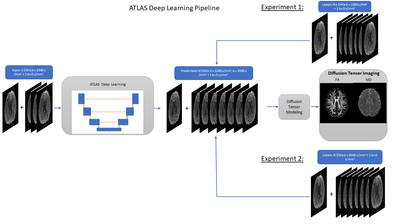
Figure 1: Experiments conducted using the proposed ATLAS framework. In these experiments, K = 6, 9, 12 DWIs on the b = 1000 s/mm2 were used as input to the ATLAS DL network. During the first experiment, ATLAS was trained to predict the remaining N-K DWIs on the b = 1000 s/mm2 shell. For the second experiment, ATLAS was trained to predict the remaining N DWIs on the b = 3000 s/mm2 shell. N=90 in both experiments. DTI metrics (FA maps and MD maps) were calculated using the predicted DWIs.
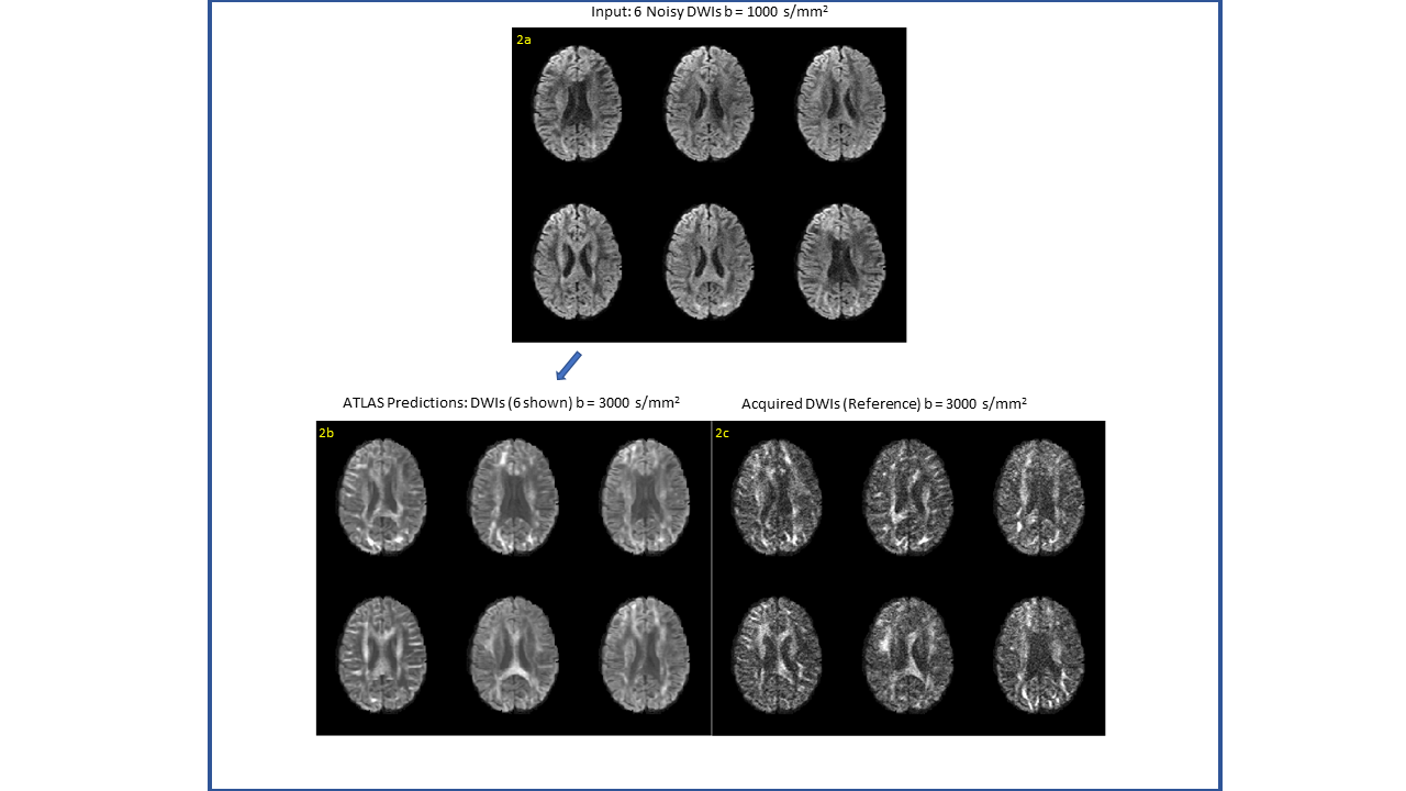
Figure 2: a) K=6 DWIs at b = 1000 s/mm2, which are used as input to the ATLAS pipeline. b) 6 sample DWIs at b = 3000 s/mm2 (out of N=90) predicted by ATLAS. c) The acquired DWIs corresponding to the same directions as those show in Figure 2(b). The ATLAS pipeline yields a significant increase in SNR in comparison to the acquired images for b = 3000 s/mm2.
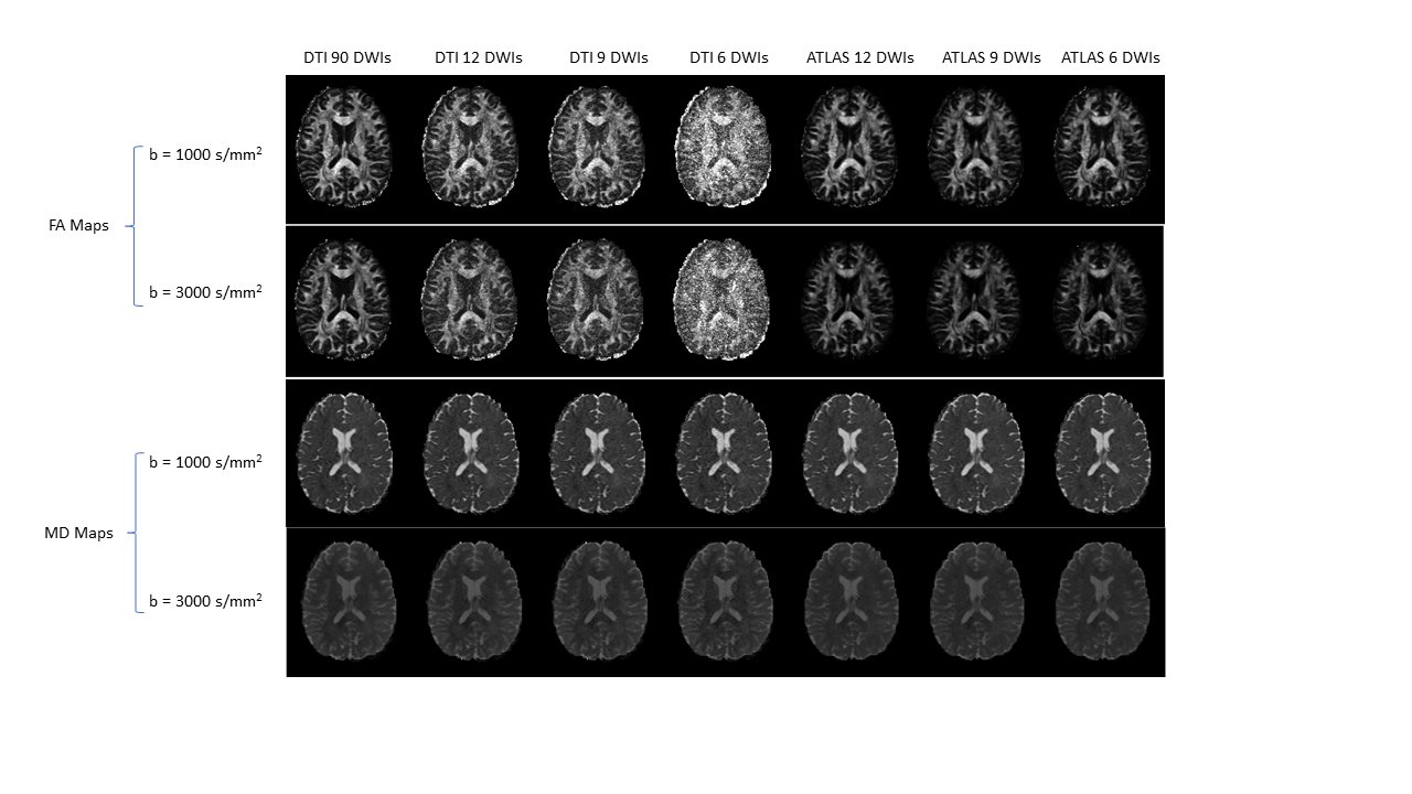
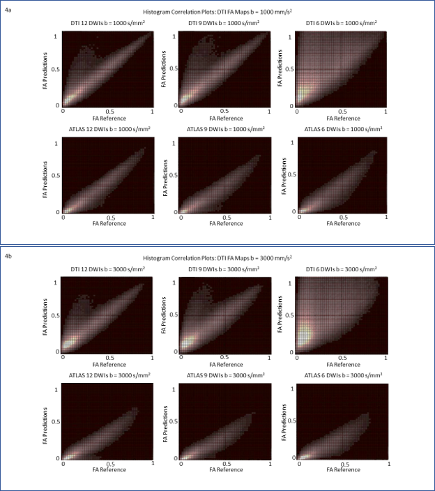
Figure 4: Figures 4a & 4b show plots of two-dimensional correlation histograms comparing the predicted FA values for each computational framework to the reference FA values over a test cohort of n=10 subjects for b = 1000 s/mm2 and b = 3000 s/mm2, respectively. The plots indicate increased overestimation of FA values with an increase in the acceleration factor for conventional DTI, while the ATLAS DL framework remains tightly distributed with respect to the reference.