3957
Feasibility of high resolution Readout-segmented echo planar imaging with simultaneous multi-slice in the assessment of rectal cancer1Sichuan Provincial People's Hospital, chengdu, China, 2MR Scientific Marketing, Siemens Healthcare, Shanghai, China
Synopsis
Keywords: Data Acquisition, Diffusion/other diffusion imaging techniques
DWI has an important role in the staging and treatment response assessment of patients with rectal cancer, but its resolution is much lower than dynamic contrast-enhanced MRI and T2W images. We achieved a high-resolution scan of the rectal cancer using SMS HR rs-EPI sequences with a resolution of 1.1×1.1×2 mm3. The SMS HR rs-EPI provided a significantly better image quality and more valuable ADC than conventional HR rs-EPI when assessing rectal cancer. The pretreatment ADC values of HR rs-EPI could be utilized to distinguish well and poorly differentiated rectal cancer.Introduction
Colorectal cancer is the third most common cancer1, MRI is the preferred diagnostic tool for local staging of rectal cancer, which provides excellent soft-tissue contrast and may be used to assess treatment response and detect postoperative local recurrence 2. Improving MRI resolution could characterize the margin of rectal cancer more accurately and show the internal heterogeneity of the lesion clearly, which is important for assessing the degree of differentiation of rectal cancer. However, high-resolution imaging of rectal cancer is mainly focused on dynamic contrast-enhanced MRI and T2W images, excluding DWI image. Previous studies have demonstrated that rs-EPI with SMS provided higher spatial resolution compared with conventional HR rs-EPI with comparable or superior image quality and stable apparent diffusion coefficient (ADC) value in breast, liver and abdominopelvic structures3-6. This study focused on the following two aims: first, to compare the image quality (IQ) of SMS HR rs-EPI and conventional HR rs-EPI in assessing rectum, and second, to determine whether the ADC values of rectal cancer measured on SMS HR rs-EPI sequence are correlated with the T stage and can predict the differentiation grade of rectal cancer.Method
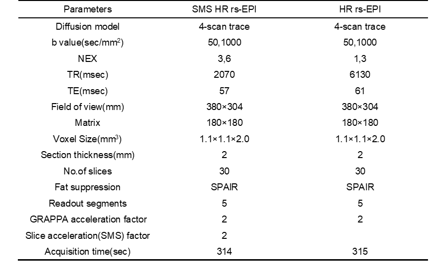
A total of eighty-three patients ( 55 males and 28 females, 22-76 years old, with a mean age of 54.6±11.9 years old) with rectal cancer were enrolled, who had undergone SMS HR rs-EPI sequence and a conventional HR rs-EPI sequence by using 3.0 T MR system with 30-channels coil (MAGNETOM VIDA, Siemens Healthcare, Erlangen, Germany). The details of parameters are shown in Table 1. The signal-to-noise ratio (SNR) and contrast-to-noise ratio (CNR) were calculated according to the following equations: SNR=Slesion /SDbackground, CNR=|Slesion-Snormal_tissue|/sqrt(SDlesion2+SDnormal_tissue2) . The region of interest (ROI) were drawn in homogeneous normal rectum tissue, which was located far from the tumor area. Subjective assessment, SNR, CNR and ADC value of the lesion were measured for comparison by paired t-test or Kolmogorov-Smirnov test. The spearman rank correlation analysis test and the receiver operating characteristic (ROC) curve was performed to evaluate the correlation between tumor ADC values, corresponding T stage and differentiation degree of rectal cancer.Results
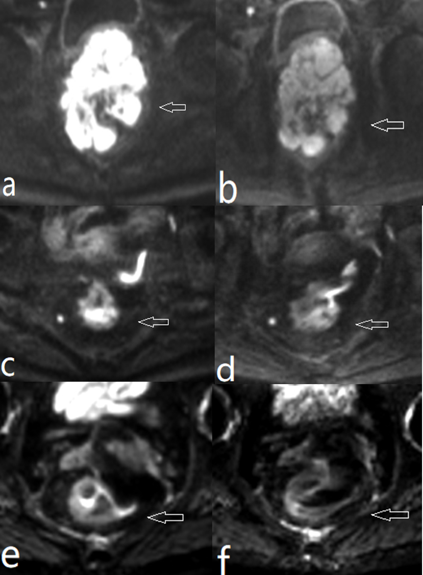
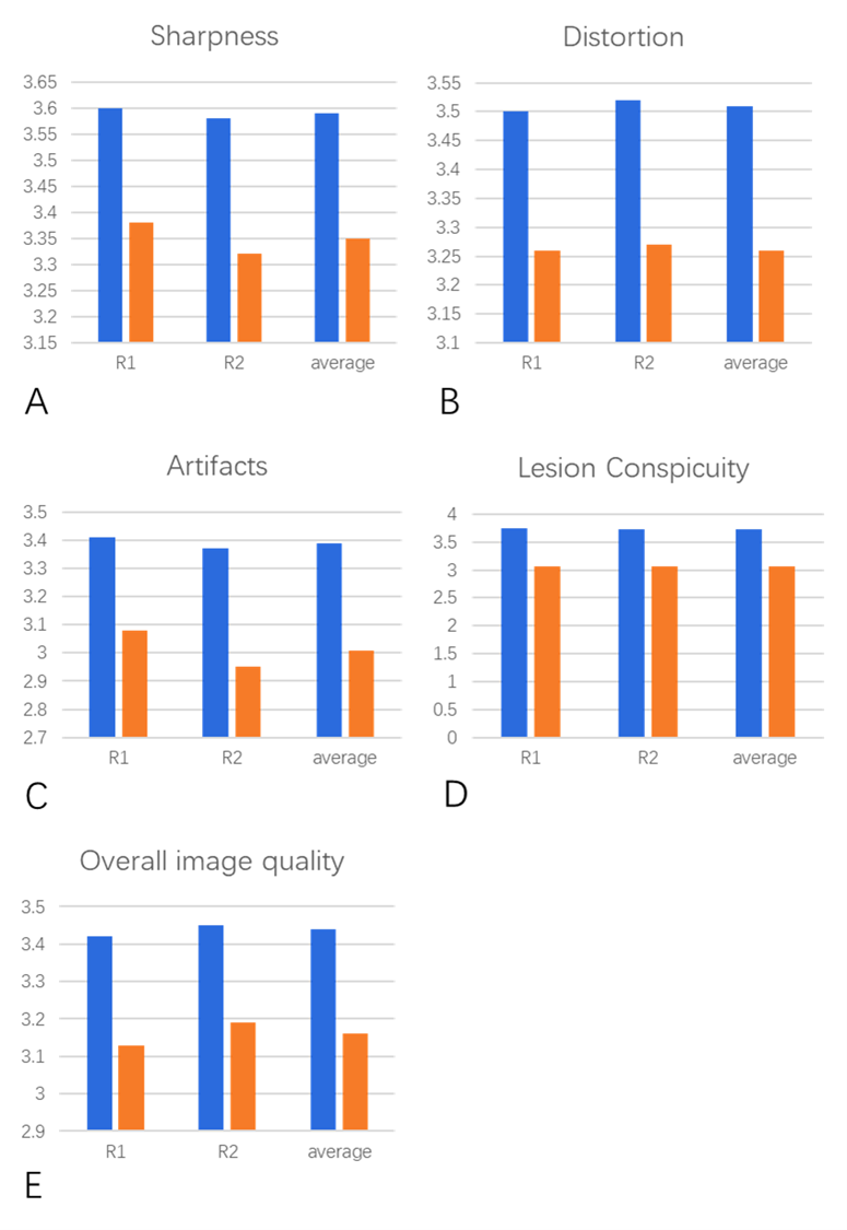
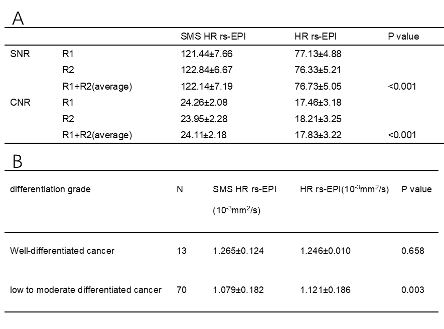
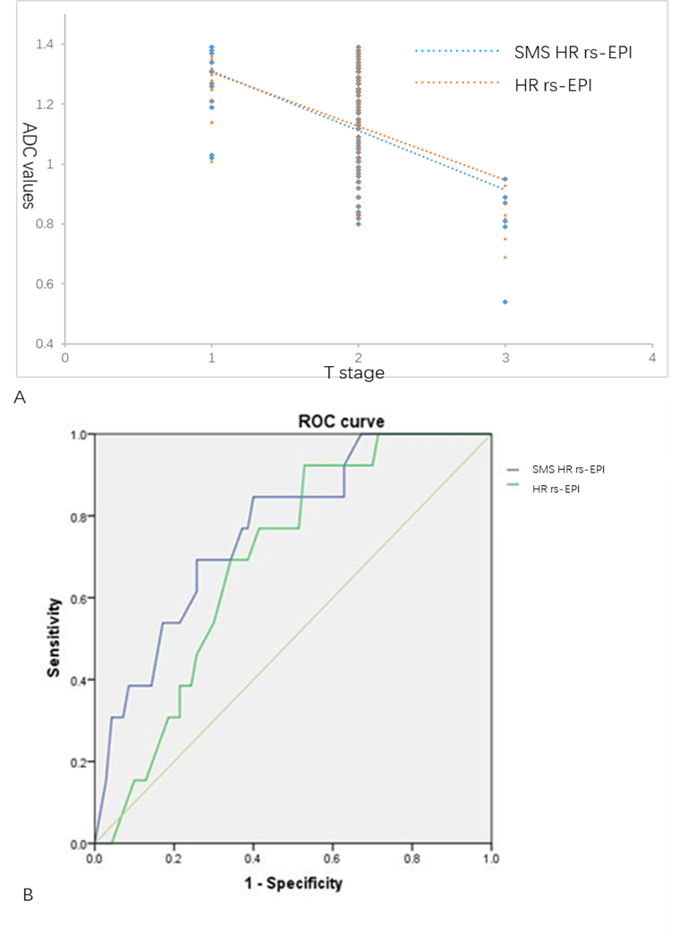
The subjective assessment of IQ of SMS HR rs-EPI were superior to conventional HR rs-EPI (p<0.001)( Figure 1,2). SNR and CNR were also significantly higher in SMS HR rs-EPI (p<0.001) and there was a significant difference between tumor ADC values on SMS HR rs-EPI and that on HR rs-EPI in the group of low to moderate differentiation degree (Table 2). T stage of rectal cancer was inversely correlated with ADC values measured on SMS HR rs-EPI (r= -0.622, p<0.001) and HR rs-EPI (r= -0.567, p<0.001) and the area under the curve (AUC) of SMS HR rs-EPI in predicting well-differentiated rectal cancer were 0.768 (Figure 3).Discussion
Better sharpness and lesion conspicuity, less distortion and image artifacts, higher SNR, CNR, and overall image scores demonstrated the feasibility of high spatial resolution rs-EPI with SMS acceleration in the clinical evaluation of rectal cancer. we achieved a higher in-plane and slice resolution DWI of 1.1×1.1×2 mm3. The high spatial resolution DWI with SMS acceleration can help increase the lesion conspicuity and sharpness of anatomical details and describe the shape and margins of rectal cancer accurately, which is greatly helpful for the staging and differentiation of rectal cancer. Regarding the relationship between ADC values and pathology T staging of rectal cancer, we observed a significant tendency for an inverse correlation between the ADC values of the two different DWI methods and the T staging. This may be due to the fact that with the increase of a tumor's T stage, its invasiveness increases, causing a continuous decrease in extracellular space, and an increase in diffusion restriction of water molecules, which results in a decline in ADC7. Our study found that the ADC values of two DWI methods were statistically different in low to moderate differentiated rectal cancer, which was mainly attributed to the fact that the higher resolution leads to a lower SNR, causing the ADC to show instability in the measurement at higher levels of cell malignancy. And ROC analysis also showed that the ADC of SMS HR rs-EPI may have the potential ability in predicting well-differentiated rectal cancer, which as a non-invasive technique, this ability of SMS HR rs-EPI should gain more attention to for predicting pathological results, and more patients with well-differentiated rectal cancer should be enrolled in future, and also we will continue to explore its treatment prediction in this cohort of patients with follow-ups.Conclusion
The SMS HR rs-EPI with SMS technique provided significantly higher IQ, SNR, and CNR and stable ADC compared to conventional HR rs-EPI. Additionally, the pretreatment ADC values of SMS HR rs-EPI could be utilized to distinguish well and poorly differentiated rectal cancer.Acknowledgements
No acknowledgement found.References
1. Lee Y, Hsieh C, Chuang J, et al. Prognostic significance of partial tumor regression after preoperative chemoradiotherapy for rectal cancer: a meta-analysis. Dis Colon Rectum. 2013; 56:1093-1101
2. Fernandes M, Gollub J, Brown G, et al. The importance of MRI for rectal cancer evaluation. Surg Oncol. 2022;10:17-39.
3. Taron J, Martirosian P, Kuestner T, et al. Scan time reduction in diffusion-weighted imaging of the pancreas using a simultaneous multislice technique with different acceleration factors: How fast can we go? Eur Radiol. 2018;28:1504-1511.
4. Peng S, Guo Y, Zhang X, et al. High-Resolution DWI with Simultaneous Multi-Slice Readout-Segmented Echo Planar Imaging for the Evaluation of Malignant and Benign Breast Lesions. Diagnostics. 2021;11:122-273.
5. Xu J, Cheng J, Wang T, et al. Simultaneous multi-slice accelerated diffusion-weighted imaging with higher spatial resolution for patients with liver metastases from neuroendocrine tumours. Clin Radiol. 2021;76(81):11-81.
6. Ciritsis A, Rossi C, Marcon M, et al. Accelerated diffusion-weighted imaging for lymph node assessment in the pelvis applying simultaneous multislice acquisition: A healthy volunteer study. Medicine. 2018;97: 11745-11755.
7. Sun Y, Tong T, Cai S, et al. Apparent Diffusion Coefficient (ADC) value: a potential imaging biomarker that reflects the biological features of rectal cancer. PLoS One. 2014; 9:109371-109377.
Figures