3949
Apparent diffusion coefficients of 31P metabolites in the human calf muscle at 7T1Animal imaging and technology core (AIT), Center for Biomedical Imaging (CIBM), Ecole Polytechnique Fédérale de Lausanne, Ecublens, Switzerland, 2Faculty of Pharmacy, University of Rennes, Rennes, France
Synopsis
Keywords: Data Acquisition, Spectroscopy, Diffusion
31P diffusion magnetic resonance spectroscopy could assess the diffusion properties of high energy metabolites. In this study, a diffusion weighted (DW) STEAM sequence was implemented, and spectra were acquired in the human calf muscle of six healthy volunteers. Frequency and phase alignments were applied prior to spectral averaging. The ADC of phosphocreatine (PCr), adenosine triphosphate (ATP), inorganic phosphate (Pi) and glycerol phosphorylcholine (GPC) were (0.24±0.02, 0.15±0.04, 0.43±0.14, 0.40±0.09)×10-3 mm2/s. To the best of our knowledge, this is the first study reporting the ADCs of ATP, Pi, and GPC, and the second study reporting the ADC of PCr in human.Introduction
31P MRS is a non-invasive technique for studying energy metabolism in vivo to track high-energy metabolites. Phosphocreatine (PCr), adenosine triphosphate (ATP), and inorganic phosphate (Pi) are the three main 31P MRS detectable metabolites essential for energy production and transport1. Combining 31P and diffusion MRS to assess their diffusion properties could provide information regarding the diffusion-related energy supply mechanism in vivo. However, due to the low concentration of phosphorous metabolites, low sensitivity of 31P nuclei, short T2 relaxation time, and J coupling effects, the measurement is difficult, especially that for ATP. In this study, we aim to investigate the diffusion coefficients of major phosphorous metabolites in the human calf muscle in vivo at 7 tesla.Methods
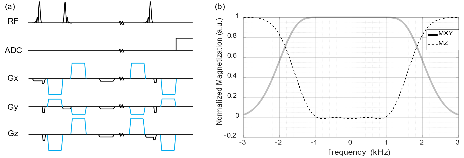
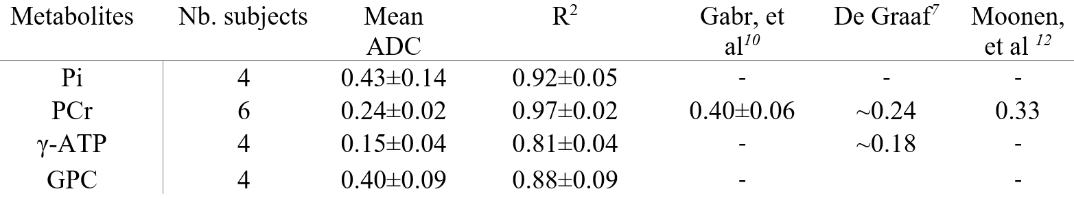
A diffusion weighted STEAM (DW-STEAM) pulse sequence was implemented (fig. 1a). A 1600 μs long asymmetric Shinnar-Le Roux RF pulse (SLR pulse) and an inverse SLR pulse were implemented2,3. Diffusion gradients were applied in three orthogonal directions [-0.5, 1, 1], [1, -0.5, 1], and [1, 1, -0.5]. To attenuate eddy currents induced by the diffusion gradients, bipolar gradients were implemented. Diffusion gradients of opposite polarities were applied in each direction to eliminate the effect of cross terms. VAPOR water suppression4 and outer volume suppression5 were applied prior to the STEAM localization. The ADC of water, Pi, PCr, and ATP were measured in vitro at room temperature (~18℃) to validate the sequence. 1H experiments were carried out with a spherical phantom containing water (pH = 7.0) using a single-channel quadrature transmit and a 32-channel receive coil (Nova Medical Inc., MA, USA), with TE/TM/TR = 15ms / 750ms / 40s, number of averages (NA) = 4. 31P experiments were carried out with a phantom containing 31mM PCr, 31mM ATP, 8mM NaH2PO4 and 106mM NaCl (pH = 7.2) using a surface coil with a single 31P loop and a quadrature 1H loops, with TE/TM/TR=15ms/750ms/60s, NA=4. Diffusion-weighted spectra at seven different b-values (0, 200, 400, 600, 800, 1000, 1200 s/mm2) were measured. Then, six healthy subjects (18-40 years old, 3 males and 3 females) were scanned for in vivo ADC measurements. Informed consent forms were obtained from all subjects before the scan. The MR spectra were acquired from a VOI of 60mm x 100mm x 120mm containing the calf muscle. Localized diffusion-weighted MR spectra at three b-values (0, 800, 1200 s/mm2) were acquired per diffusion direction (TE/TM/TR = 15ms / 750ms / 5s, NA = 32). The total in vivo data acquisition time was 40 min. All experiments were performed at a 7T/68cm MR scanner (Siemens Medical Solutions, Erlangen, Germany). The scanner is equipped with a gradient coil capable of reaching 80mT/m in total, and 50mT/m along each axis simultaneously. Phase and frequency drift correction, outlier removal were implemented prior to spectra averaging. The averaged spectra were quantified by LCModel.Results
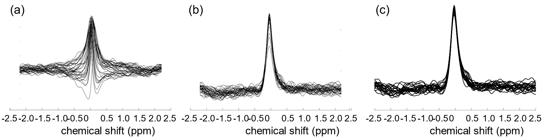
The measured ADC of water, PCr, Pi, γ-ATP and α-ATP were shown in table 1, coherent with the reported values6. The in vivo ADC values of Pi, PCr, ATP, and GPC were measured in the human calf muscle. High spectral SNR was achieved at all b-values with average linewidth being around 13.7 Hz. PCr was reliably quantified at all b-values with mean CRLB less than 1%, while γ-ATP, α-ATP, Pi and GPC less than 10%. The ADCs of PCr, Pi, γ-ATP and GPC were (0.24 ± 0.02, 0.43 ± 0.14, 0.15 ± 0.04, 0.40 ± 0.09) × 10-3 mm2/s respectively with average R2 being 0.97, 0.92, 0.81, 0.88 (table 2).Discussion
The high sensitivity of 7T enabled the successful measurement of PCr, ATP, Pi, and GPC from the in vivo spectra at the high b-value, allowing for the first measurement of ADC of ATP, Pi, and GPC in human calf muscles. In this study, the ADCs of PCr and ATP in human calf muscle were in a good agreement with the values reported in rat skeletal muscles7,8. However, the ADC of PCr was lower than that reported by Gabr, et al.9 in human calf muscles under similar diffusion time (~755 ms). The disagreement could lie in the differences during the post-processing process. Diffusion MRS is very sensitive to motions, which could lead to phase dispersion and thus the overestimation of ADC. Due to the low SNR of the individual scan in the study performed by Gabr, et al.10 at 3T, no phase correction was implemented and thus the PCr’s ADC was likely to be overestimated9. In the current study, scan-to-scan phase and frequency correction were implemented to mitigate the problem (fig. 2). The measured ADC of Pi was higher than that reported by K. Yoshizaki, et al. in bullfrog muscles ex vivo at room temperature (24℃)10. This discrepancy could be due to the different experimental conditions (human vs bullfrogs and in vivo vs ex vivo, 37℃ vs 24℃, respectively).Conclusion
We conclude that it is feasible to measure the ADC of four major 31P metabolites (PCr, ATP, Pi and GPC) in vivo in the human calf muscle at 7T. This study paves the way to investigate 31P metabolite diffusion properties in health and disease on the clinical MR scanner.Acknowledgements
This work was partially supported by the Swiss National Science Foundation (n° 320030_189064). We acknowledge accessto the facilities and expertise of the CIBM Center for Biomedical Imaging, a Swiss research center of excellence foundedand supported by Lausanne University Hospital (CHUV), University of Lausanne (UNIL), Ecole polytechnique fédérale deLausanne (EPFL), University of Geneva (UNIGE) and Geneva University Hospitals (HUG).References
(1) Rietzler, A.; Steiger, R.; Mangesius, S.; Walchhofer, L.-M.; Gothe, R. M.; Schocke, M.; Gizewski, E. R.; Grams, A. E. Energy Metabolism Measured by 31P Magnetic Resonance Spectroscopy in the Healthy Human Brain. Journal of Neuroradiology 2022, 49 (5), 370–379.
(2) Le Roux, P. Introduction to the Shinnar-Le Roux Algorithm; 1995.
(3) Pauly, J.; Le Roux, P.; Nishimura, D.; Macovski, A. Parameter Relations for the Shinnar-Le Roux Selective Excitation Pulse Design Algorithm (NMR Imaging). IEEE Transactions on Medical Imaging 1991, 10 (1), 53–65.
(4) Tkáč, I.; Starčuk, Z.; Choi, I.-Y.; Gruetter, R. In Vivo 1H NMR Spectroscopy of Rat Brain at 1 Ms Echo Time. Magnetic Resonance in Medicine 1999, 41 (4), 649–656.
(5) Tkáč, I.; Andersen, P.; Adriany, G.; Merkle, H.; Uǧurbil, K.; Gruetter, R. In Vivo 1H NMR Spectroscopy of the Human Brain at 7 T. Magnetic Resonance in Medicine 2001, 46 (3), 451–456.
(6) de Graaf, R. A.; van Kranenburg, A.; Nicolay, K. In Vivo 31P-NMR Diffusion Spectroscopy of ATP and Phosphocreatine in Rat Skeletal Muscle. Biophysical Journal 2000, 78 (4), 1657–1664.
(7) van Gelderen, P.; DesPres, D.; van Zijl, P. C.; Moonen, C. T. Evaluation of Restricted Diffusion in Cylinders. Phosphocreatine in Rabbit Leg Muscle. J Magn Reson B 1994, 103 (3), 255–260.
(8) Yoshizaki, K.; Watari, H.; Radda, G. K. Role of Phosphocreatine in Energy Transport in Skeletal Muscle of Bullfrog Studied by 31P-NMR. Biochimica et Biophysica Acta (BBA) - Molecular Cell Research 1990, 1051 (2), 144–150.
(9) Gabr, R. E.; El-Sharkawy, A.-M. M.; Schär, M.; Weiss, R. G.; Bottomley, P. A. High-Energy Phosphate Transfer in Human Muscle: Diffusion of Phosphocreatine. Am J Physiol Cell Physiol 2011, 301 (1), C234-241.
(10) Bogner, W.; Chmelik, M.; Schmid, A. i.; Moser, E.; Trattnig, S.; Gruber, S. Assessment of 31P Relaxation Times in the Human Calf Muscle: A Comparison between 3 T and 7 T in Vivo. Magnetic Resonance in Medicine 2009, 62 (3), 574–582.
Figures