3931
Mechanical Stiffness and Anisotropy Measured by MR Elastography during Brain Development in the Minipig1Washington University in St. Louis, St. Louis, MO, United States, 2University of Delaware, Newark, DE, United States, 3Dartmouth College, Hanover, NH, United States
Synopsis
Keywords: Elastography, Tissue Characterization
The relationship between brain development and mechanical properties of brain tissue is important but remains incompletely understood. Here we use data from magnetic resonance elastography (MRE) and diffusion tensor imaging (DTI) to estimate anisotropic mechanical properties in six female Yucatan minipigs at ages from 3 to 6 months with a transversely- isotropic nonlinear inversion (TI-NLI) algorithm. Our results show that white matter is more dissipative and anisotropic than gray matter, and reveal effects of brain development on brain stiffness and structural anisotropy. Changes in brain mechanical properties may be a fundamental biophysical signature of brain development.Introduction
Adolescence is a critical period for human brain development, including the emergence of neuropsychiatric disorders1, which are often accompanied by structural changes in the brain2, may affect the mechanical properties of brain tissue3. Thus, characterization of mechanical properties during brain development is important for understanding the normal brain structure, function, and health. Brain white matter (WM) is mechanically anisotropic due to the alignment of axon fibers, whose development is analogous in juvenile minipigs and adolescent humans, but brain development in the minipig occurs over a compressed time period relative to humans4. Accordingly, longitudinal imaging studies in the minipig are logical and feasible for investigating changes in the brain during development. The primary objective of this research is thus to measure anisotropic mechanical properties in the minipig brain during brain development and characterize changes that may occur over this period.Methods
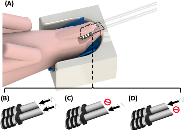
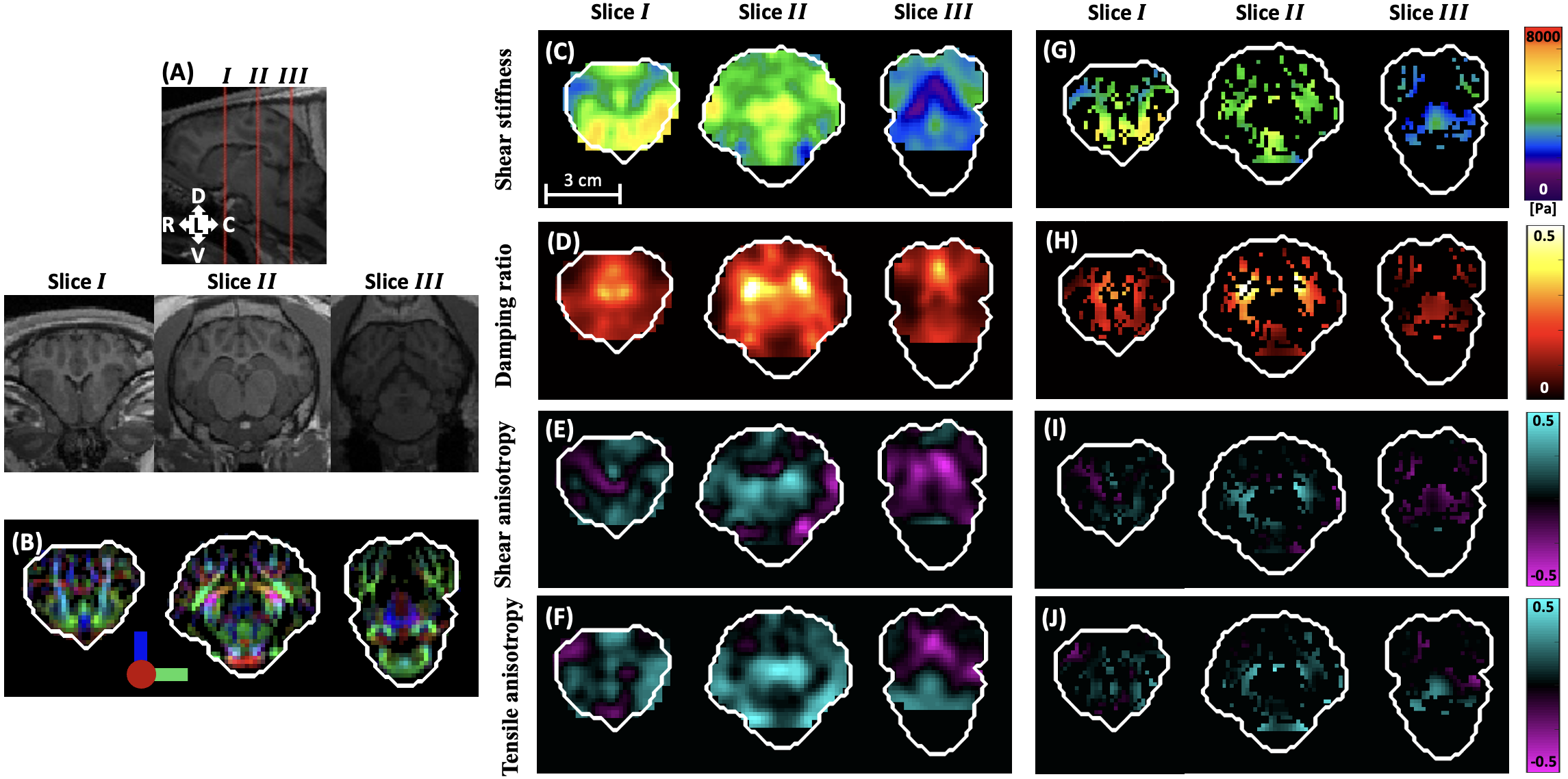
Six juvenile female Yucatan minipigs were scanned using a Siemens Prisma® 3T scanner once per month for a total of four scans per animal. Ages at initial scan were 2.93 ± 0.12 months (weights 11.20 ± 1.13) and at final scan were 5.82 ± 0.15 months (25.76 ± 2.37). The anesthetized minipig was positioned supine on the scanner table with head placed in the bottom half of the Siemens Head/Neck 20 coil (Figure 1A). Shear waves in the brain were induced at a frequency of 100 Hz using a pneumatic driver (ResoundantTM) and custom jaw actuator5 in three different configurations: “left”, “right” and “both” (Figure 1,B-D). MRE imaging was performed with a 2D multishot spiral sequence with OSCILLATE acceleration6; phase contrast proportional to displacement (1.4977 microns/rad); 8 temporal samples acquired per period of harmonic motion; TR/TE = 4800/60 ms; and FOV = 180 x 180 x 72 mm3 with 1.5 mm isotropic spatial resolution. T1- and T2-weighted anatomical MR images were also acquired at 0.8 mm isotropic resolution for volume of 205 x 205 x 154 mm3. Diffusion tensor imaging (DTI) was performed to estimate WM fiber direction. DTI parameters included: single-shot echo-planar imaging acquisition; 30 diffusion-weighted directions with two averages; and FOV = 192 x 192 x 72 mm3 with 1.5 mm isotropic spatial resolution. Fiber directions and fractional anisotropy (FA) maps were determined using Tortoise v3.27, WM fiber direction were assigned in voxels with FA >0.1. A transversely isotropic (TI) material model has been incorporated into the non-linear inversion (NLI) framework8, this TI-NLI estimation inversion was performed using MRE displacement data from the three actuator configurations and DTI-derived fiber directions to estimate anisotropic mechanical properties of the minipig brain. FA from DTI and four estimated mechanical properties (baseline shear stiffness, damping ratio, shear anisotropy, tensile anisotropy) were analyzed using a repeated one-way analysis of variance (ANOVA) and a repeated two-way ANOVA, respectively.Results and Discussion
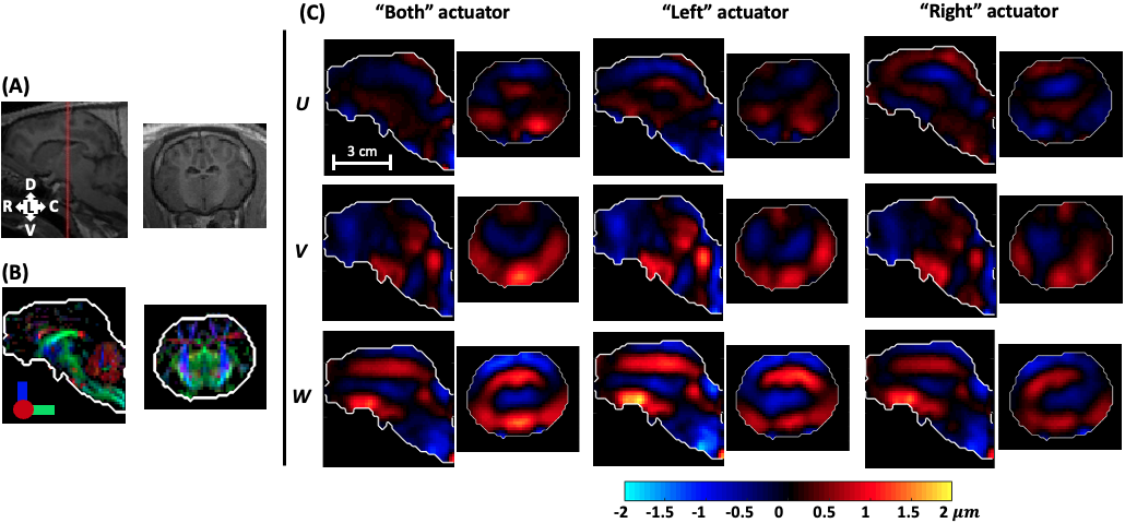
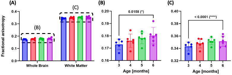
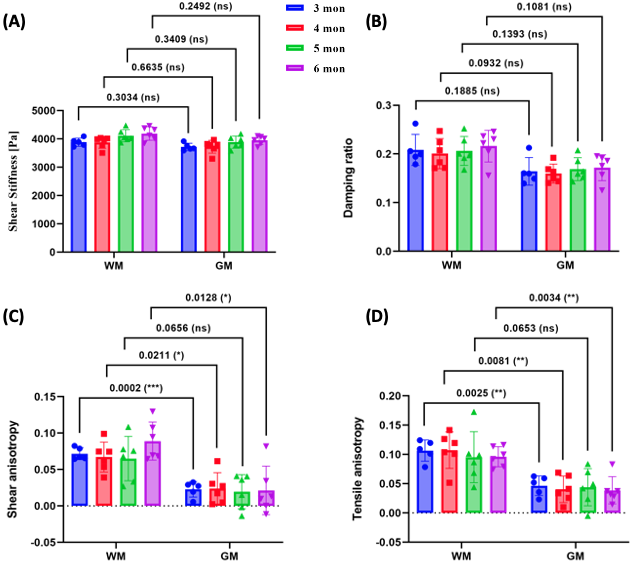
Figure 2 shows examples of anatomical structure, fiber direction, and wave displacement fields for one sagittal and axial plane. The three configurations of the MRE jaw actuator imparted different types of wave motion to the brain, providing four combinations of input data for the TI-NLI algorithm. The rostral-caudal (W) component of wave motion is the dominant component, and the maximum displacement amplitude is about 2 µm. Figure 3 shows the mean FA values calculated from diffusion tensors of all six animals at different ages. The mean FA values increase slightly with age and a significant effect of age on FA was observed in whole brain and the WM. Figure 4 displays representative TI-NLI-estimated material properties in both the whole brain volume and the WM sub-volume. For damping ratio, 𝜉, shear anisotropy, 𝜙, and tensile anisotropy, 𝜁, estimated values appear generally higher in WM voxels, but no obvious difference is apparent among the baseline shear stiffness, 𝜇, in brain, WM, and GM. The means and standard deviations of the parameters in WM and GM are plotted versus age in Figure 5. At all ages, shear stiffness differences were similar in WM and GM. The damping ratio appears generally higher in WM, but the differences in damping ratio between brain region did not reach statistical significance. However, for shear anisotropy and tensile anisotropy, the differences between WM and GM were statistically significant. The shear stiffness of both WM and GM increases with significantly with age, but no consistent trends were observed in the other three parameters.Conclusion
This study is the first to investigate anisotropic mechanical properties in a large animal brain during development. The current results reveal that WM is more anisotropic than GM, both structurally and mechanically, and WM is more dissipative than GM in the minipig. Baseline shear stiffness of WM and GM are similar, so WM does not appear stiffer than GM when deformed in the plane normal to the fiber axis. However, WM appears stiffer than GM when deformed in all other planes due to WM anisotropy in shear and tension. Brain development is associated with increasing tissue stiffness and structural anisotropy (diffusion anisotropy). Anisotropic mechanical properties of brain tissue are of fundamental biophysical significance, and future studies with anisotropic MRE may improve our understanding of normal brain structure and function during development, aging, injury, and disease.Acknowledgements
NIH Grant R01EB027577 and ONR Grant N00014-22-1-2198.References
1. McGorry PD, Purcell R, Goldstone S, Amminger GP. Age of onset and timing of treatment for mental and substance use disorders: implications for preventive intervention strategies and models of care. Curr Opin Psychiatry. 2011; 24(4): 301-306.
2. Opel N, Goltermann J, Hermesdorf M, et al. Cross-Disorder Analysis of Brain Structural Abnormalities in Six Major Psychiatric Disorders: A Secondary Analysis of Mega- and Meta-analytical Findings From the ENIGMA Consortium. Biol Psychiatry. 2020; 88(9): 678-686.
3. Sack I, Johrens K, Wurfel J, Braun J. Structure-sensitive elastography: on the viscoelastic powerlaw behavior of in vivo human tissue in health and disease. Soft Matter. 2013; 9(24): 5672-5680.
4. Swindle MM, Makin A, Herron AJ, Clubb FJ Jr, Frazier KS. Swine as models in biomedical research and toxicology testing. Vet Pathol. 2012; 49(2): 344-356.
5. Guertler CA, Okamoto RJ, Schmidt JL, et al. Mechanical properties of porcine brain tissue in vivo and ex vivo estimated by MR elastography. J Biomech. 2018; 69: 10-18.
6.McIlvain G, Cerjanic AM, Christodoulou AG, et al. OSCILLATE: A low-rank approach for accelerated magnetic resonance elastography. Magn Reson Med. 2022; 88(4): 1659-1672.
7.Irfanoglu MO, Nayak A, Jenkins J, Pierpaoli C. TORTOISE v3: Improvements and new features of the NIH diffusion MRI processing pipeline. InProgram and proceedings of the ISMRM 25th annual meeting and exhibition, Honolulu, HI, USA 2017.
8. McGarry M, Van Houten E, Sowinski D, et al. Mapping heterogenous anisotropic tissue mechanical properties with transverse isotropic nonlinear inversion MR elastography. Med Image Anal. 2022; 78: 102432.
Figures