3909
Remote detection MRI using a flexible non-selective metamaterial at 7 T1Departamento de Fisica, UNAM, Mexico City, Mexico, 2Department of Physical Intelligence, Max Planck Institute for Intelligent Systems, Stuttgart, Germany, 3Electrical Engineering, UAM Iztapalapa, Mexico City, Mexico
Synopsis
Keywords: Non-Array RF Coils, Antennas & Waveguides, RF Arrays & Systems, travelling-wave
We propose a method to remotely acquire MR images at 7 T with a preclinical imager using a bio-inspired surface coil, a parallel-plate waveguide and a non-selective metamaterial. Phantom images were acquired with and without a flexible metasurface inserted in cylindrical phantom filled with saline solution and a birdcage coil. Uniformity profiles were computed for all cases showing that the birdcage coil and metasurface profiles have a very good correspondance. An improvement was observed for the images obtained with the metasurface. This approach offers a convenient alternative to obtained high quality images.Introduction
The travelling-wave MRI (twMRI) approach offers the advantage of acquiring images with larger field-of-view. We have previously shown that the use of a parallel-plate waveguide (PPWG) together with a surface coil is a viable way to acquire MR images, because it has no limitations of cut-off frequency [1]. However, images have lower signal-to-noise ratio (SNR) values [2]. Metamaterials are an alternative to increase the performance of RF coils [3]. Erni et. al. have proposed a numerical method for RF excitation with multiple rings inserted in a cylindrical waveguide [4]. We proposed a much simpler approach consisting of a PPWG together with a metasurface and a bio-inspired surface coil for twMRI at 7 T.Method
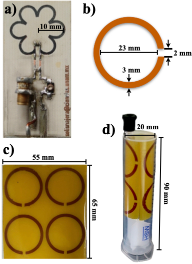
A prototype of the bio-inspired surface coil was built, including six circular slots (0.45 cm dimeter) and total coil radius of 20 mm (see Fig. 1.a). The coil prototype was constructed using copper sheets laminated onto a nonconductive board. Tuning and matching capacitors (0–15 pF: Voltronics Co. Salisbury, MD, USA) were soldered directly onto the surface: two parallel ceramic capacitors (ATC, Huntington Station, NY, USA) were placed as shown in Fig. 1.a). The coil prototype was then matched and tuned to 50 Ω and 300 MHz, respectively (proton frequency for 7 T). The metasurface was formed of an array of 4x4 C-shape (see Fig. 1.c) units and constructed using flexible hydrocarbon ceramic laminates (RO4003C3: 𝜖 = 3.55 and tan(𝛿) = 0.0027, thickness = 0.508 mm, 65 mm long and 55 mm wide). The C-shaped unit had a 23 mm diameter and a gap of 2 mm and a 3 mm strip width. The resonant frequency of the C-shaped unit cell is [5]:$$f= \frac{1} {2 \pi\sqrt{L_{C}C_{gap}}}= \frac{1} {\sqrt {\mu_0 2\pi I d\ln\left( \frac {8d} {w} -1.75 \right) C_{gap}}} \,\, (1)$$
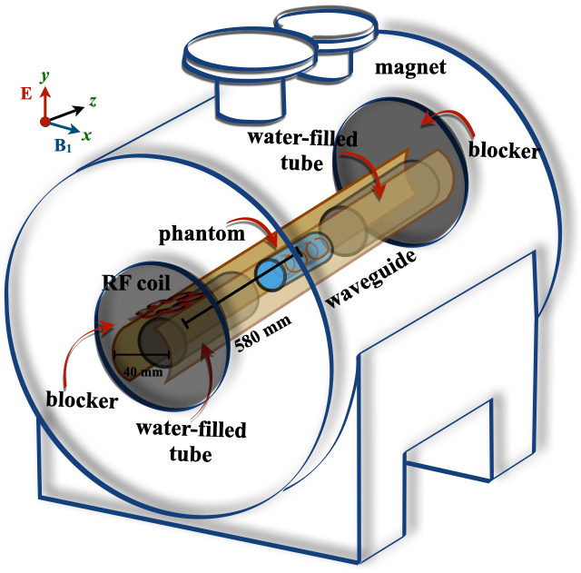
where LC is the inductance and Cgap is the capacitance of the C-shaped unit cell, w is the strip width, d is the coil diameter. The inductance was theoretically computed using the parameters in Fig. 1.b), and the capacitance of the gap, Cgap was obtained according the experimental method reported in [6]. The resonant frequency of the unit cell in the metasurface (see Fig. 1.b) was measured as the loss return (S11), and using a 20 mm-diameter loop and a network analyzer (Model 4396A, Hewlett Packard, Agilent Technologies, CA). Phantom images were obtained using a cylindrical phantom (20 mm diameter and 90 mm) filled with saline solution and the metasurface surface was inserted in the phantom (Fig. 1.e). Then, the phantom and the metasurface were inserted in the cylindrical phantom for the imaging experiments (Fig. 1.d). A bio-inspired surface coil located outside the waveguide and parallel to the plates (see Fig. 2). A standard gradient echo sequence was used for the MRI experiments. The acquisition parameters were: TE/TR = 4.39/200 ms, FOV = 40 mm x 40 mm, matrix size = 256 x 256, Flip angle = 450, slice thickness = 2 mm, NEX = 1. Additionally, phantom images without the metasurface and a quadrature birdcage coil (4 cm diameter, 64 cm long and 4 rungs) were also acquired for comparison purposes. All MRI experiments were performed on a 7T/30cm Bruker imager (Bruker, BioSpin MRI, GmbH, Germany).
Results and Discussion
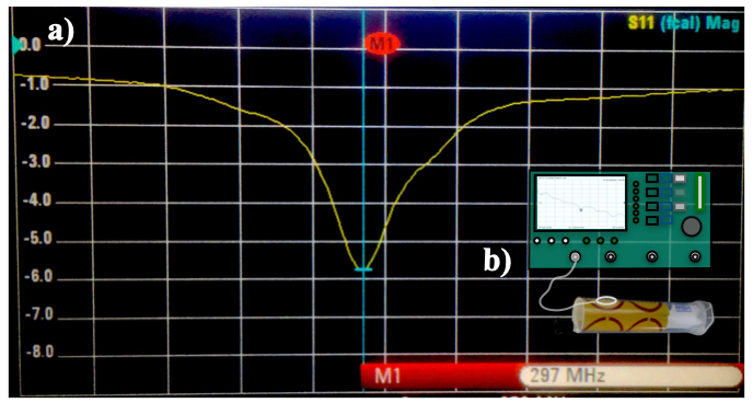
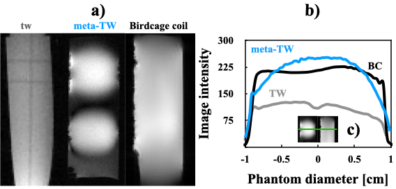
The Cgap was 6.45 pH, and replacing this value and the corresponding dimensions of the C-shaped unit cell in Eq. (1), the theoretical resonant frequency of the cell is 302.29 MHz. The experimental S11-parameter of the C-shaped unit cell was 297 MHz as shown in Fig. 3. These resonant frequency values show a very good concordance. Phantom images were acquired with and without the metasurface to demonstrate the feasibility of this approach as shown in Fig. 4.a). With the image data, the SNR values and uniformity profiles were computed and shown in Fig. 4.b). The SNR values are: SNRmetasurface = 35.71, SNRBC = 32.42 and SNRstd = 23.52. Transversal Image profiles were taken to avoid distortion due to PPWG modes, the metasurface profile has slightly greater intensity values at the central region. Those images obtained without the metasurface show really low profile values compared to the other two profiles. This corroborates previous results obtained at different resonant frequencies [1-2]. This is rather encouraging to continue investigating this approach because allows us to obtain a remarcable improvement over the standard twMRI approach. There was no need to use electronic components for tuning the metasuraface the resonant frequency of the MR imager. From these results, we can conclude that high quality images can be obtained with this easy-to-implement method.Conclusions
We have experimentally demonstrated that the use of flexible metasurfaces can produce high SNR images via the travelling-wave approach. We have shown that this approach overcomes the standard twMRI method at 7 T with a preclinical MR imager.Acknowledgements
This project received funding from the UAM Division of Basic Science and Engineering under the Special Program for Education and Research (DCBI-190-2022).References
1. Vazquez, F. et. al. (2021). Remote RF excitation for small-bore MR imager at 15.2 T. Journal of Magnetic Resonance, 323, 106896. https://doi.org/10.1016/j.jmr.2020.106896.
2. Vazquez, F. et. al. (2016). Travelling-wave transmitted with a simple waveguide for rodents Magnetic Resonance Imaging at 9.4 T. 33rd Ann. Meet. ESMRMB, 32, S31-S32.
3. Wiltshire, M. C. K. (2007). Radio frequency (RF) metamaterials. Physica Status Solidi (b), 244(4), 1227-1236. https://doi.org/10.1002/pssb.200674511.
4. Erni D. et. al. (2011). Highly adaptive RF excitation scheme based on conformal resonant CRLH metamaterial ring antennas for 7-Tesla traveling-wave magnetic resonance imaging. 2011 IEEE EMBS (pp. 554-558). 10.1109/IEMBS.2011.6090102.
5. Grover, F. W., Inductance Calculations, Chs. 2 and 13, Dover, New York, 1964.
6. Marrufo O. et. al. ( 2011) Slotted cage resonator for high-field magnetic resonance imaging of rodents. J Phys D: Appl Phys, 44(15), 155503. https://doi.org/10.1088/0022-3727/44/15/155503.
Figures