3901
Transceiver 8Tx/32Rx array with L-shaped loop elements for accelerated cardiac MRI at 7T: initial optimization and experimental testing.1Chair of Cellular and Molecular Imaging,Comprehensive Heart Failure Center, University Hospital Würzburg, Wuerzburg, Germany, 2Department of Internal Medicine I, Cardiology, University Hospital Würzburg, Wuerzburg, Germany
Synopsis
Keywords: RF Arrays & Systems, Parallel Transmit & Multiband, cardiovascular
The concept of the L-shaped loop elements architecture with the antisymmetric arrangement was applied to develop a 8Tx/32Rx transceiver array for cardiac MRI at 7T. This work aimed to check B1+ shimming and parallel imaging capabilities of the array and to perform the initial testing in human thorax phantom and pig cadaver. Both phase-only and pTX optimization modes demonstrated the possibility for efficient shaping of B1+ with an optimized spatial homogeneity. Despite the sub-optimal geometry of the pig thorax, the array demonstrated a good B1+ coverage of the heart region with no apparent destructive interferences.Introduction
Ultrahigh-field (UHF) (≥ 7T) MRI demonstrates a significant gain in signal-to-noise ratio (SNR) compared to clinical systems (e.g., ≤ 3T). To overcome the problem of forming standing-wave patterns of transmitted B1+ a parallel transmit (pTx) approach in combination with optimized multi-channel transceiver arrays is used. This allows RF shimming and shaping of a uniform field distribution within a region-of-interest (ROI) by manipulating the magnitude/phase of the driving voltage of each Tx-channel. Multiple studies have demonstrated the successful application of 7T MRI in cardiac imaging using transceiver arrays with pTX support [1]. In our earlier works, the potential of using L-shaped antisymmetric loops to enable efficient B1-shimming and accelerated parallel imaging have been demonstrated for both human [2] and large animal 8Tx/16Rx arrays for 7T cardiac MRI [3]. The concept of the L-shaped loops was extended further to develop a transceiver array based on the 32 elements' architecture. This work aimed to check B1+ shimming capabilities of the array on EM simulation data using both phase-only and pTX-based approaches and to perform the initial testing of the array in human thorax phantom and fresh pig cadaver.Method
All EM-simulation have been done in CST MW-studio software using “Duke” and “Ella” human voxel models. The postprocessing and B1+ optimization were done using an in-house developed Matlab toolbox [4]. All measurements were done using the Magnetom “Terra” 7T scanner supplied with 8-channel RFPA for pTX support. To connect the array to the scanner, the in-house developed interface with 32 TxRx switches and 8 power-deviders (1:4 ways) is used. The interface comprises 32 BNC sockets for connecting coaxial cables to adjust the phase of each element.The “coil-utility” vendor protocol was used to measure g-factor and SNR maps using a GRE sequence with the following parameter: TR/TE = 9.1/4.8 ms, matrix = 256×256, slice thickness = 6 mm, FOV=400mm2. Following the “3R-principle” and German National Animal Care regulations (approval #55.2 DMS 2532-2-664), an 42kg pig cadaver (30-60 minutes after euthanasia) was used for testing the functionality of the array. Acute myocardial infarction was induced in the pig before the MRI study. “Gadovist”-enhanced T1w MR images were acquired at high spatial resolution using a GRE-based cardiac pulse sequence. Measurement parameters were: TR/TE= 440/2.1 ms, FA = 35°, matrix= 336 × 336, FOV=330mm2, slicе= 4 mm, GRAPPA acceleration R = 3, 4 averages.
Results
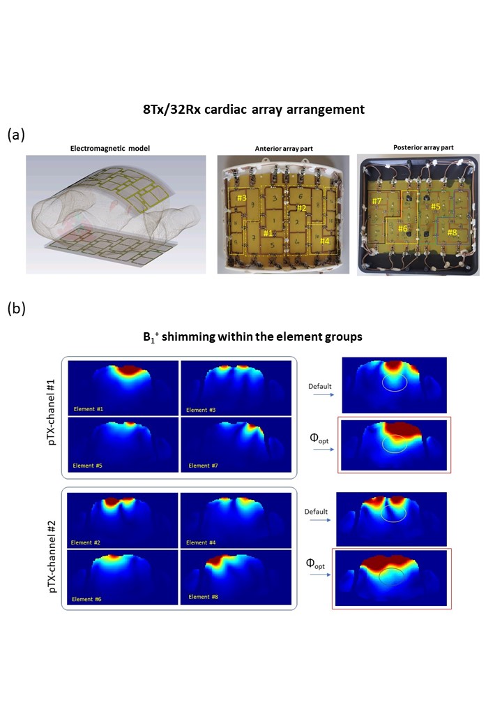
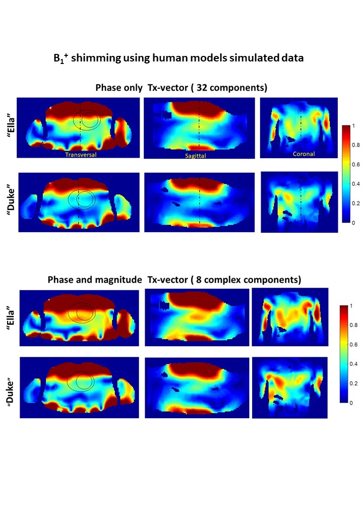
Figure 1 (a) shows the sketch of the array EM model and physical implementation. Two parts (anterior and posterior) each include 16 rectangular elements partially arranged in L-shaped substructures to minimize coupling. This allows shaping of the constructive B1+ pattern (i) using phase-only (sTX) B1+ shimming for 32 elements and (ii) shimming of B1+ for the groups of 4 elements connected to the individual channel of the scanner RFPA ( panel (b)) to perform further a pTX-based B1+ shimming.Figure 2 (a and b ) demonstrates the results of the B1+ shimming using sTX B1+ optimization in two human models using phases of 32 elements and static pTX using phases and magnitudes of 8 driving voltages. Both optimization modes demonstrate the possibility for efficient shaping B1+ in the region of the heart avoiding destructive interferences.
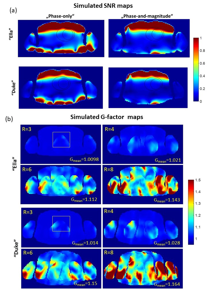
Figure 3 shows the simulated SNR maps(a) and computed g-factors of the array for the parallel imaging acceleration factor up to 8 (b). The simulated g-factor value in human models for left-to-right PE direction shows that theoretical noise correlation penalty can be nearly negligible ( Gmean<1.03 in the heart region ) for the acceleration up to factor 4 with a moderate increase for the higher acceleration factors.
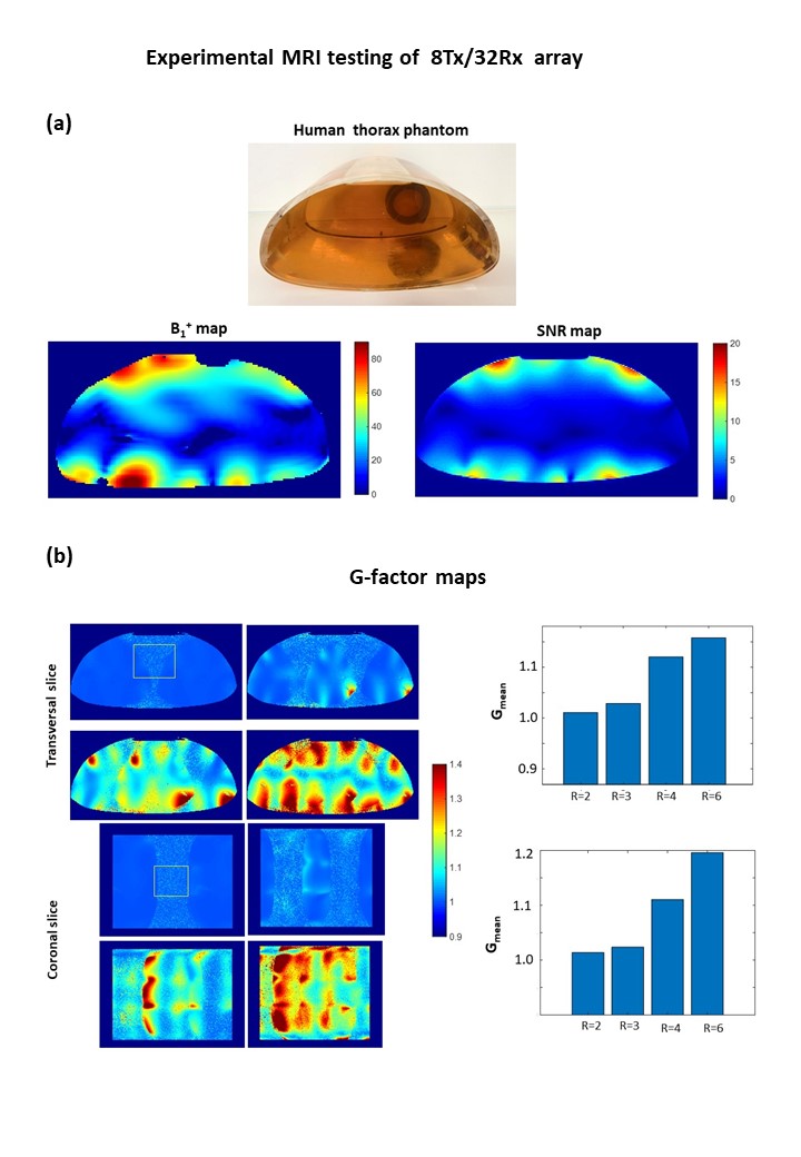
Figure 4 shows the results of the experimental tests of the array in human thorax phantom filled with PVP solution mimicking human tissue electrical properties. The phase-only B1-vector optimized for the “Ella” model was used for B1 shimming using connected coaxial cables of a specific length. Results of g-factor mapping confirm that parallel imaging reconstruction with a moderate noise correlation penalty (<20%) is feasible for acceleration factors up to R=6.
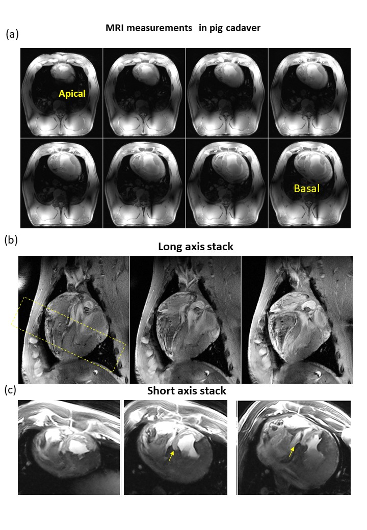
Figure 5 shows the proof-of-the-principle application of the new array for cardiac MRI using a fresh pig cadaver (42kg). The transversal stack demonstration penetration of B1+ and coverage of the heart is demonstrated together with anatomical oblique slices ( long and short axis stacks). Despite of deformed post-mortem heart geometry and contrast different from the one typical for in-vivo-cardiac MRI the remained late-gadolinium enhancement in the infarcted area could be clearly identified.
Discussion
We have demonstrated that the designed 8Tx/32Rx array allows for efficient B1+ shimming for both sTX applications using pre-defined phase-only vectors, and using pTX mode with 8 Tx channels RFPA. Despite the sub-optimal geometry of the pig thorax (which is essentially more narrow in the coronal plane than a human), the array demonstrated a relatively homogeneous coverage of B1+ with no apparent destructive interferences in the heart of the pig cadaver.Conclusion
The initial results show that L-shaped antisymmetric architecture can be successfully scaled up to design and implement an array with a larger number of transceiver elements. This expands the capability of this array design for usage in both parallel transmit and accelerated parallel receive for cardiac MRI at 7T.Acknowledgements
We are grateful to Dr. Eduardo Mauro and Anja Stadtmüller for their veterinary support. The work is partially supported by Collaborative Research Centre (CRC) 1525 "Cardio-immune Interfaces" funded by the German Research Foundation (DFG)References
1. Graessl A, Renz W, Hezel F, Dieringer MA, Winter L, Oezerdem C, Rieger J, Kellman P, Santoro D, Lindel TD, Frauenrath T, Pfeiffer H, Niendorf T (2014) Modular 32-channel transceiver coil array for cardiac MRI at 7.0T. Magnetic resonance in medicine 72 (1):276-290.
2. Elabyad I, Terekhov, M., Bille, M., Schreiber, L.M. (2021) Design and Implementation of Two 16-Element Antisymmetric Transceiver Coil Arrays for Parallel Transmission Human Cardiac MRI at 7 T. IEEE Transactions on Microwave Theory and Techniques 7 (69): 3540-3557.
3. Elabyad IA, Terekhov M, Lohr D, Stefanescu MR, Baltes S, Schreiber LM (2020) A Novel Mono-surface Antisymmetric 8Tx/16Rx Coil Array for Parallel Transmit Cardiac MRI in Pigs at 7T. Sci Rep 10 (1):3117.
4. Terekhov M, Elabyad IA, Schreiber LM (2021) Global optimization of default phases for parallel transmit coils for ultra-high-field cardiac MRI. PLoS One 16 (8):e0255341.
Figures