3896
Knee MRI Using a Wireless and Passive Metasurface Coil1Department of Radiology, Beijing Tsinghua Changgung Hospital, School of Medicine, Tsinghua University, Beijing, China, 2State Key Laboratory of Tribology, Department of Mechanical Engineering, Tsinghua University, Beijing, China, 3Center for Biomedical Imaging Research, Department of Biomedical Engineering, School of Medicine, Tsinghua University, Beijing, China, 4State Key Laboratory of New Ceramics and Fine Processing, School of Materials Science and Engineering, Tsinghua University, Beijing, China
Synopsis
Keywords: New Devices, Non-Array RF Coils, Antennas & Waveguides, Metasurface
Metasurface is an effective solution to enhance SNR of MRI images. Here, we propose a wireless and passive metasurface coil and evaluate its performance in knee MRI. Phantom studies suggest improved image uniformity compared to the conventional phased-array radio-frequency coils. In vivo knee MRI of volunteers suggests higher image quality scores and better contrast of knee cartilage in metasurface groups. When used in conjunction with the spine coil, the metasurface coil provides a favorable SNR between that of the 4- and 12-channel phased-array coil. It is more convenient, cost-effective, and applicable to 1.5T MRI systems from different manufacturers.
Introduction
MRI is vital to the diagnosis of various knee conditions. Improving image quality contributes to higher efficiency in examinations and better diagnostic accuracy. Metasurfaces have been proven as a novel method to improve the SNR of MRI images1-3. Our recent work demonstrated a cylindrical and compact metasurface device that provides homogeneous SNR enhancement4. The metasurface device can significantly amplify the RF receiving field and achieve two to three folds SNR enhancement compared to a 4-channel flexible coil, thus can be used as an alternative surface coil in clinical wrist MRI. Inspired by metasurfaces, this study was performed to design a wireless and passive surface coil and evaluate its application in knee MRI at 1.5T.Methods
Coil construction: The metasurface coil consists of four parts (Figure 1): a 156 mm inner diameter cylindrical support, sixteen array elements made of printed circuit boards on the surface, the copper rings connecting the array elements, and the control circuit including capacitors and diodes. The control circuit is designed to ensure that the metasurface automatically switched its resonant modes during the RF transmitting and receiving periods. Its detailed working principle was reported in our previous work4.MRI protocols: All experiments were performed using the uMR570 1.5T MR system (United Imaging Healthcare, China), unless indicated otherwise. For phantom studies, T1-weighted spin-echo, T2-weighted fast spin-echo, and gradient-recalled echo images were acquired using the following parameters: FOV, 120 mm × 120 mm; matrix, 256 × 256; slice thickness, 4 mm. In the metasurface groups, 3 identical metasurface coils were used to ensure the reliability of imaging performance. For in vivo studies, 10 healthy volunteers were included and subjected to a standard clinical knee MRI protocol in the supine position. Sagittal sections were acquired using the following parameters: FOV, 160 mm × 160 mm; matrix, 256 × 256; slice thickness, 4 mm.
Image analysis: Image SNR, CNR, and uniformity calculations were performed according to the National Electrical Manufactures Association Standards Publication MS 1-2008 (R2014). Knee MRI images were reviewed by two experienced radiologists. Images were reviewed independently, and coil information was blinded during interpretations. Image quality was assessed using a five-point Likert scale scored from 1 to 5.
Statistical analysis: Data are presented as the mean ± SEM and analyzed using Prism (GraphPad Software). Pairwise comparisons were performed using the Student's t-test. Multiple comparisons were tested using the two-way ANOVA. The results were considered significantly different at p-values ≤ 0.05.
Results
Phantom experiment: The SNR analysis showed that metasurface coils presented image SNR equivalent to the 4-channel flexible coil when received by the body coil, and equivalent to the 12-channel knee coil when received by the spine coil (Fig 2B). Both metasurface groups showed better image uniformity (Fig 2C). Spoke-shaped artifacts were observed in the images acquired using phased-array coils, but not visible in metasurface groups (Fig 2A). The waved curves of the SNR distribution imply that the SNRs were higher near the surface and lower in the center in images obtained using phased-array coils, while not in metasurface groups (Fig 2, D to F).Volunteer experiment: In the knee MRI scans involving 10 volunteers, all four groups obtained images with good quality (Fig 3A). Compared with conventional coils used in clinical knee MRI (12-channel knee coil and 4-channel flexible coil), the image quality scores were higher when the metasurface coil was applied (Fig 3B). In combination with the spine coil, the metasurface coil yielded MRI images with SNRs between that afforded by the 4-channel flexible coil and the 12-channel knee coil (Fig 4, A to D). The metasurface groups showed higher CNRs of cartilage to muscle than the conventional coils on the proton density-weighted fat-saturated spin-echo images (Fig 4E), indicating their better contrast in the observation of knee cartilages.
Knee MRI on multiple 1.5T scanners: One volunteer’s knee was imaged using the same metasurface coil in 1.5T MRI systems of three different manufacturers without any interfaces or imaging protocol modifications. All images presented good quality (Fig 5).
Discussion and conclusion
In this study, we assessed the pilot clinical use of a metasurface-based surface coil for knee MRI. The metasurface coil can work in conjunction with the body coil or spine coil and achieves better image quality scores and CNRs in preclinical knee MRI of volunteers. Improved image homogeneity was observed in both phantom and in vivo studies. Image SNR was between the 4-channel flexible coil and 12-channel knee coil when received by the spine coil, meets the clinical use.The metasurface coil is lightweight, wireless, and passive, which are three important properties for future RF coil design highlighted by recent investigations5,6. Without the limitations of coil interfaces and power sources, the metasurface coil was successfully operated in multiple MRI systems by different manufacturers; hence, increased convenience of clinical use and cost reduction were realizable.
In conclusion, the proposed metasurface coil can improve the image quality of knee MRI. At the application level, the coil is convenient, friendly, and cost-effective for both patients and medical facilities. Its diagnostic efficacy in knee MRI of patients needs to be further investigated by clinical trials.
Acknowledgements
None.
References
1. Wiltshire MC, Pendry JB, Young IR, et al. Microstructured magnetic materials for RF flux guides in magnetic resonance imaging. Science. 2001;291(5505):849-51.2. Slobozhanyuk AP, Poddubny AN, Raaijmakers AJ, et al. Enhancement of Magnetic Resonance Imaging with Metasurfaces. Adv Mater. 2016;28(9):1832-8.
3. Zhao X, Duan G, Wu K, et al. Intelligent Metamaterials Based on Nonlinearity for Magnetic Resonance Imaging. Adv Mater. 2019;31(49):e1905461.
4. Chi Z, Yi Y, Wang Y, et al. Adaptive Cylindrical Wireless Metasurfaces in Clinical Magnetic Resonance Imaging. Adv Mater. 2021;33(40):e2102469.
5. Deller TW, Mathew NK, Hurley SA, et al. PET Image Quality Improvement for Simultaneous PET/MRI with a Lightweight MRI Surface Coil. Radiology. 2021;298(1):166-172.
6. Darnell D, Truong TK, Song AW. Recent Advances in Radio-Frequency Coil Technologies: Flexible, Wireless, and Integrated Coil Arrays. J Magn Reson Imaging. 2022 Apr;55(4):1026-1042.
Figures
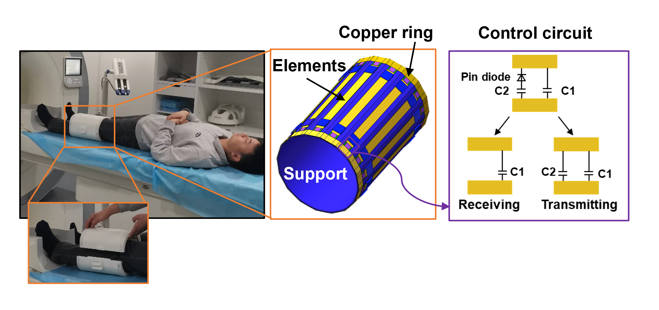
Figure 1. Coil design and experiment setup. During image acquisition, the metasurface coil was placed between the subject and the receiver coil, which was right above the examination bed.
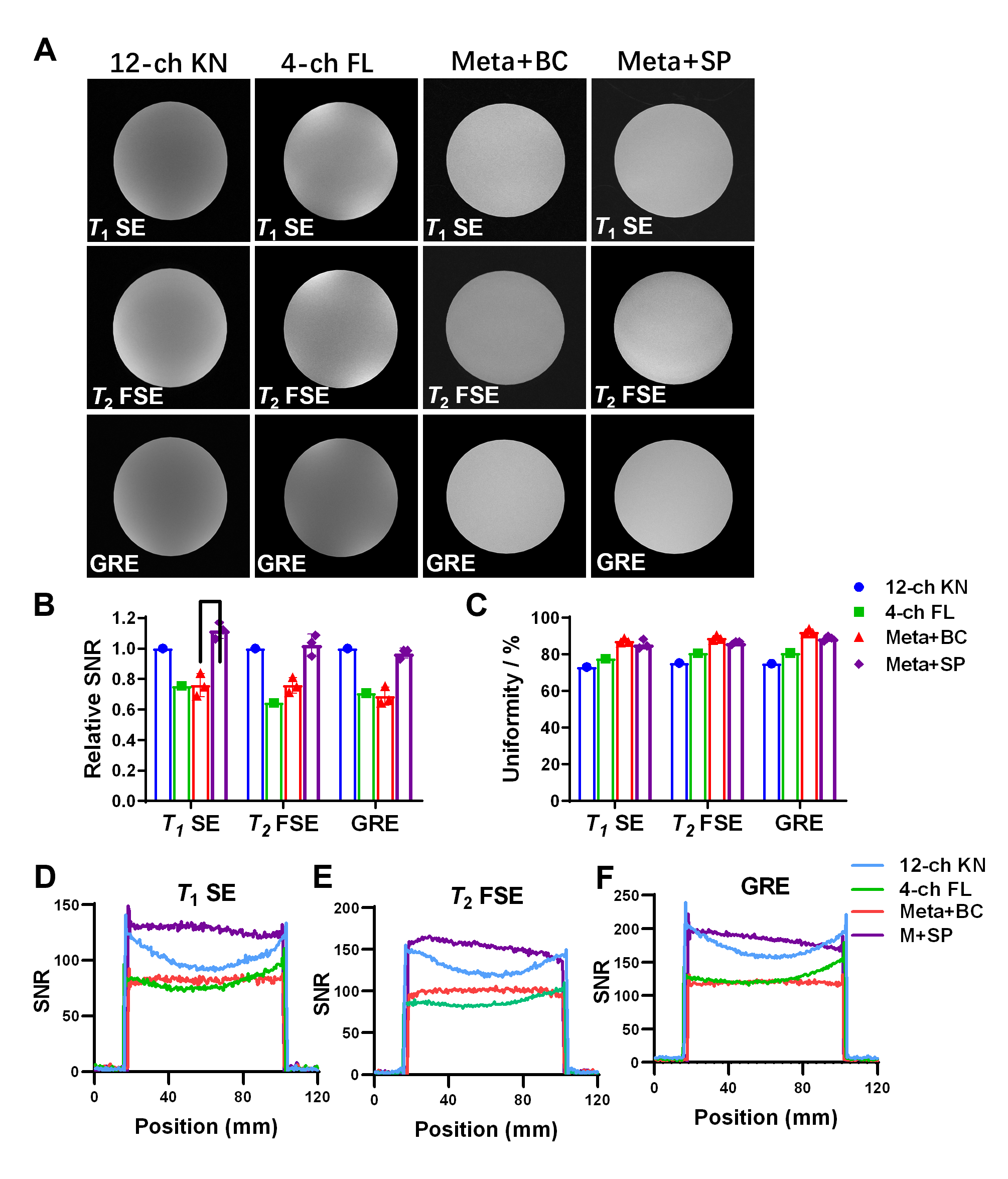
Figure 2. Phantom experiments. A. Representative axial images of a cylindrical water phantom using 12-channel knee coil (12-ch KN), 4-channel flexible coil (4ch-FL), and metasurface coil. Meta+BC refers to where metasurface coil was used with the body coil serving as receiver coil. Meta+SP refers to where metasurface coil was used with the spine coil serving as receiver coil. B and C. SNR and uniformity analysis of axial phantom MRI images. D–F. SNR distribution on midline of images in A.
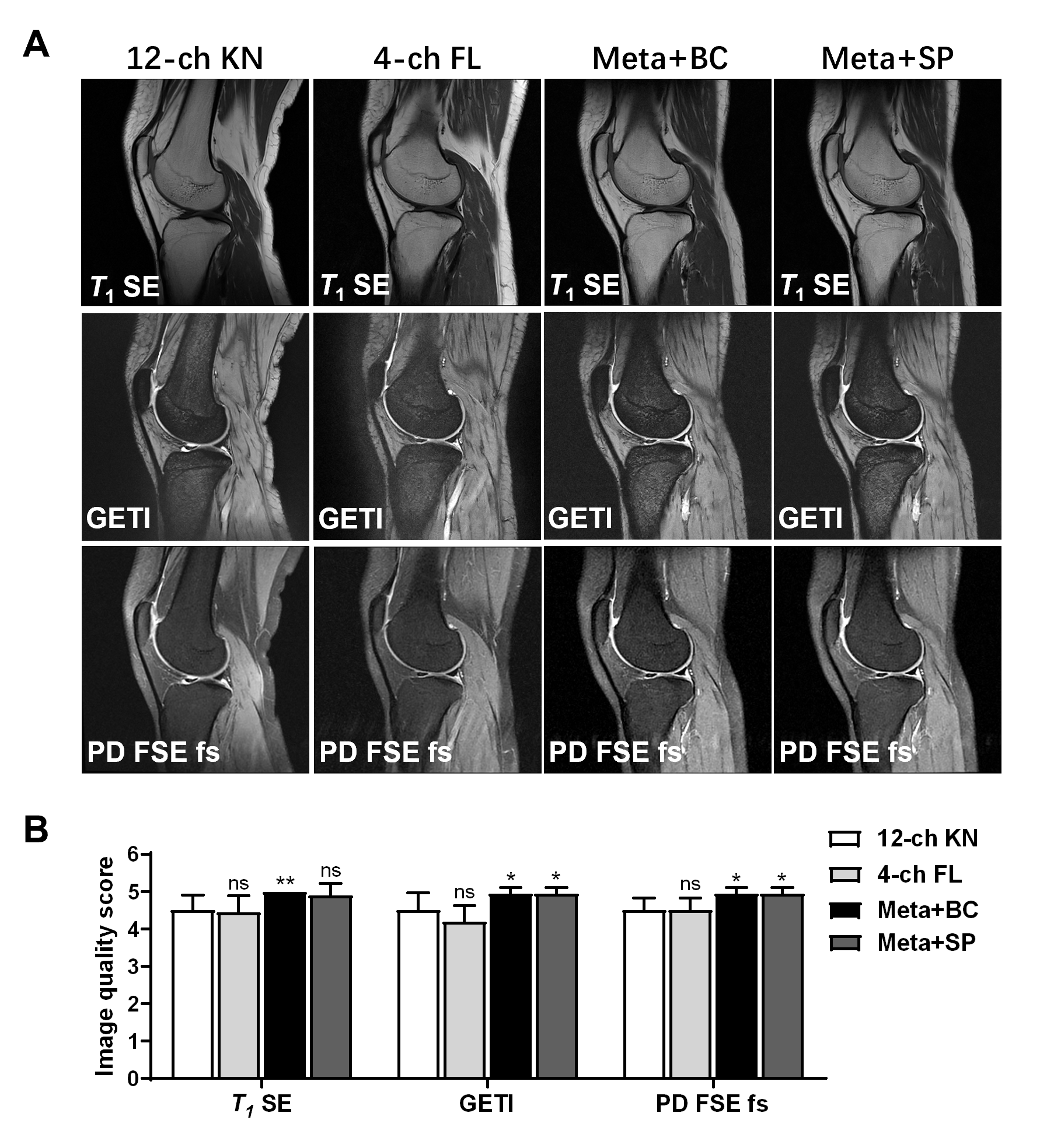
Figure 3. Knee MRI of volunteers and image quality analysis. A. Representative sagittal images of human knee MRI. GETI=gradient echo train image. B. Image quality scores of knee MRI scans of volunteers (n=10). All error bars represent standard error of mean (S.E.M.); *p < 0.05, **p < 0.01.
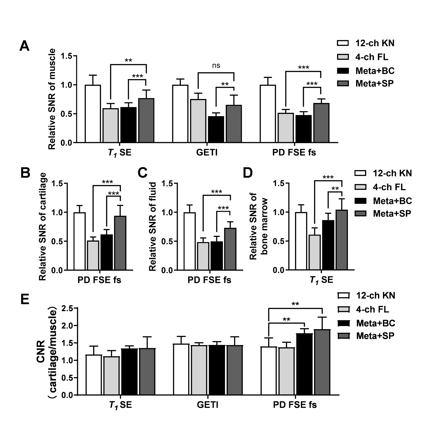
Figure 4. SNR and CNR analysis of in vivo knee MRI. A–D. SNR analysis of knee MRI scans of volunteers (n=10). E. CNR analysis of cartilage to muscle. All error bars represent standard error of mean (S.E.M.); **p < 0.01, ***p < 0.001.
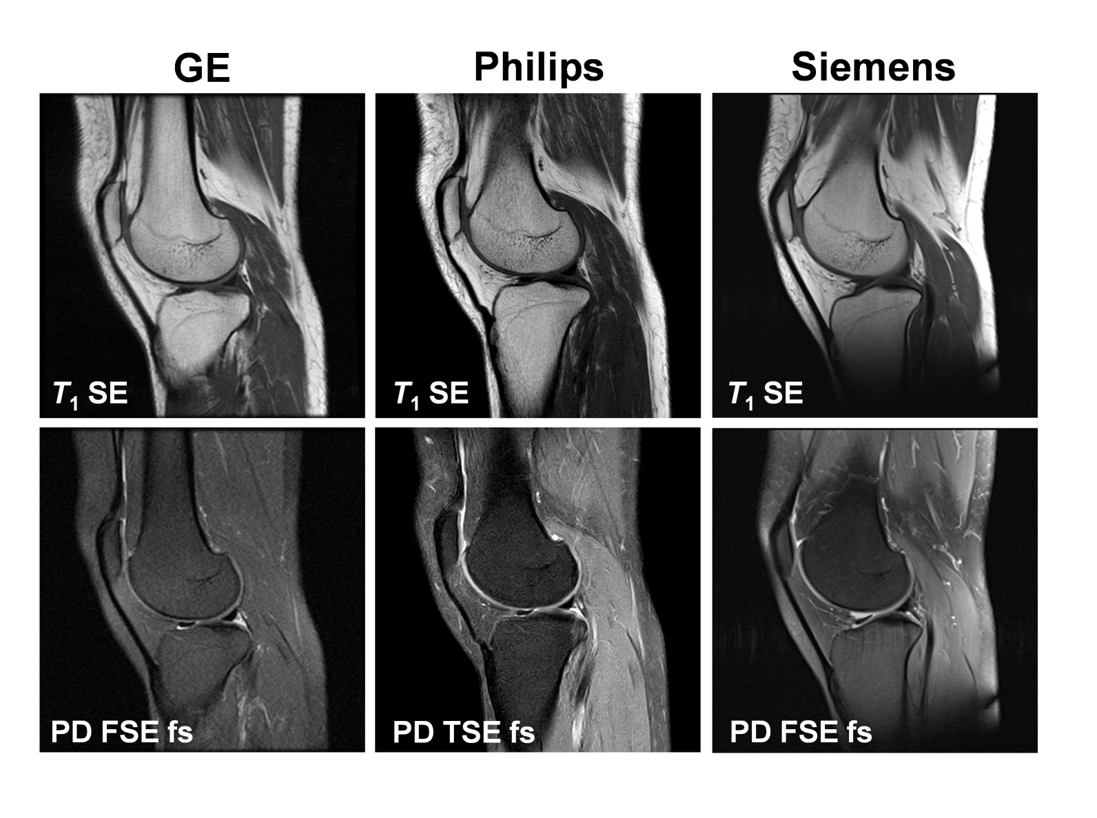
Figure 5. In vivo performance of metasurface in multiple MRI systems. Knee MRI scans of a healthy volunteer were obtained using metasurface in multiple MRI systems, including GE Signa Explorer 1.5T, Philips Ingenia 1.5T, and Siemens Magnetom Aera 1.5T (in alphabetical order).