3890
Deep Learning Reconstruction in MRI: Comparison of Image Quality in Patients With Hepatic Malignancy1West China Hospital of Sichuan University, Chengdu, China, 2GE Healthcare, MR Research, Beijing, China
Synopsis
Keywords: Machine Learning/Artificial Intelligence, Image Reconstruction
Improving image resolution by denoising is an important research goal in MRI reconstruction. An emerging technique, deep learning reconstruction (DLR), has shown great potential in MRI denoising. In this study, we included 37 patients with pathologically diagnosed hepatic malignancy, and compared the image quality of DLR and original reconstruction regarding dual echo T1 weighted sequence, diffusion weighted imaging (DWI) and fat-suppressed T1 weighted gadolinium-enhancement. It was shown that DLR significantly improved the image quality by reducing background noise, thus making hepatic malignancy more conspicuous.Background and Purpose
Magnetic resonance imaging (MRI) plays a vital role in disease diagnosis. However, low spatial resolution and long scan time of MRI limited its clinical application. Generally, higher spatial resolution and less acquisition time lead to a reduced signal-to-noise ratio (SNR), probably degrading image quality and diagnostic performance1. Thus, denoising is crucial for MRI to accomplish better image quality while reducing the scan time2.Deep learning techniques have been widely used in the medical imaging field3. Deep learning-based reconstruction has been introduced to improve MRI image quality while maintaining a reasonable scan duration. One of the novel deep learning reconstruction algorithms (AIRTM Recon DL [ARDL], GE Healthcare, Waukesha, WI) shows the powerful ability to denoise images and eliminate truncation artifacts. Unlike other algorithms that may change images details, ARDL takes raw k-space data as an input before Fourier transform, and uses a convolutional neural network (CNN) with over 4.4 million parameters and over 10.000 kernels to learn the characteristics of noise, and eventually produces ARDL images along with original images (Non-ARDL). Recent studies have demonstrated the improvement of image quality by ARDL in liver4, brain5,6, cardiac7,8 and prostate9 MRI.
Although the feasibility of ARDL in normal liver have been confirmed, its superiority in patient cohorts, especially in patients with hepatic malignancy, remains unknown. The potential altered MRI information of hepatic malignancy puts a huge challenge on radiologists to achieve faithful diagnosis10. Hence, our study aimed to evaluate the performance of ARDL in hepatic malignancy with 2D axial sequences.
Methods
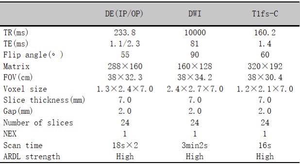
In this study, we collected consecutive abdominal enhanced examinations with ARDL in a 3.0 T MRI scanner (SIGNA Architect, GE Healthcare) using a 30-channel AIRTM anterior array from June 2022 to October 2022. Patients with pathologically diagnosed hepatic malignancy were included. Images with severe motion artifacts or magnetic susceptibility artifacts related to abdominal metallic devices were excluded. Dual echo (DE) sequence (ARDL and Non-ARDL), diffusion weighted imaging (DWI) sequence (ARDL and Non-ARDL, b=1000) and fat-suppressed T1 weighted gadolinium enhanced (T1fs-C) sequence (ARDL and Non-ARDL) were analyzed. The imaging parameters of each sequence are listed in Figure 1.In objective analysis, three sequences were evaluated by one junior radiologist (8-year experience) and one senior radiologist (20-year experience), who both were aware of the study concept but had been blinded to all clinical information in order to avoid bias. Mean signal intensity of normal hepatic parenchyma, malignant lesions, muscle and the standard deviation (SD) of the background were measured by the junior radiologist, and then verified by the senior radiologist. The SNR and contrast-to-noise ratio (CNR) of normal hepatic parenchyma and malignant lesions of each sequence were calculated with the following formulas4, where the “liver” represents the normal hepatic parenchyma and the “lesion” represents the hepatic malignancy.
$$SNR(liver)=\frac{S(liver)}{SD(background)}$$
$$CNR(liver)=\frac{|S(liver)-S(muscle)|}{SD(background)}$$
$$SNR(lesion)=\frac{S(lesion)}{SD(background)}$$
$$CNR(lesion)=\frac{|S(lesion)-S(muscle)|}{SD(background)}$$
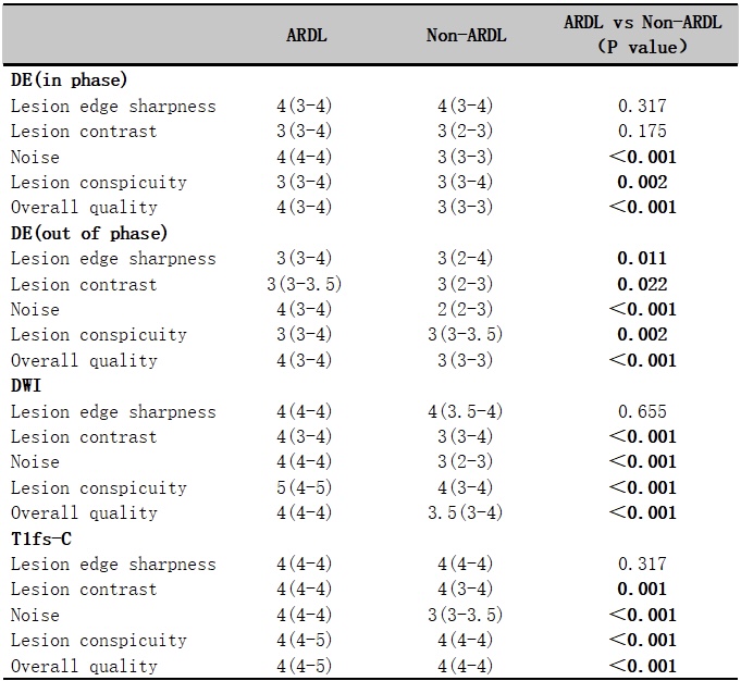
Moreover, the two above-mentioned radiologists assessed the image quality in ARDL and Non-ARDL images and reached a consensus for disagreement after discussion. Subjective image quality had been assessed with a five-point Likert scale containing: (a) lesion edge sharpness: 1=poor; 2=mild; 3=moderate; 4=good; 5=very good. (b) lesion contrast: 1=insufficient; 2=mild; 3=moderate; 4=good; 5=very good. (c) noise: 1= severe (hindering diagnosis); 2=acceptable; 3=moderate; 4=mild; 5=absence of noise; (d) lesion conspicuity: 1=unable to see; 2=blurry but visualized; 3=moderate; 4=good; 5=excellent. (e) overall quality: 1=poor; 2=mild; 3=moderate; 4=good; 5=excellent.
Continuous variables are compared using the paired-samples t-test or the Wilcoxon test. The normality of data distribution is assessed by Shapiro-Wilk test. A two-sided P value < 0.05 was considered statistically significant. All statistical analyses will be conducted using SPSS 21.0.
Results
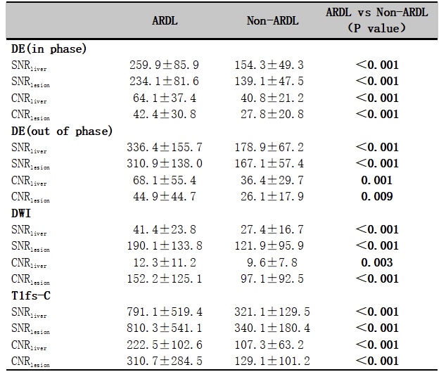
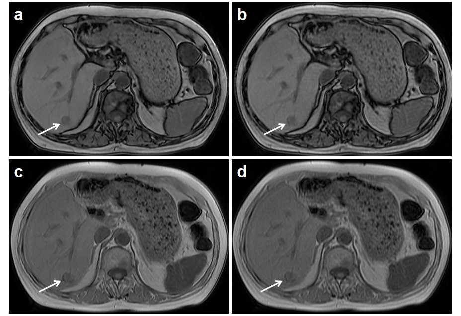
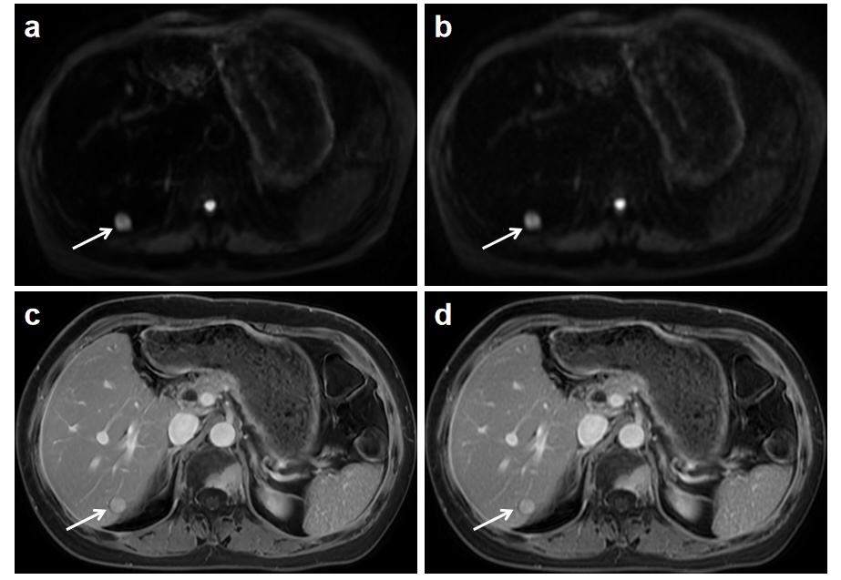
A total of 37 patients (25 men and 12 women, 55.2±12.7 years old) were included. There were 17 patients with hepatocellular carcinoma, 19 patients with hepatic metastases and 1 patient with intrahepatic cholangiocarcinoma. The SNRliver, CNRliver, SNRlesion and CNRlesion of all the sequences with ARDL were all higher than those without ARDL (P<0.05). We found the SNR and CNR of both normal hepatic parenchyma and malignant lesions in DE (in phase) and DWI would increase by approximately 50%, and as for DE (out of phase), T1fs-C, the SNR and CNR would increase by approximately 100%. Detailed results for all the objective evaluation are listed in Figure 2. Lesion conspicuity, noise and overall quality of ARDL were significantly higher than Non-ARDL in all sequences (P<0.001). The sharpness between ARDL and Non-ARDL images was not significant in DE (in phase), T1fs-C and DWI (P=0.317, P=0.317 and P=0.655). The contrast was not significant in DE (in phase) with P=0.175. Detailed results of subjective evaluation are listed in Figure 3. The representative pictures are displayed in Figure 4 and Figure 5.Discussion and Conclusion
In this study, all the objective indexes were higher as ARDL denoised the background, meaning that ARDL may achieve a new balance between scan time and spatial resolution. According to subjective evaluation, with the enhanced visualization of lesions, ARDL is likely to improve the diagnostic performance in hepatic malignancy. In conclusion, deep learning reconstruction can significantly improve image quality by denoising, providing better overall quality and making the hepatic malignancy more conspicuous. ARDL has great potential in routine 2D axial sequences of hepatic malignancy.Acknowledgements
NoneReferences
1. Machado-Rivas, F., Jaimes, C., Kirsch, J.E. & Gee, M.S. Image-quality optimization and artifact reduction in fetal magnetic resonance imaging. Pediatr Radiol 50, 1830-1838 (2020).
2. You, X., Cao, N., Lu, H., Mao, M. & Wanga, W. Denoising of MR images with Rician noise using a wider neural network and noise range division. Magn Reson Imaging 64, 154-159 (2019).
3. Gore, J.C. Artificial intelligence in medical imaging. Magn Reson Imaging 68, A1-a4 (2020).
4. Zerunian, M., et al. Artificial intelligence based image quality enhancement in liver MRI: a quantitative and qualitative evaluation. Radiol Med 127, 1098-1105 (2022).
5. Kim, M., et al. Thin-Slice Pituitary MRI with Deep Learning-based Reconstruction: Diagnostic Performance in a Postoperative Setting. Radiology 298, 114-122 (2021).
6. Kim, S.H., et al. Deep learning reconstruction in pediatric brain MRI: comparison of image quality with conventional T2-weighted MRI. Neuroradiology (2022).
7. Ogawa, R., et al. Reconstruction of cardiovascular black-blood T2-weighted image by deep learning algorithm: A comparison with intensity filter. Acta Radiol Open 10, 20584601211044779 (2021).
8. van der Velde, N., et al. Improvement of late gadolinium enhancement image quality using a deep learning-based reconstruction algorithm and its influence on myocardial scar quantification. Eur Radiol 31, 3846-3855 (2021).
9. Wang, X., et al. Novel deep learning-based noise reduction technique for prostate magnetic resonance imaging. Abdom Radiol (NY) 46, 3378-3386 (2021).
10. Costa, A.F., Clarke, S.E., Stueck, A.E., McInnes, M.D.F. & Thipphavong, S. Benign Neoplasms, Mass-Like Infections, and Pseudotumors That Mimic Hepatic Malignancy at MRI. J Magn Reson Imaging 53, 979-994 (2021).
Figures