3883
Evaluation of deep learning-based reconstruction for qualitative and quantitative DW-MRI in head and neck cancers
Ramesh Paudyal1, Akash Deelip Shah2, Amaresha Shridhar Konar1, Jaemin Shin3, Eve LoCastro1, Nisha Bagchi4, Maggie Fung3, Suchandrima Banerjee5, Nancy Lee6, and Amita Shukla-Dave1,2
1Medical Physics, Memorial Sloan Kettering Cancer Center, New York, NY, United States, 2Radiology, Memorial Sloan Kettering Cancer Center, New York, NY, United States, 3GE Health Care, New York, NY, United States, 4Kimmel Medical College, Thomas Jefferson University, Philadelphia, PA, United States, 5GE Health Care, Menlo Park, CA, United States, 6Radiation Oncology, Memorial Sloan Kettering Cancer Center, New York, NY, United States
1Medical Physics, Memorial Sloan Kettering Cancer Center, New York, NY, United States, 2Radiology, Memorial Sloan Kettering Cancer Center, New York, NY, United States, 3GE Health Care, New York, NY, United States, 4Kimmel Medical College, Thomas Jefferson University, Philadelphia, PA, United States, 5GE Health Care, Menlo Park, CA, United States, 6Radiation Oncology, Memorial Sloan Kettering Cancer Center, New York, NY, United States
Synopsis
Keywords: Machine Learning/Artificial Intelligence, Tumor
The head and neck (HN) region have complex anatomical structures that affect the image quality of diffusion-weighted MRI. Therefore deep learning (DL)-based Reconstruction (Recon) for DW-MRI could be a promising method that can help improve image sharpness and signal-to-noise ratio (SNR) without increasing signal averaging. The present study aimed to evaluate the performance of qualitative and quantitative multiple b-value DW-MRI powered by DL-based Recon for tumors in the HN region. The DL-based recon method improved the DW image quality and SNR compared to those without DL recon.Purpose
Diffusion-weighted (DW)-MRI has been used for tumor characterization and treatment response assessment in tumors of the head and neck (HN) region.1,2 New data acquisition and post-processing methods have shown incremental value in reducing image distortion on DW-MRI images.3 Recently, a novel vendor-developed deep learning (DL)-based DW-MRI reconstruction (Recon) method, AIRTM Recon DL, has shown promise in enhancing the image signal-to-noise ratio (SNR) and sharpness in data obtained from prostate cancer patients.4,5,6 The DL-based Recon can be clinically useful for detecting and delineating tumor extent in DW images, allowing for the accurate apparent diffusion coefficient (ADC) measurement.2 The present study is the first to evaluate the performance of qualitative and quantitative multiple b-value DW-MRI powered by DL-based Recon for tumors in the HN region.Methods
Phantom: NIST/QIBA ice water phantom DWI data were acquired on a 3T MRI scanner (SIGNA Premier, GE Healthcare) using a 21-channel head and neck unit. MRI data acquisition: The multi b-value (b=0, 500, 900, 2000 s/mm2) DW images were acquired using a single shot spin echo planar imaging (SS-SE-EPI) sequence with TR/TE=15000/99 (minimum) ms, the field of view (FOV)=20 cm, slices=15, slice thickness=4mm, number of excitation (NEX)=1.Patient: Multiple b-value DW-MRI data were acquired from six male, HN cancer patients (median age 59 years, 2 HPV (+) positive, 1 HPV (-), and 3 with unknown primary tumor status) in this retrospective study between December 2021 and June 2022. These patients underwent chemo-radiation therapy (CRT).
MRI data acquisition: MRI protocol consisted of multi-planar T1/T2 weighted imaging followed by multi-b-value at pre-treatment. The multi b-value DW images were acquired using a SS-SE-EPI sequence with TR/TE=4000/80 (minimum) ms, FOV=20-24 cm, matrix=128×128, slices=8-10, slice thickness=5mm, number of excitation (NEX)=2, and b=0,20,50,80,200,300,500,1000,1500,2000 s/mm2. The raw data from the DW-MRI scans were transferred and retro-reconstructed using the AIRTM Recon DL method in the GE reconstruction pipeline (Orchestra SDK, GE Healthcare), and finally labeled as DL recon DW images.4,5
Region of Interest Contouring and DWI Data Analysis: Regions of Interest (ROIs) were drawn on the DWI phantom images by the imaging scientist, and the primary tumors and neck nodal metastases were delineated by an experienced neuroradiologist on DL-Recon DW images (b = 0 s/mm2) using ITK-SNAP. Data analysis was performed using the MRI-QAMPER tool.7 Mean ADC values were calculated for all b- values, and we compared the DW images with and without DL recon using the Wilcoxon signed rank test (WSRT). A P <0.05 was considered significant. The rΔADC (%) =(ADCwithDL-ADCwithoutDL)/ADCwithDL×100 was calculated, where ADCwithDL and ADCwithoutDL represent ADC values with and without DL-Recon, respectively. Signal noise ratio (SNR) was calculated (SNR=µ/σ, where µ is the mean of the signal and σ is the standard deviation) for both primary tumor and metastatic neck nodes.
Qualitative image rating for patient data was performed for both DW images with and without DL recon (b= 0, 1000, and 200 s/mm2) on standard workstations. The overall diagnostic image quality was rated on a five-point scale as follows: 5 = excellent; 4 = good; 3 = acceptable (acceptable for diagnostic use but with minor issues); 2 = poor (not acceptable for diagnostic use); or 1 = unacceptable for diagnostic use as detailed elsewhere.8
Results
Phantom: The NIS/QIBA diffusion phantom ADC maps with and without DL-Recon were analyzed, and ADC values exhibited no difference for all vials, whereas the standard deviation between them varied between 2-33 % (Figure 1A).Patient: Qualitative analysis (image rating score) for b=0, 1000, and 2000 s/m2 is shown in Figure 2. For b=1000 s/mm2, DL recon DW images exhibited higher scores than those without DL (4.2± 0.4 vs. 3.7±0,5, P= 0.1). The DL recon method improved overall SNR by 50% (143.0 vs. 71.0, P = 0.002, for b= 0 s/mm2) and 45% (63.0 vs. 34.0, P = 0.048, for b= 1000 s/mm2), compared to those without DL-Recon, the ROIs size ranging between 21- 81 mm2. The present study evaluated a total of 10 tumor ROIs (i.e., 4 primary lesions and 6 metastatic nodes) from 6 HN cancer patients. Mean ADC values with and without DL for tumors in the HN region were not significantly different (P>0.05, Table 1, Figure 3). Figures 4 show the ADC maps generated with and without DL-Recon from patients with tumors in the HN region.
Discussion and Conclusion:
The image rating scores of DL DW images were higher than those without DL (Excellent 17% and good 83% vs. excellent 0% and good 67%, for b= 1000 s/mm2). The calculated SNR values of DL recon DW images were 45% higher than those without DL-Recon. Mean ADC values between with and without DL showed a minimal difference (0.04- 1.35%), while the maximum standard deviation was 33% between them. The results suggest that DL recon on DW images improved the image sharpness, allowing better tumor delineation at a higher b-value. In summary, DW images exhibit underlying hindered and restricted diffusion. The implementation of DL recon for diffusion in the HN region significantly improved image quality and, after validation, could be included in the HN imaging workflow.Acknowledgements
Support: Funding support from National Institutes of Health Grant: U01 CA211205 (ASD)References
1. Paudyal R, Chen L, Oh JH, et al. Nongaussian Intravoxel Incoherent Motion Diffusion Weighted and Fast Exchange Regime Dynamic Contrast-Enhanced-MRI of Nasopharyngeal Carcinoma: Preliminary Study for Predicting Locoregional Failure. Cancers (Basel). Mar 6 2021;13(5)doi:10.3390/cancers13051128 2. Riaz N, Sherman E, Pei X, et al. Precision Radiotherapy: Reduction in Radiation for Oropharyngeal Cancer in the 30 ROC Trial. JNCI: Journal of the National Cancer Institute. 2021;113(6):742-751. doi:10.1093/jnci/djaa184 3. Konar AS, Fung M, Paudyal R, et al. Diffusion-Weighted Echo Planar Imaging using MUltiplexed Sensitivity Encoding and Reverse Polarity Gradient in Head and Neck Cancer: An Initial Study. Tomography. Jun 2020;6(2):231-240. doi:10.18383/j.tom.2020.00014 4. Lebel RM. Performance characterization of a novel deep learning-based MR image reconstruction pipeline. arXiv preprint arXiv:200806559. 2020; 5. Choi M, Figee M, Lebel R, et al. Evaluation of the efficacy of a Deep Learning-based Reconstruction in the Connectomic Deep Brain Stimulation. Proc. Intl. Soc. Mag. Reson. Med. 30 (2022); 2022:3357. 6. Ueda T, Ohno Y, Yamamoto K, et al. Deep Learning Reconstruction of Diffusion-weighted MRI Improves Image Quality for Prostatic Imaging. Radiology. 2022;303(2):373-381. 7. Paudyal R, Konar AS, Obuchowski NA, et al. Repeatability of Quantitative Diffusion-Weighted Imaging Metrics in Phantoms, Head-and-Neck and Thyroid Cancers: Preliminary Findings. Tomography. Mar 2019;5(1):15-25. doi:10.18383/j.tom.2018.00044Figures
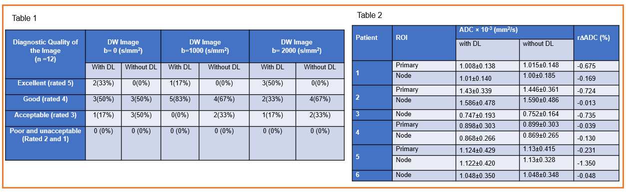
Table 1. Summary of the ADC values obtained with and without DL-Recon of DWI patient data. Table 2: Diagnostic qualitative image quality rating for DW images with and without DL-Recon.
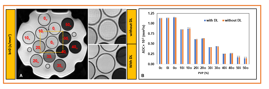
Figure 1. (A). Left: The diffusion-weighted image (b= 0 s/mm2) of the QIBA phantom at 00C. The images were reconstructed with and
without the deep learning-based reconstruction (DL-Recon) method. The vials
represent the different concentrations of chemical PVP in an aqueous solution. Right:
The magnified DL-Recon DWI image shows the image's sharpness and reduced ringing
artifact compared to images without DL. (B) Bar plot showing the ADC values for
all the thirteen vials measured with and without DL recon.
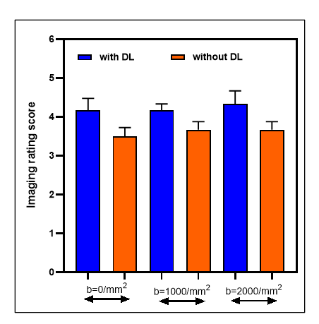
Figure 2. Bar plot showing the image rating score from six
patients' DW images with and without DL-Recon. Error bars are SEM of the mean.
The DL Recon showed a higher image rating than without DL for all b-values.
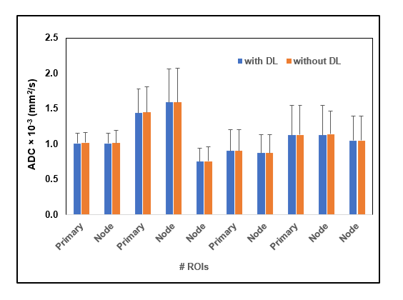
Figure 3. Bar plot showing the ADC values obtained with and without DL-Recon for ten ROIs from the tumor in the head and neck region. The standard deviation
shows variation across the primary tumors and metastatic nodes in HN patients.
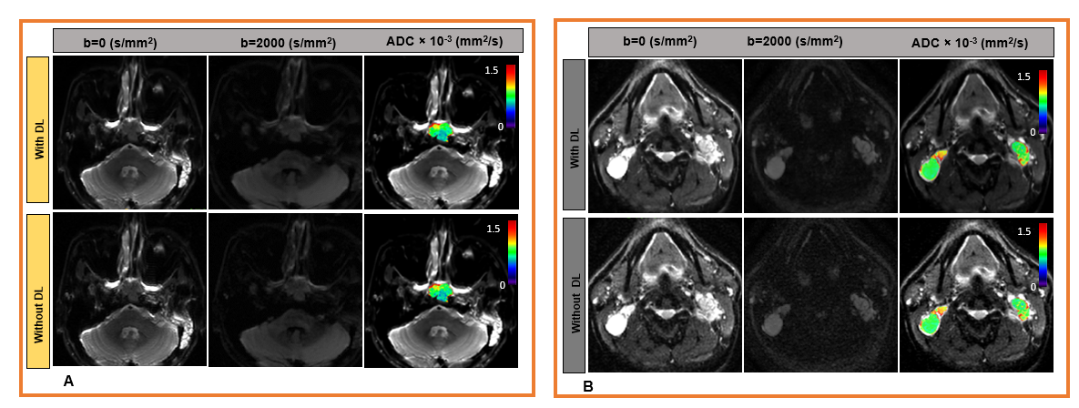
Figure 4. Representative DW images and apparent diffusion
coefficient (ADC) maps with and without DL-Recon. Exhibiting (A) the primary tumors
of a nasopharyngeal cancer patient (38 years, male). (B). Bilateral metastatic nodes in nasopharyngeal
cancer patient (40 years, male).
DOI: https://doi.org/10.58530/2023/3883