3856
Changes in Aspartate Metabolism in the medial-prefrontal cortex of Nicotine Addicts Based on J-edited Magnetic resonance spectroscopy1Magnetic Resonance Imaging, The First Affiliated Hospital of Zhengzhou University, Zhengzhou, China, 2Clinical and Technical Support, Philips Healthcare, Beijing, China
Synopsis
Keywords: fMRI, Metabolism, Hydrogen proton magnetic resonance spectroscopy (1H MRS)
Cigarette smoking harms nearly every organ of the body, causes many diseases, and reduces the health of smokers in general. Our study aims to explore the changes of aspartate (Asp) levels in the medial prefrontal cortex of patients with nicotine addiction using the J-edited 1H MRS technique. Results showed that the Asp level in medial prefrontal of nicotine addicts is relatively increased, suggesting that the metabolism of aspartate may play a key role in nicotine dependence.Introduction
According to statistics, there are currently more than 1 billion smokers in the world[1]. Long-term smoking not only causes cardiovascular and respiratory diseases, but also damages the cognitive and nervous systems. Nicotine dependence is the leading cause of smoking addiction, and treatment and intervention for nicotine addiction are urgent. A wealth of studies has shown that nicotine addiction causes changes in brain structure and function[2, 3], but there is very few on the altered biochemical metabolism in the brain of smokers. Hydrogen proton magnetic resonance spectroscopy (1H MRS) can non-invasively detect brain metabolites and has been widely used in clinical neuro metabolism related research. Some neurotransmitters with trace amounts in the brain can also be specifically and reliably detected by using the recently developed J-edited 1H MRS. Our study aims to explore the changes of aspartate (Asp) levels of in the medial prefrontal cortex of patients with nicotine addiction (NA) using the J-edited 1H MRS technique, which may provide a positive imaging evidence for the therapy and intervention of nicotine addiction.Method
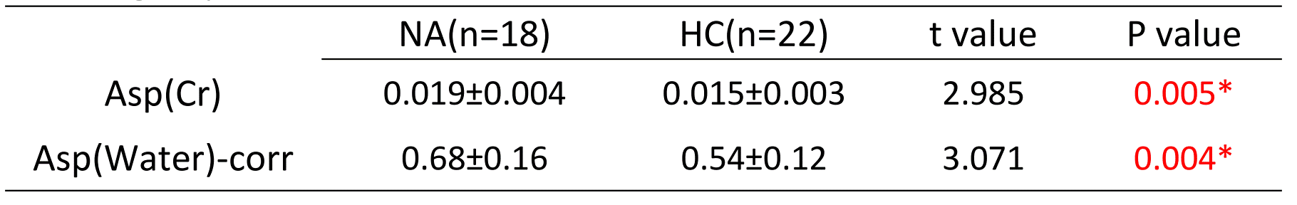
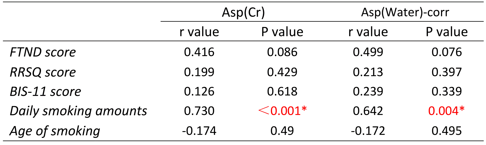
A total of 45 men aged 40 to 60 years were initially recruited in Henan province, whose have not taken any drugs recently, including 21 patients in the NA group and 24 patients in the healthy control (HC) group. All subjects underwent routine MRI and the edited MRS (MEGA-PRESS) scans on a 3.0T MRI scanner (Ingenia Meta, Philips Healthcare, Best, the Netherlands). After excluding patients with underlying diseases, alcohol dependence, and insufficient data quality, 18 patients with NA and 22 cases of age- and sex-matched HCs were finally included. The MEGA-PRESS sequence was implemented with parameters as follow: repetition time (TR) = 2000 ms; echo time (TE) = 90 ms; number of averages = 96; on/off frequencies = 3.89/5.21 ppm; scan time = 6 min 30 s. The VAPOR scheme was used for water suppression. The edited spectra were post-processed and quantitatively analyzed using the Gannet tools on the MATLAB software (Figure 1). The structural MPRAGE images were then segmented into gray matter, white matter, and cerebrospinal fluid using SPM12, and the voxel for MRS acquisition was aligned to the MPRAGE image for tissue segmentation. The Asp levels were quantified with reference to the creatine (Asp/Cr) and water signal (Asp/Water, corrected for brain tissue segmentation), respectively. Two independent samples t-test was used to analyze the differences in Asp/Cr and Asp/Water levels between the two groups. Finally, the correlation analysis of Asp levels with the clinical characteristic assessment scales (Including Daily smoking amounts, Age of smoking, FTND score, RRSQ score and BIS-11 score) was performed using the Spearman criteria.Result
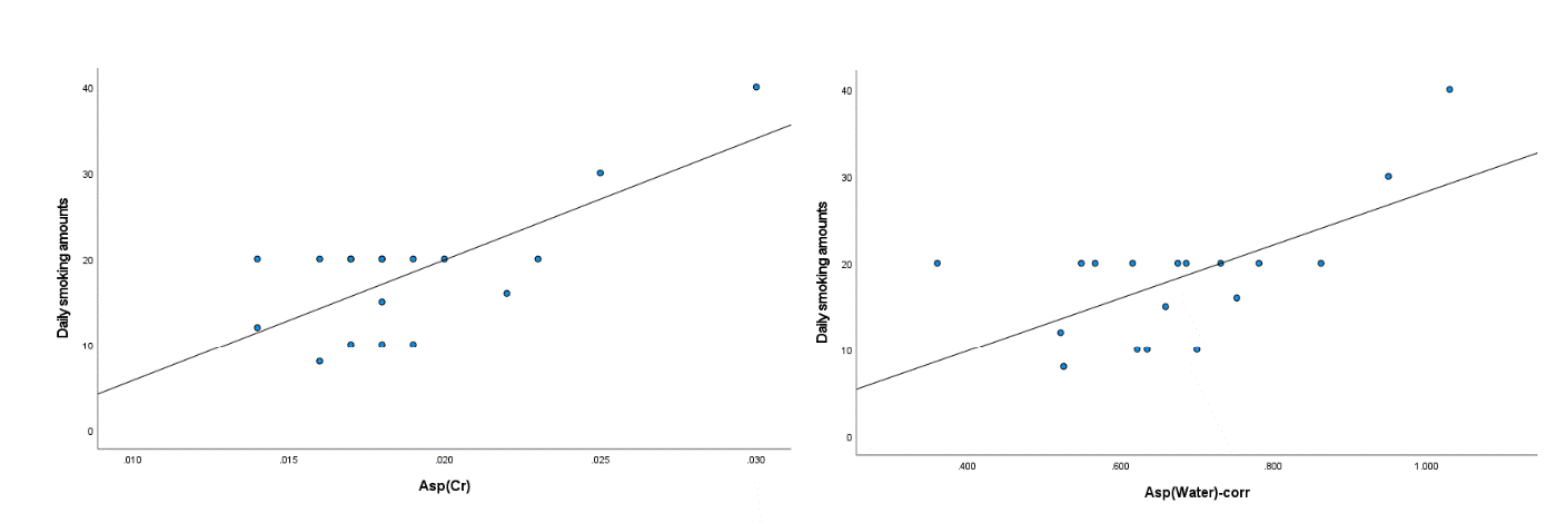
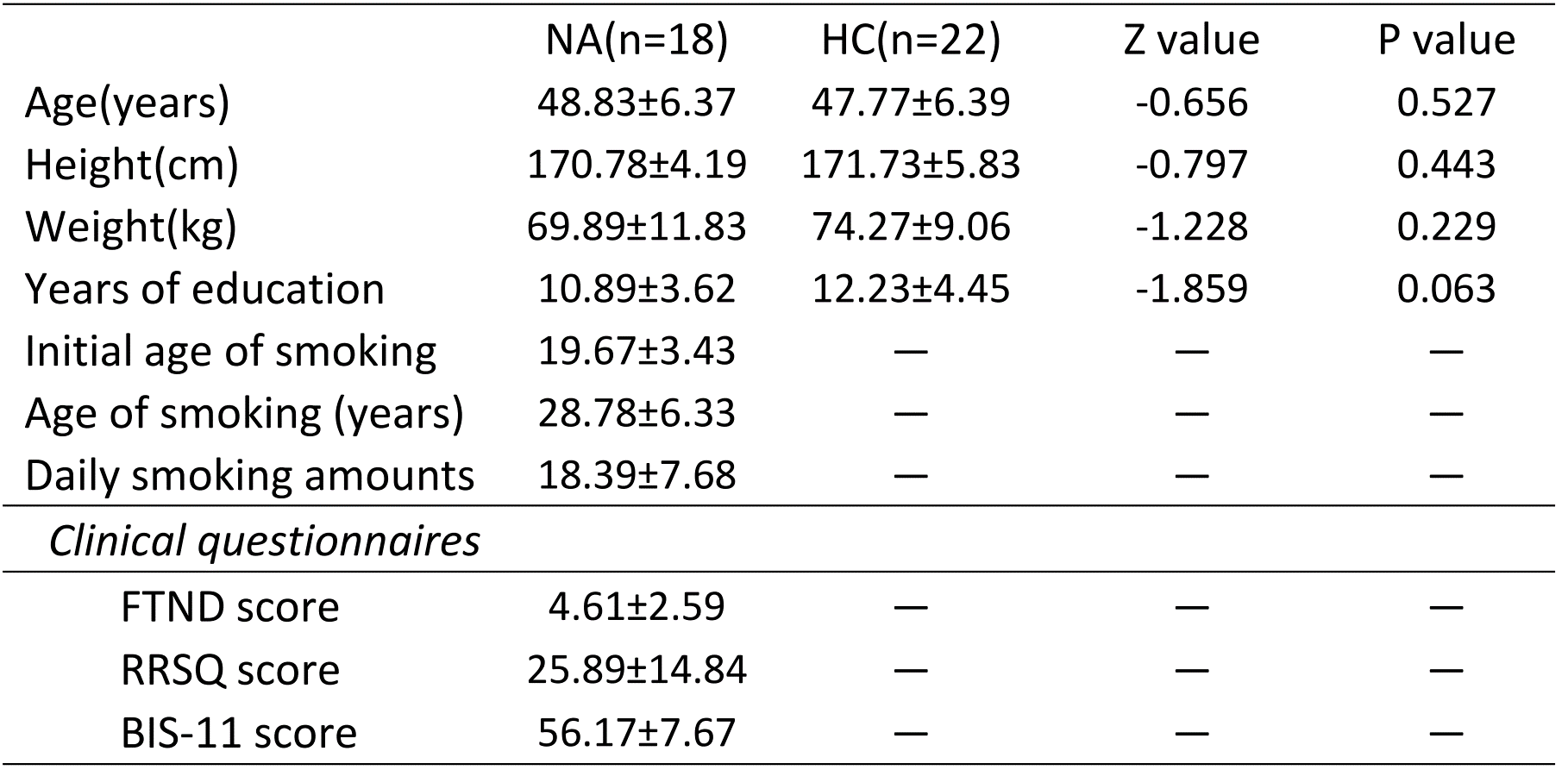
The demographic data are shown in Table 1. Compared to the HC group, Asp/Cr and Asp/water levels in the medial prefrontal cortex were higher in nicotine addicts (Table 2). In addition, Asp/Cr and Asp/water levels were positively associated with daily smoking amounts (Table 3 and Figure 2). No significant correlation was found between the Asp levels and the FIND score, the BIS-11 score or the RRSQ score.Discussion
J-edited MRS can be used to separate overlapped signals of metabolites, which allows us to directly and accurately detect the isolated Asp signal in specific brain regions[4]. Aspartate is one of the excitatory neurotransmitters in the brain, and its level changes can regulate the metabolic function of the brain and nerves. Currently, disorder of Asp metabolism has been found in some neurological diseases and mental diseases[5, 6]. Previous studies showed that the medial prefrontal lobe of function and structure were altered in nicotine addicts[2, 3]. In this work, the differential of Asp concentrations in the medial prefrontal brain region between the NA patients and the HCs were compared using the J-edited 1H-MRS technique for the first time. We found that aspartate levels in the medial prefrontal lobe of NA patients were elevated relative to the healthy group, and were positively correlated with daily smoking volume. This suggests that the metabolism of aspartate is changed in the pathogenesis of nicotine dependence patients, and the change of Asp levels may be an important factor in the formation of addictive behaviors in nicotine addicts.Conclusion
The aspartate level in medial prefrontal of nicotine addicts is relatively increased, suggesting that the metabolism of aspartate may play a key role in nicotine dependence patients. The alteration in aspartate level may provide a new basis for the future development of cell-specific drugs for intervention and treatment in nicotine dependence.Acknowledgements
No acknowledgements found.References
1. Reitsma, M.B., et al., Spatial, temporal, and demographic patterns in prevalence of smoking tobacco use and attributable disease burden in 204 countries and territories, 1990–2019: a systematic analysis from the Global Burden of Disease Study 2019. The Lancet, 2021. 397(10292): p. 2337-2360.
2. Weng, J.-C., et al., Association between functional brain alterations and neuropsychological scales in male chronic smokers using resting-state fMRI. Psychopharmacology, 2021. 238(5): p. 1387-1399.
3. Zhong, J., et al., Voxelwise meta-analysis of gray matter anomalies in chronic cigarette smokers. Behav Brain Res, 2016. 311: p. 39-45.
4. Menshchikov, P.E., T.A. Akhadov, and N.A. Semenova, Quantification of cerebral aspartate concentration in vivo using proton magnetic resonance spectroscopy. Bulletin of the Lebedev Physics Institute, 2017. 44(3): p. 56-60.
5. Errico, F., et al., Decreased levels of D-aspartate and NMDA in the prefrontal cortex and striatum of patients with schizophrenia. J Psychiatr Res, 2013. 47(10): p. 1432-7.
6. Nuzzo, T., et al., Decreased free d-aspartate levels are linked to enhanced d-aspartate oxidase activity in the dorsolateral prefrontal cortex of schizophrenia patients. NPJ Schizophr, 2017. 3: p. 16.
Figures
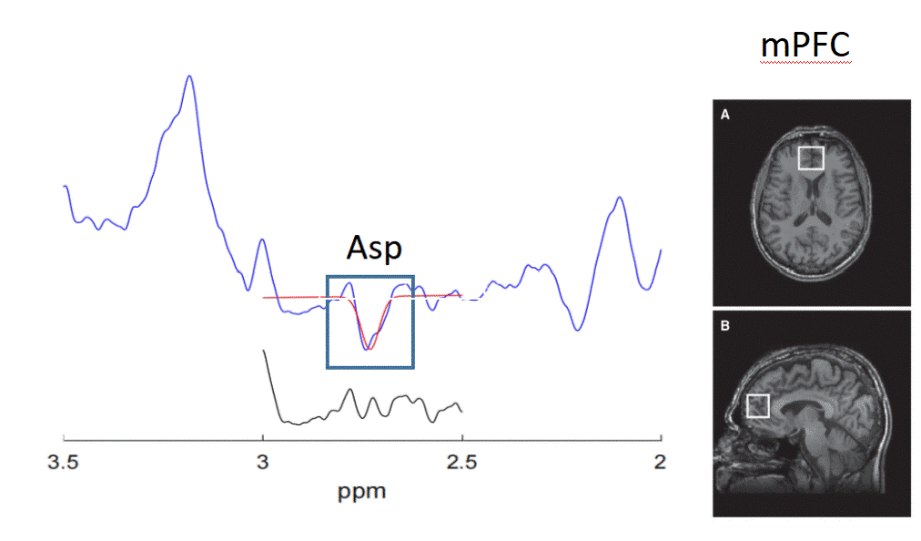
Figure 1. Aspartate signal fitting by Gannet tools
Note: mPFC: medial prefrontal cortex