3851
Cerebrovascular reactivity mapping using intermittent breath modulation: temporal resolution dependence and comparisons with CO2 inhalation1Department of Diagnostic Radiology and Nuclear Medicine, University of Maryland Baltimore, Baltimore, MD, United States
Synopsis
Keywords: fMRI, Neuro
Cerebrovascular reactivity (CVR) is typically measured using a carbon dioxide (CO2) stimulus combined with BOLD fMRI. However, this requires considerable subject cooperation. Although resting-state BOLD fMRI has shown its potential to generate CVR maps, the CVR results could be unreliable due to little fluctuation in some subjects’ spontaneous breathing. A new method utilizing intermittent breath modulation requires no gas-inhalation and presents higher sensitivity than resting-state CVR mapping. In this study, we investigated the effect of temporal resolution on CVR mapping obtained from breath modulation BOLD data. Our results showed that good CVR quality can be achieved with TR of 0.72s.INTRODUCTION:
Cerebrovascular reactivity measures the dilatory function of cerebral blood vessels1, and it has been reported to be a sensitive biomarker in various cerebrovascular conditions such as aging2, small vessel and large vessel diseases3,4. CVR mapping is typically performed using hypercapnic gas inhalation or breath-holding as a vasoactive challenge1, but both methods require considerable subject cooperation and could be uncomfortable for some subjects. There have also been attempts to use resting-state BOLD data to map CVR by utilizing spontaneous fluctuations in breathing patterns5,6. However, in subjects who have little fluctuation in their spontaneous breathing, the CVR results could be noisy or even undetectable. More recently, we have developed a new technique for CVR mapping using intermittent breath modulation during resting-state BOLD scans7, which does not require gas inhalation yet provides substantially higher sensitivity than resting-state CVR mapping. In the present study, we aim to investigate the effect of temporal resolution of the BOLD acquisition on CVR maps obtained with intermittent breath modulation, and compare them with the CVR maps obtained from the conventional CO2 inhalation method.METHODS:
Subjects and imaging protocols26 healthy subjects (40.1±18.0 years, 11F/15M) were studied on a 3T Siemens scanner. Each subject had three 7-min BOLD scans for CVR mapping: 1) intermittent breath modulation acquired at TR of 0.72s, 2) intermittent breath modulation acquired at TR of 1.5s, and 3) CO2 inhalation acquired at TR of 0.72s. During the two breath modulation scans, the subjects were asked to breathe through their nose at their own pace except for the periods when pacing instructions, e.g. “breathe in”, “breathe out”, appear on the screen. The pacing instructions appeared for 12 seconds after every 30-60s free breathing period, with a pacing frequency at 4s/breath (2s in/2s out). In the CO2 inhalation scan, the subjects were fitted with a nose clip, and breathed room air and hypercapnia gas (5%CO2) in an interleaved fashion (50s CO2, 70s room air, repeated three times) through a mouthpiece. The BOLD imaging parameters of the three scans were identical: FOV=208x208x144mm3, voxel size=2x2x2mm3, TE/FA=37ms/52º, multiband factor=8, axial slices with whole-brain coverage.
Data analysis
After motion correction, BOLD images from the two breath modulation scans were coregistered to the CO2-inhalation scan, smoothed by four Gaussian kernels (2mm, 4mm, 6mm, and 8mm), and filtered by a low-path filter (fcutoff= 0.08Hz 7). Next, a whole-brain-averaged BOLD time course was obtained and voxel-wise general linear model analyses were performed to obtain a CVR map at each smoothness, where the voxel-wise BOLD signal was the dependent variable, and the filtered whole-brain time course was the independent variable along with six motion vectors as covariates. The calculated CVR map in units of BOLD percentage change was further normalized to the whole-brain average to get a relative CVR map7. The BOLD images of the CO2-inhalation scan were also smoothed by four gaussian kernels, and CO2-CVR was obtained for each smoothness following the standard analysis described previously1. Spatial correlations between the CO2 CVR map and the breath modulation CVR maps were assessed using Pearson’s Linear Correlation Coefficient. Sensitivity of each breath modulation CVR map was evaluated using Z-score maps yielded from GLM analysis and mean Z scores between the two temporal resolutions were compared using paired t-test.
RESULTS and DISCUSSION:
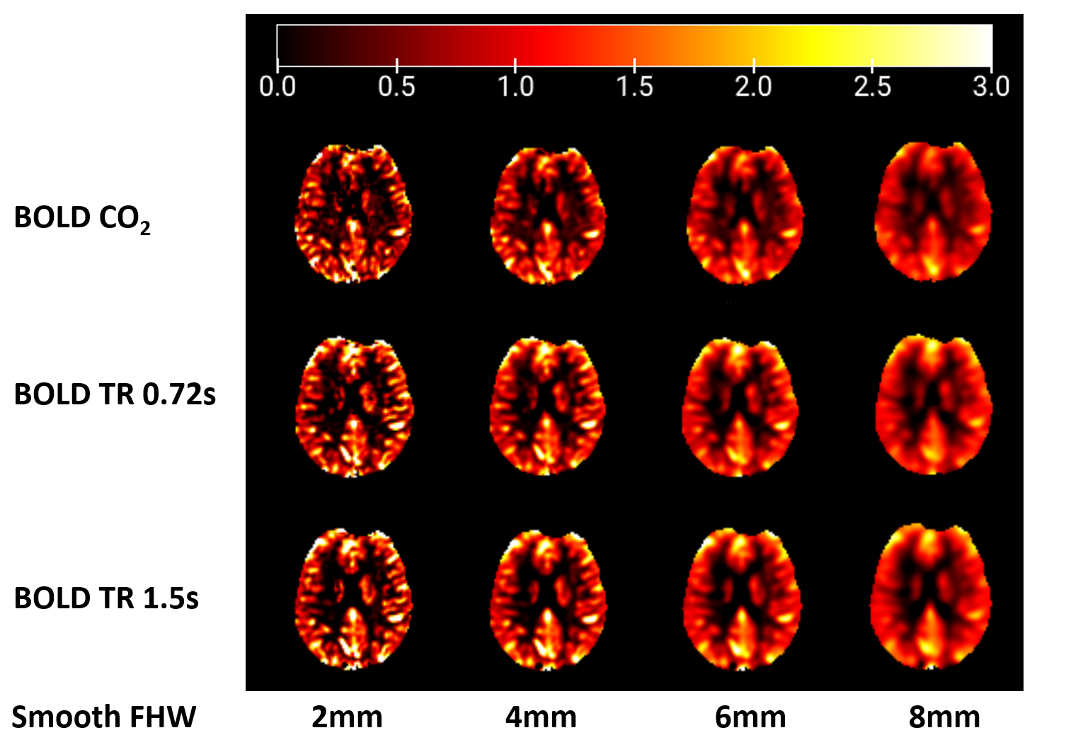
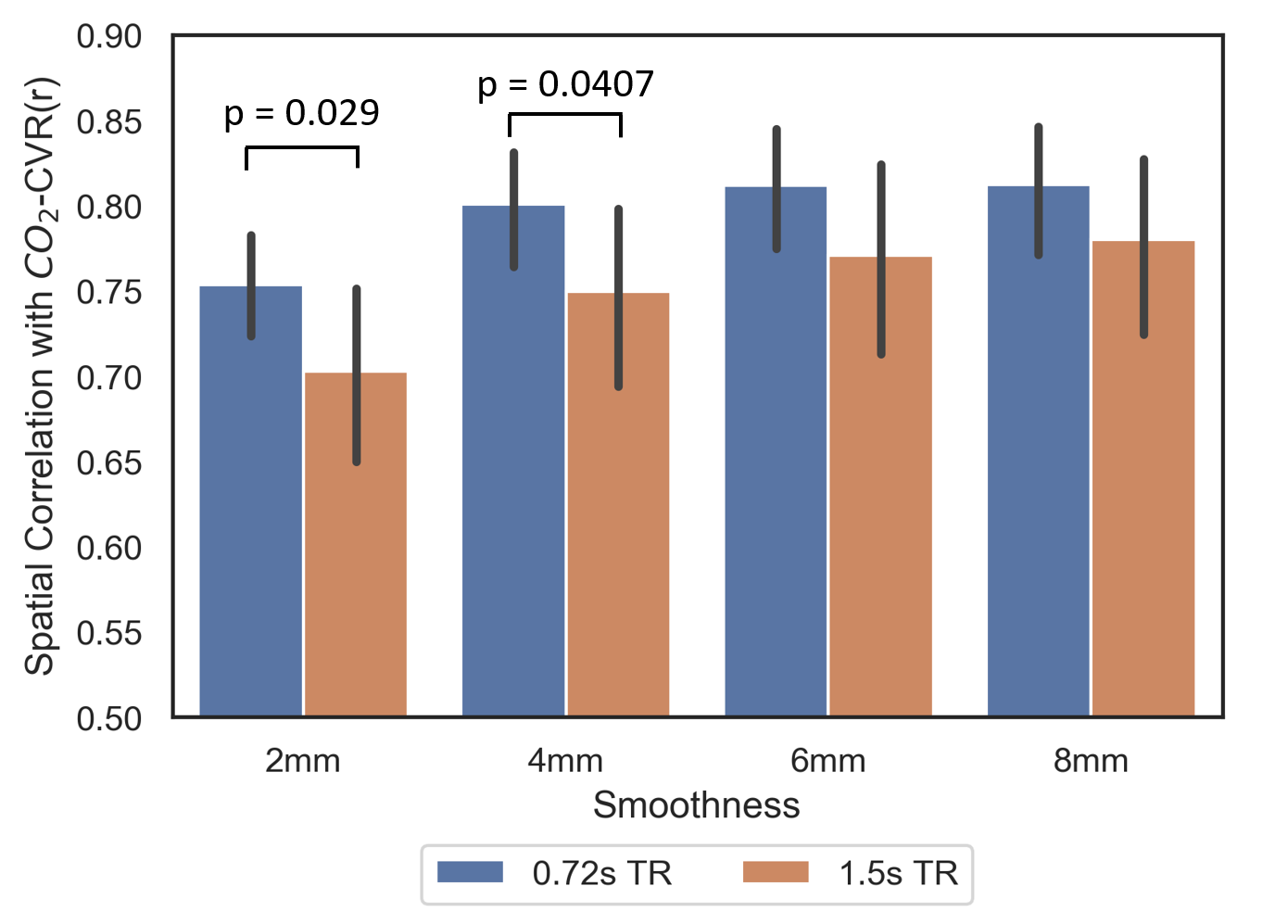
Figure 1 shows the resulting CVR maps from a representative subject. Through visual inspection, good CVR map quality can be seen in all scans, with the maps being less noisy with larger smoothing kernels.Figure 2 demonstrates the spatial correlations between CO2 CVR map and breath modulation CVR maps obtained at two different temporal resolutions. For both temporal resolutions, the spatial correlations with CO2 CVR were above 0.7, suggesting an overall good consistency of CVR mapping between the intermittent breath modulation method and the conventional CO2 inhalation method. CVR maps from the TR of 0.72s scans showed significantly higher spatial correlation with the CO2 CVR maps when using smoothing kernel of 2mm and 4mm (p < 0.03 and p < 0.041 respectively), suggesting higher accuracy of breath modulation CVR mapping when using a shorter TR. As the size of smoothing kernel increases, spatial correlation coefficients increased for both breath modulation scans, and the significant difference between the two modulation scans disappeared. This observation suggested that with good SNR (achieved by increasing the spatial smoothness of the BOLD data), the effect of TR on breath modulation CVR mapping is reduced.
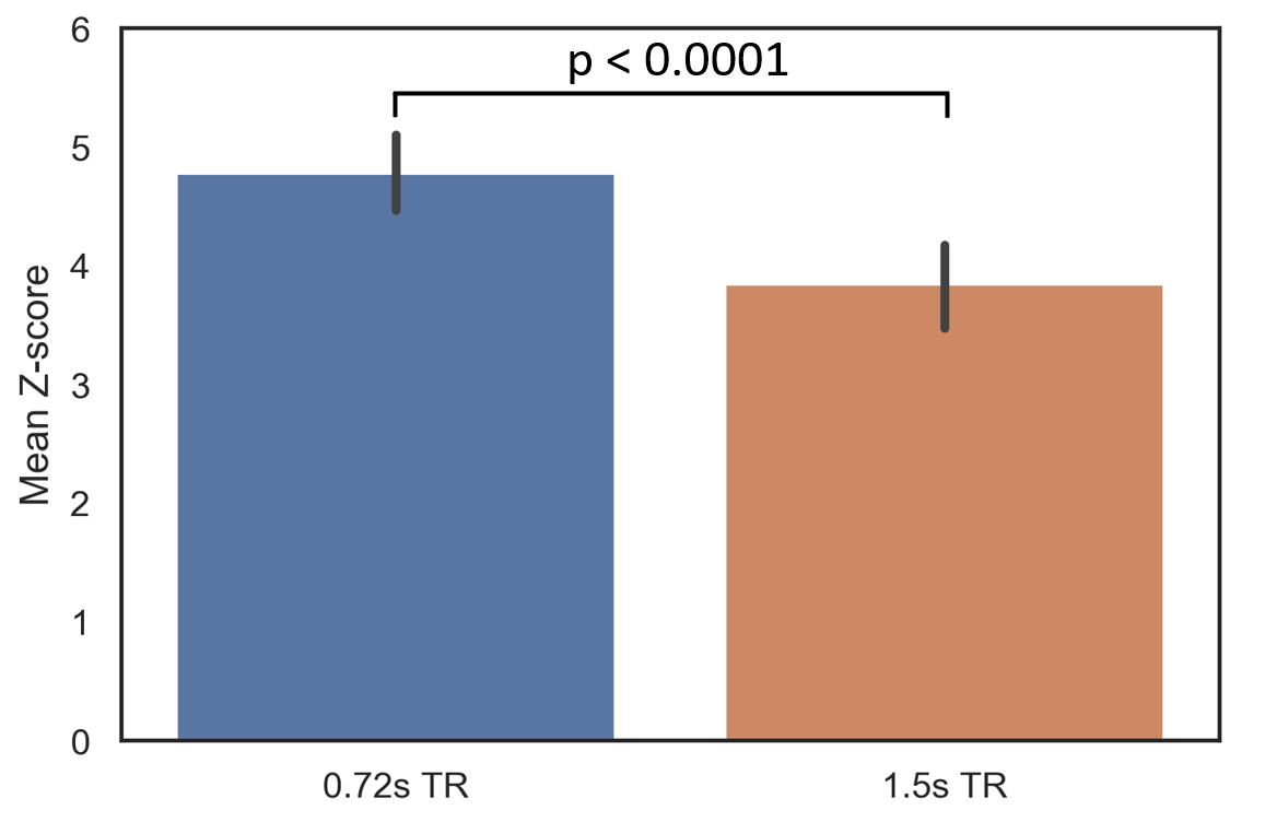
A comparison of the CVR mapping sensitivity (measured as the mean Z scores) of the two temporal resolutions is shown in Figure 3. CVR maps from the TR of 0.72s scans had significantly higher Z scores than those from the TR of 1.5s scans (p=3.9x10⁻⁷). This result confirmed the benefit of sampling at higher frequencies with the same total scan duration.
CONCLUSION:
CVR mapping with intermittent breath modulation can provide relative CVR maps in good agreement with the conventional CO2-inhalation method with typical BOLD temporal resolutions, even though scans acquired with higher temporal resolution may have better accuracy and sensitivity. Further work will be performed to evaluate the absolute quantification of CVR using the intermittent breath modulation method.Acknowledgements
No acknowledgement found.References
1. Liu P, De Vis JB, Lu H. Cerebrovascular reactivity (CVR) MRI with CO2 challenge: A technical review. Neuroimage. 2019;187:104-115.
2. Lu H, Xu F, Rodrigue KM, Kennedy KM, Cheng Y, Flicker B, Hebrank AC, Uh J, Park DC. Alterations in cerebral metabolic rate and blood supply across the adult lifespan. Cereb Cortex. 2011;21(6):1426-34.
3. Sur S, Lin Z, Li Y, Yasar S, Rosenberg P, Moghekar A, Hou X, Kalyani R, Hazel K, Pottanat G, Xu C, van Zijl P, Pillai J, Liu P, Albert M, Lu H. Association of cerebrovascular reactivity and Alzheimer pathologic markers with cognitive performance. Neurology. 2020;95(8):e962-e972.
4. Mandell DM, Han JS, Poublanc J, Crawley AP, Stainsby JA, Fisher JA, Mikulis DJ. Mapping cerebrovascular reactivity using blood oxygen level-dependent MRI in Patients with arterial steno-occlusive disease: comparison with arterial spin labeling MRI. Stroke. 2008;39(7):2021-8.
5. Golestani AM, Chang C, Kwinta JB, Khatamian YB, Chen JJ. Mapping the end-tidal CO2 response function in the resting-state BOLD fMRI signal: spatial specificity, test-retest reliability and effect of fMRI sampling rate. Neuroimage. 2015;104: 266-277.
6. Liu P, Li Y, Pinho M, Park DC, Welch BG, Lu H. Cerebrovascular reactivity mapping without gas challenges. Neuroimage. 2017;146:320-326.
7. Liu P, Xu C, Lin Z, Sur S, Li Y, Yasar S, Rosenberg P, Albert M, Lu H. Cerebrovascular reactivity mapping using intermittent breath modulation. Neuroimage. 2020;215:116787.
Figures