3836
Effect of age on the evaluation of left ventricular kinetic energy from 4d-flow CMR1The First Affiliated Hospital of Dalian Medical University, Dalian, China
Synopsis
Keywords: Heart, Heart
Four-dimensional flow cardiovascular magnetic resonance imaging (4d-flow CMR) left ventricular kinetic energy (LVKE) parameter Peak A- wave KEiEDV increased, KEiEDV E/A ratio decreased with age. This suggests that 4d-flow CMR LVKE parameters can respond to age-induced changes in LV diastolic function.Introduction
Four-dimensional flow cardiovascular magnetic resonance imaging (4d-Flow CMR) enables simultaneous velocity encoding in all three directions and provides time-resolved (4D = 3 spatial dimensions and time) 3D volumetric information for visualizing and quantifying blood flow within the heart and major blood vessels. 4d- flow CMR yields ventricular hemodynamic parameters such as left ventricular kinetic energy (LVKE). LVKE is an important part of the energy generated by cardiac exercise that is transferred to the blood, which is of interest for the evaluation of cardiac exercise function1. This study aimed to explore the effect of age on LVKE.Methods
We prospectively recruited 11 health participants. All participants with 4d-flow CMR scans were performed on a 3.0T MRI scanner (Philips Ingenia, Philips Healthcare, Cleveland, Ohio, USA) by 16 coil. The orientation of the acquisition of 4D flow data was coronal. Velocity-encoding of 120 cm/s in all three directions was used in a standard four-point encoding scheme, spatial resolution 2.5 × 2.5 × 2.5 mm3, field of view 240-300×240-300 mm, 90-110 slices, flip angle 8°, echo time (TE) 2.3 ms, repetition time (TR) 4.1 ms, accelerate factor was CS-SENSE 6. Free-breathing was allowed. Scan time is about 8-11 minutes2. Cine two-dimensional (2D) left 3-chamber was acquired using steady-state free-precession (SSFP) sequences. Commercially available dedicated software CVI42 (version 5.14, Circle Cardiovascular Imaging Inc., Calgary, Canada) was used to analyze LVKE. All LVKE parameters were normalized to LV end-diastolic volume (KEiEDV) and expressed in μJ/ml. KEiEDV parameters for physiologically relevant time points (global [whole cycle], systolic, and diastolic mean KEiEDV) and cardiac cycle time points (peak systolic, peak E-wave, and peak A-wave) were extracted from KEiEDV time curves. The Shapiro-Wilk test assessed the normality of the data. Analysis of variance using independent samples t-test and Mann-Whitney U test. Pearson correlation and Spearman rank correlation were used for correlation analysis.Results
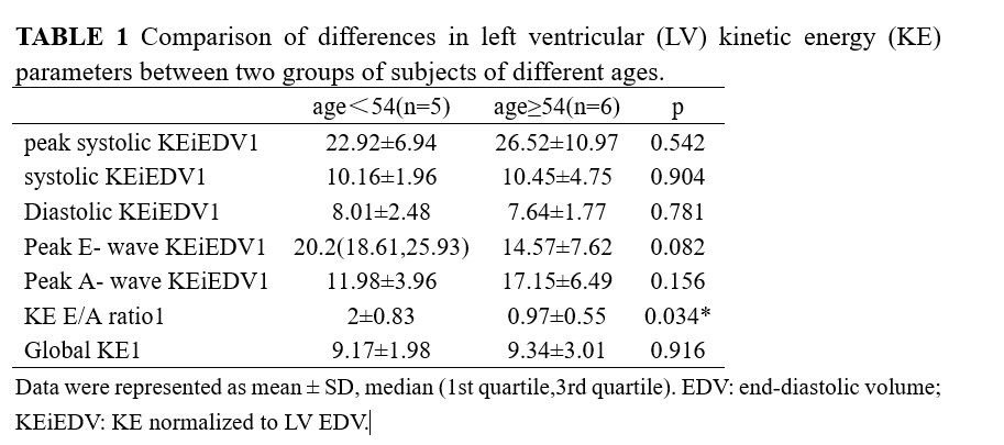
The median age of 54 was used as the cut-off point to divide the subjects into two groups, less than 54 years old(range: 45-53) and greater than or equal to 54 years old(range: 54-70) for five and six subjects, respectively. KEiEDV E/A ratio (2±0.83 vs. 0.97±0.55 p=0.034) significant difference between the two age groups. Additionally, with age increasing, the KEiEDV E/A ratio(r=-0.729) significantly decreased, and Peak A- wave KEiEDV (r=0.823) significantly increased.Discussion
We found no differences in global, peak systolic, systolic, and diastolic, and Peak E-wave KEiEDV between the two age groups. However, Peak A-wave KEiEDV increased, and the KEiEDV E/A ratio decreased with age. In the echocardiogram, The Doppler velocity of peak E gradually decreases with age below peak A3. We get a similar finding in our study, Peak E-wave KEiEDV and Peak A-wave KEiEDV respectively, reflect LV early and late diastolic function. The KEiEDV E/A ratio decrease means that increased active contractility of the left atrium is used to compensate for the decreased early diastolic capacity of the left ventricle.Conclusion
The 4d flow CMR left ventricular kinetic energy parameter is a valuable indicator to evaluate left ventricular diastolic function in different age populations.Acknowledgements
No acknowledgement found.References
1. Demirkiran A, van Ooij P, Westenberg JJM, et al. Clinical intra-cardiac 4D flow CMR: acquisition, analysis, and clinical applications. Eur Heart J Cardiovasc Imaging. 2022 Jan 24;23(2):154-165.
2. Zhong L, Schrauben EM, Garcia J, Uribe S, Grieve SM, Elbaz MSM, Barker AJ, Geiger J, Nordmeyer S, Marsden A, Carlsson M, Tan RS, Garg P, Westenberg JJM, Markl M, Ebbers T. Intracardiac 4D Flow MRI in Congenital Heart Disease: Recommendations on Behalf of the ISMRM Flow & Motion Study Group. J Magn Reson Imaging. 2019 Sep;50(3):677-681.
3. L. Caballero, S. Kou, R. Dulgheru, et al., Echocardiographic reference ranges for normal cardiac Doppler data: results from the NORRE Study, Eur. Heart J. Cardiovasc. Imaging 16 (2015) 1031–1041
Figures