3835
Quantitative Mitral Valvular Regurgitation Hemodynamics Analysis Using 4D flow MRI and Echocardiography: Characteristics of Flow Momentum1Radiology, Northwestern University, Chicago, IL, United States, 2Northwestern University, Chicago, IL, United States, 3Cardiology, Northwestern Memorial Hospital, Chicago, IL, United States, 4Radiology, Northwestern University, chicago, IL, United States
Synopsis
Keywords: Valves, Velocity & Flow, Mitral valvular regurgitation, 4D flow MRI, Jet momentum
Quantitative hemodynamic assessment of mitral valvular regurgitation (MVR) is critical for evaluating severity of the disease, but has been challenging with conventional echocardiographic and cardiac MRI techniques. 4D flow MRI is a promising tool that enables direct retrospective quantification of MVR hemodynamics. This study proposes a regurgitant flow momentum as a novel and reliable fluid dynamic parameter for MVR assessment using 4D flow MRI. The conservation characteristics of MVR jet flow momentum and its relationship with LA enlargement were investigated in 44 patients using 4D flow MRI and echocardiography.Purpose
Non-invasive quantitative assessment of mitral valvular regurgitation (MVR) hemodynamics is crucial for determining MVR severity.1 4D flow MRI has been shown to be a promising tool for MVR assessment in that it enables quantification of regurgitant flow volume (RVol) directly across the MVR flow jet using retrospective valve-tracking or flow-tracking.2-4 However, a previous in-vitro study using pulsatile MVR jet flow models has shown that the flow rate of a jet is not equal to a regurgitant flow rate in most part of the jet and is, in fact, higher than the regurgitant flow rate due to flow entrainment (surrounding fluid is drawn into the jet as the jet propagates) (Figure 1).5 On the other hand, the flow momentum of a regurgitant flow is conserved throughout a jet due to the momentum conservation principle and is proportional to RVol.5 In addition, using control volume analysis, the flow momentum of a jet is directly related to a wall-impinging force exerted by the jet. This suggests that the jet flow momentum, which can be easily quantified by 4D flow MRI, may be a more reliable fluid dynamic parameter to assess MVR hemodynamics and may provide useful insights into the hemodynamic impact of MVR to the left atrial (LA) enlargement frequently seen in severe MVR patients. This study aims to evaluate whether MVR jet flow momentum in patients satisfies the conservation principle and its relationship with the LA volume assessed with clinical standard echocardiography. We hypothesized that the flow momentum is a conserved quantity of MVR flow jets in patients and is associated with dilated LA volume.Methods
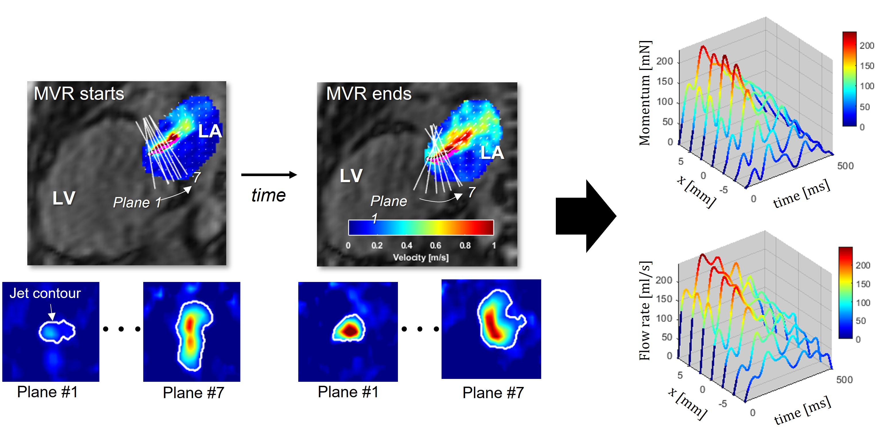
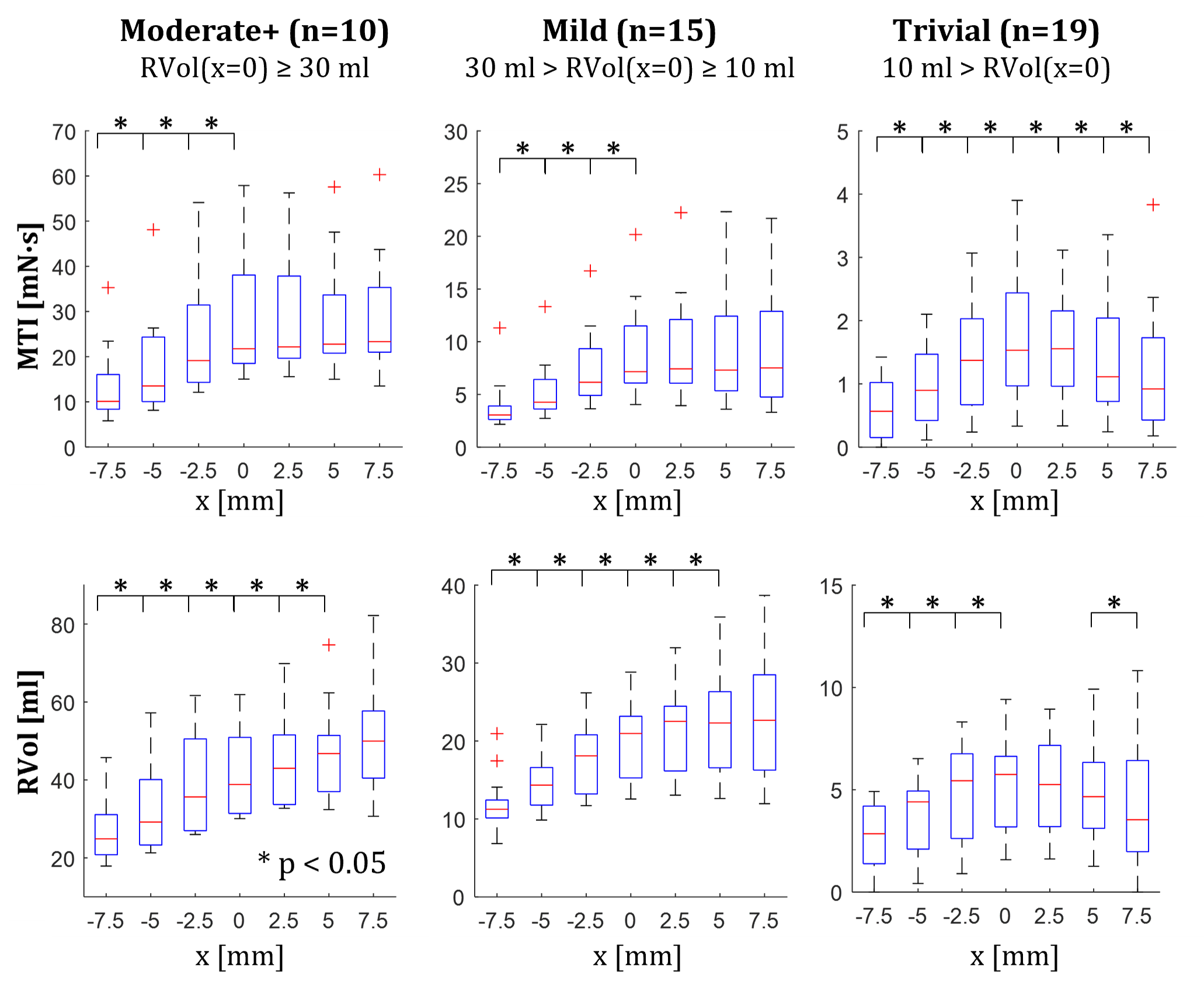
Forty-four MVR patients (age 62±12, 17 female) underwent standard-of-care echocardiography and 4D flow MRI within 3 months apart. A free-breathing prospectively ECG-gated 4D flow MRI was acquired using 1.5T Siemens system (Aera or Avanto) with following sequence parameters: TE=2.2–2.4ms, TR=4.5−4.8ms, temporal resolution=36–38ms, spatial resolution=3.8–4.2 x 2.4–2.5 x 2.6–3mm3, field-of-view=309–375x380–400x84–108mm3 and encoding velocity=150/250cm/s. An in-house MATLAB code was used to analyze the MVR flow jet in the LA (Figure 2). The flow momentum and the flow rate were quantified by placing a plane at 7 equi-distant (2.5 mm) locations along the jet where the middle reference plane (x=0) was set at the peak jet velocity location at each time point. Reformatted through-plane velocity images were generated to contour jet area and quantify momentum ($$$\rho\int_{contour-area}^{}V^2dA$$$) and flow rate ($$$\int_{contour-area}^{}VdA$$$) which were then integrated over the duration of MVR to compute momentum-time-integral (MTI) and RVol, respectively. Patients were sorted into three severity groups based on RVol measured at the middle plane; (1) Trivial: RVol<10ml, (2) Mild: 10ml≤RVol<30ml, and (3) Moderate+: RVol≥30ml. LA volume was assessed following American Society of Echocardiography guidelines and normalized with body surface area to account for gender differences.6Results
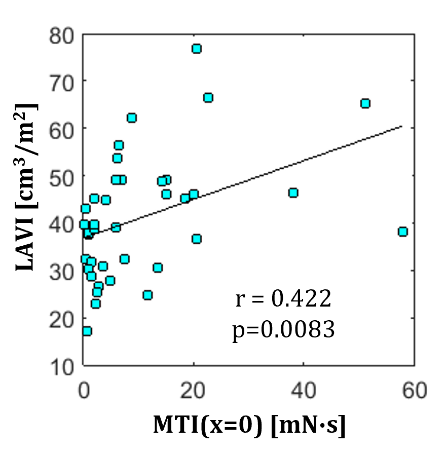
Figure 3 shows the distribution of MTI and RVol at 7 locations for each severity group. MTI was the highest in the moderate+ group and decreased with improved severity. In both moderate+ (n=10) and mild (n=15) group, MTI and RVol demonstrated a consistent increase (p < 0.05) from x=-7.5 to 0mm but MTI remained relatively unchanged from x=0 to 7.5mm (p > 0.05) while RVol continued to increase until x = 5 mm (p < 0.05). In trivial MVR patients, MTI and RVol increased from x=-7.5 to 0 mm and decreased afterward. Using MTI at x=0 as a representative value, a higher MTI was associated with a larger LAVI (r=0.422, p=0.0083) (Figure 4).Discussion
In line with a previous in-vitro study5, the time-integral form of momentum (MTI) of MVR flow in patients remained relatively unchanged, suggesting the momentum conservation principle holds true for in-vivo MVR flow jets. On the other hand, the consistent increase in RVol along the jet indicates the non-negligible impact of flow entrainment on RVol quantification. RVol and MTI are likely to be underestimated near the orifice due to a large velocity gradient and insufficient voxels within the jet, which may explain the decreasing trend of RVol and MTI with decreasing x. This issue can be avoided in momentum by placing a plane away from the origin where the jet becomes wider. The association between MTI and LAVI (r=0.422) may imply a potential relationship between momentum and LA enlargement, although a longitudinal study is needed to elucidate its impact on the progression of LA remodeling.Conclusion
We have demonstrated that the regurgitant flow momentum is a conserved hemodynamic quantity throughout an MVR flow jet associated with RVol and LA volume. The flow momentum can be easily quantified with 4D flow MRI and is robust to variation of measurement location along the jet.Acknowledgements
No acknowledgement found.References
1. Bonow RO, O'Gara PT, Adams DH, et al. 2020 Focused Update of the 2017 ACC Expert Consensus Decision Pathway on the Management of Mitral Regurgitation: A Report of the American College of Cardiology Solution Set Oversight Committee. J Am Coll Cardiol. 2020;75(17):2236-2270.
2. Gupta AN, Avery R, Soulat G, et al. Direct mitral regurgitation quantification in hypertrophic cardiomyopathy using 4D flow CMR jet tracking: evaluation in comparison to conventional CMR. Journal of Cardiovascular Magnetic Resonance. 2021;23(1):1-13.
3. Blanken CP, Westenberg JJ, Aben J-P, et al. Quantification of mitral valve regurgitation from 4D flow MRI using semiautomated flow tracking. Radiology: Cardiothoracic Imaging. 2020;2(5).
4. Garg P, Swift AJ, Zhong L, et al. Assessment of mitral valve regurgitation by cardiovascular magnetic resonance imaging. Nature Reviews Cardiology. 2020;17(5):298-312.
5. Lee J, Gupta AN, Ma LE, et al. Valvular regurgitation flow jet assessment using in vitro 4D flow MRI: Implication for mitral regurgitation. Magnetic resonance in medicine. 2022;87(4):1923-1937.
6. Lang RM, Badano LP, Mor-Avi V, et al. Recommendations for cardiac chamber quantification by echocardiography in adults: an update from the American Society of Echocardiography and the European Association of Cardiovascular Imaging. European Heart Journal-Cardiovascular Imaging. 2015;16(3):233-271.
Figures