3808
Higher Field Reduced-FOV Diffusion-Weighted-Imaging for Abdominal Imaging at 5.0 Tesla: Image Quality Evaluation Compared with 3.0 Tesla
Yunfei Zhang1, Yongming Dai1, and Mengsu Zeng2
1MR Collaboration, Central Research Institute, United Imaging Healthcare, Shanghai, China, 2Zhongshan Hospital of Fudan University, Shanghai, China
1MR Collaboration, Central Research Institute, United Imaging Healthcare, Shanghai, China, 2Zhongshan Hospital of Fudan University, Shanghai, China
Synopsis
Keywords: New Devices, Diffusion/other diffusion imaging techniques
Reduced field-of-view (rFOV) technique is a feasible approach to counter the constraints of conventional DWI. Most recently, the 5.0 T whole-body MRI system was developed, which provides an alternative choice for high field abdominal DWI. This research aims to evaluate the image quality of 5.0 T rFOV-DWI with the 3.0 T rFOV-DWI as the reference. The results indicated that 5.0 T rFOV-DWI had better overall image quality and improved SNR compared to 3.0 T rFOV-DWI, which holds clinical potential for identifying the abdominal abnormalities in routine practice.Introduction
Reduced field-of-view (rFOV) technique is a feasible approach to counter the constraints of conventional DWI. Utilizing a spatially selective localized radiofrequency (RF) excitation pulses or/and an outer-volume suppression (OVS), the sampling density of K-Space is reduced, which results in a smaller dataset size for given resolution. Therefore, the acquisition time, motion artifacts, susceptibility effects and distortion will be diminished.(1, 2) Previous findings unveiled that the combination of rFOV and high-field DWI allows ultra-high-resolution imaging for human brain.(3, 4)Most recently, the 5.0 T whole-body MRI system was developed, which provides an alternative choice for high-field abdominal DWI. Besides, it was hypothesized by us that: 1) The 5.0 T DWI may be feasible and valuable for abdominal imaging. 2) The combination of 5.0 T high field DWI and rFOV technique is beneficial for providing a relatively good image quality. 3) The image quality of 5.0 T rFOV-DWI is better than 3.0 T rFOV-DWI for abdominal applications. Therefore, this research aims to evaluate the image quality of 5.0 T rFOV-DWI with the 3.0 T rFOV-DWI as the reference. To the best of our knowledge, hardly has the high field abdominal DWI been performed.
Methods
In total, 15 volunteers were included. All subjects underwent the 3.0 T- and 5.0 T-MRI examinations. The 5.0 T MRI examinations were performed with a prototype whole-body MRI scanner (uMR Jupiter, United Imaging Healthcare). Free-breathing (FB), respiratory-triggered (RT) and navigator-triggered (NT) spin-echo echo-planner imaging (SE-EPI) based three imaging protocols were performed subsequently. Except the difference in the strategies of countering the breathing motion, three sequences (5.0 T-FB-DWI (FB5.0 T), 5.0 T-RT-DWI (RT5.0 T) and 5.0 T-NT-DWI (NT5.0 T)) were configured to as the same imaging parameters as possible. The detailed imaging protocols were as follows: repetition time (TR): ~4500 ms (Influenced by the respiratory cycle), echo Time (TE): 50.5 ms, flip angle (FA): 90°, field of view (FOV): 120×280 mm2, matrix: 96×224, slice thickness: 6 mm, b values: 0 s/mm2 (two averages) and 800 s/mm2 (8 averages). The 3.0 T MRI examinations were performed with a commercial MRI scanner (uMR 790, United Imaging Healthcare, Shanghai, China). Similar to 5.0 T examinations, FB, NT and RT SE-EPI based three DWI acquisitions (3.0 T-FB-DWI (FB3.0 T), 3.0 T-RT-DWI (RT3.0 T) and 3.0 T-NT-DWI (NT3.0 T)) were performed subsequently. The SNR of DWI images were independently measured and compared. The image quality of each DWI acquisition was evaluated based on the overall quality of DWI images (b = 0 and 800 s/mm2) and ADC maps in terms of sharpness, distortion and artifacts. Specifically, the sharpness, distortion and artifact were scored based on the 5-point scaling criteria.Results
The representative DWI images of six acquisition approaches were displayed in Figure 1-3. For four upper abdominal organs (liver, pancreas, spleen and kidney) in DWI images of b = 0 s/mm2 and b = 800 s/mm2, there existed the significant differences among the SNRs of six DWI examinations (p < 0.05) (Table 1, Figure 4). The SNRs of liver were relatively low compared to those of pancreas, spleen and kidney. The post-hoc multiple comparisons suggested that the SNRs of any 5.0 T DWI images were significantly higher than those of any 3.0 T DWI images for same anatomic location in DWI images of b = 0 s/mm2 and 800 s/mm2. The results regarding the image quality analysis were displayed in Figure 5. 5.0 T DWI examinations yielded the significantly higher sharpness scores than their counterparts at 3.0 T. With respect to geometric distortion, six imaging protocols have the similar distortion scores (min: 2.9±0.4, max: 3.7±0.4). The severest artifacts were observed in FB3.0 T and FB5.0 T followed by NT3.0 T, NT5.0 T, RT3.0 T and RT5.0 T (FB3.0 T = 2.3±0.8, FB5.0 T = 2.5±0.5, NT3.0 T = 3.2±0.9, NT5.0 T = 3.3±0.5, RT3.0 T = 3.3±0.7 and RT5.0 T = 3.4±0.5, p < 0.001). Finally, the RT5.0 T displayed the best overall IQ followed by NT5.0 T, FB5.0 T, RT3.0 T, NT3.0 T and FB3.0 T (RT5.0 T = 3.9±0.3, NT5.0 T = 3.8±0.3, FB5.0 T = 3.4±0.3, RT3.0 T = 3.2±0.4, NT3.0 T = 3.1±0.4 and FB3.0 T = 2.7±0.4, p < 0.001).Discussion
In this study, the results involving the SNR comparison showed that the increase in B0 field strength provided a remarkable SNR gain. For four upper abdomen organs containing the liver, pancreas, spleen and kidney, the SNRs of both b0 and b800 DWI images at 5.0 T were significantly higher than those at 3.0 T. the overall IQs of six sequences were quantified based on the sharpness, distortion and artifacts. Our results showed that the RT5.0 T yielded the best image quality score followed by NT5.0 T, FB5.0 T, RT3.0 T, NT3.0 T and FB3.0 T. Besides, no significant difference was observed between the IQ of RT5.0 T and NT5.0 T, demonstrating that the RT5.0 T and NT5.0 T were recommended for abdominal DWI imaging.Conclusion
In view of the results that 5.0 T rFOV-DWI showed an improved image quality for upper abdomen compared to 3.0 T rFOV-DWI, 5.0 T rFOV-DWI holds clinical potential for visualizing the abdominal abnormalities.Acknowledgements
NoneReferences
1. Wilm BJ, Svensson J, Henning A, Pruessmann KP, Boesiger P, Kollias SS. Reduced field‐of‐view MRI using outer volume suppression for spinal cord diffusion imaging. Magnetic Resonance in Medicine: An Official Journal of the International Society for Magnetic Resonance in Medicine. 2007;57(3):625-30.2. Tanabe M, Higashi M, Benkert T, Imai H, Miyoshi K, Kameda F, et al. Reduced Field‐of‐View Diffusion‐Weighted Magnetic Resonance Imaging of the Pancreas With Tilted Excitation Plane: A Preliminary Study. Journal of Magnetic Resonance Imaging. 2021.
3. Wargo CJ, Gore JC. Localized high-resolution DTI of the human midbrain using single-shot EPI, parallel imaging, and outer-volume suppression at 7 T. Magn Reson Imaging. 2013;31(6):810-9.
4. von Morze C, Kelley DA, Shepherd TM, Banerjee S, Xu D, Hess CP. Reduced field-of-view diffusion-weighted imaging of the brain at 7 T. Magn Reson Imaging. 2010;28(10):1541-5.
Figures
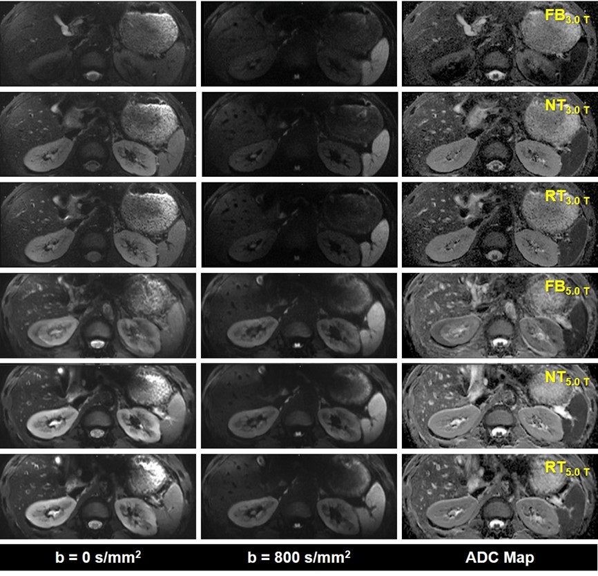
Figure 1 Representative MR images of an included subject. Note: 1) The DWI images obtained by six acquisition approaches are listed in different rows; 2) The DWI images of b = 0 s/mm2, b = 800 s/mm2 and ADC parametric maps are displayed in different columns.

Figure 2 Representative MR images of an included subject. Note: 1) The DWI images obtained by six acquisition approaches are listed in different rows; 2) The DWI images of b = 0 s/mm2, b = 800 s/mm2 and ADC parametric maps are displayed in different columns.
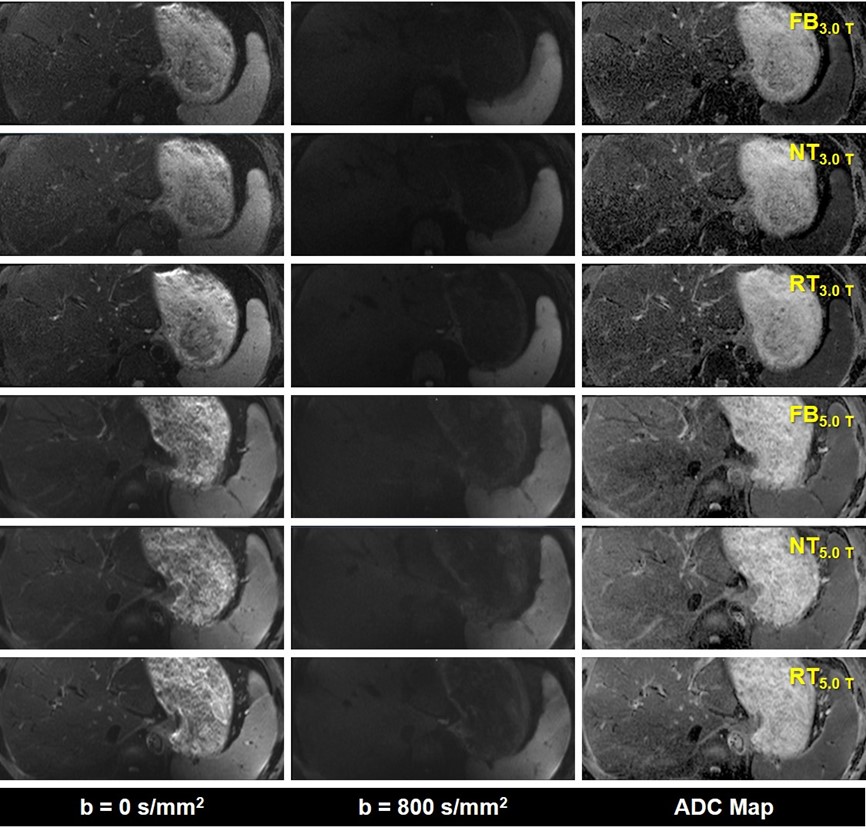
Figure 3 Representative MR images of an included subject. Note: 1) The DWI images obtained by six acquisition approaches are listed in different rows; 2) The DWI images of b = 0 s/mm2, b = 800 s/mm2 and ADC parametric maps are displayed in different columns.
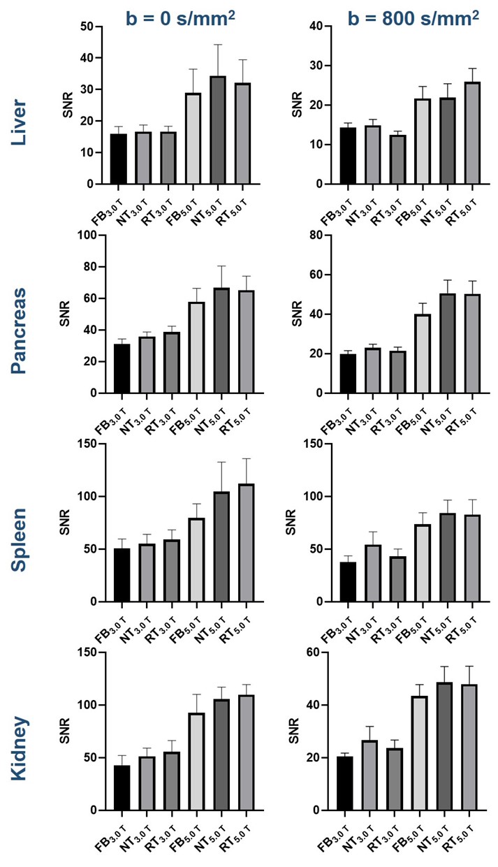
Figure 4 Box-plots show the SNRs of different anatomical structures in DWI images obtained with six acquisition approaches.
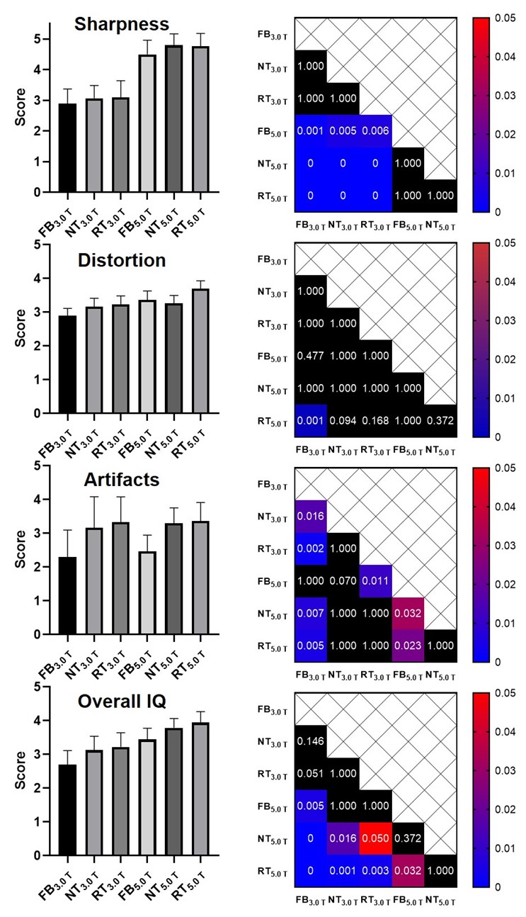
Figure 5 Image quality evaluation. Note: 1) The heat maps in the right of box-plots show the corresponding post-hoc comparison results. For example, the heat map exhibited in the first row demonstrates the results in terms of sharpness scores. 2) The values labeled in each cell represent the p values. 3) The black filled cells indicate no significant difference. 4) The values of 0 mean that p values are less than 0.001.
DOI: https://doi.org/10.58530/2023/3808