3796
Noise and distortion reduction for Reduced FOV diffusion utilizing Compressed SENSE framework for single shot EPI (EPICS)1Philips Healthcare, Shenzhen, Ltd., Shenzhen, China, 2Philips Health Technology, Suzhou, China, 3BU MR Application, Philips Health Technology, Suzhou, China, 4BU MR Clinical Science, Philips Healthcare, Tokyo, Japan, 5Philips Healthcare, Beijing, China, 6MR Clinical Application, Philips Healthcare, Beijing, China, 7BU MR R&D, Philips Health Technology, Suzhou, China
Synopsis
Keywords: Data Acquisition, Diffusion/other diffusion imaging techniques, Reduced FOV, Compressed SENSE
DWI is very important for MRI examination, but it has limited resolution due to distortion, blurring, and signal loss caused by B0 inhomogeneity. Reduced FOV imaging could decrease these impacts. However, due to the coil geometry penalty, it’s hard to combine it with parallel imaging to further improve the image quality, it will suffer from noise breakthrough issues and unfolding artifacts. We propose a framework that combines reduced FOV imaging, Compressed SENSE framework simultaneously to overcome these issues. This framework allows a new solution for reduced FOV based diffusion imaging with high resolution, low distortion, and without noise breakthrough issue.Introduction
Diffusion weighted imaging (DWI) based on single shot EPI is a very helpful sequence for oncological applications and microstructure study. But it suffers severe geometry distortion, image blurring, and signal loss which are caused by the local B0 inhomogeneity1. The distortion could be represented by the following equation:△y=△f(x,y)⋅FOVpe⋅ESP
where △y means the distortion in phase encoding direction, △f(x,y) means the local B0 inhomogeneity, FOVpe means the FOV at phase encoding direction, ESP means the echo spacing for EPI based sequence.
Reduced FOV imaging (rFOV) based on 2D RF pulse2,3 or Out Volume Suppression4 could be used to improve resolution and reduce the distortion for DWI. However, rFOV is still suffered from the distortion and signal loss when the local B0 inhomogeneity is large.
Parallel imaging, such as SENSE could also be used to decrease the echo train length and improve the image quality5. However, it will increase noise-like artifacts on the center of the images due to the high geometry factor when large reduction factors are used due to the coil geometry penalty5, it will be more severe if it was used for rFOV. Compressed SENSE could be useful to achieve an optimal balance between noise reduction and data consistency6. Recent studies demonstrated Compressed SENSE reconstruction framework could improve image quality for single shot EPI based DWI7,8,9,10, it’s called EPICS.
We combine reduced FOV imaging and EPICS simultaneously for the first time to achieve high acceleration factor to produce DWI images with high resolution, low distortion and relative high SNR.
Methods
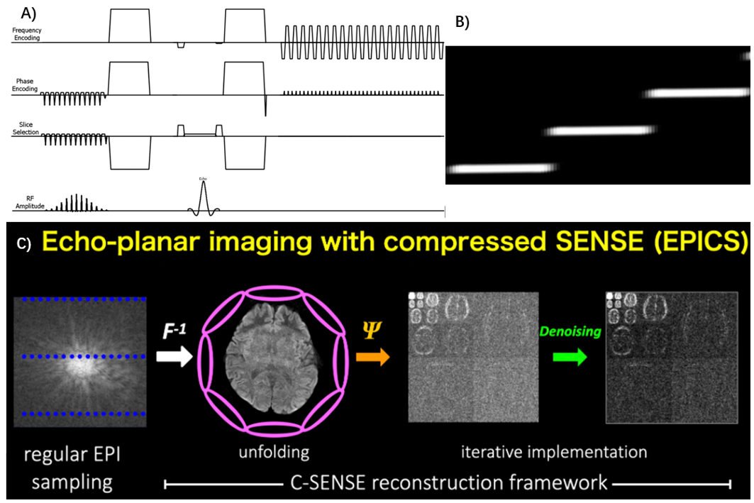
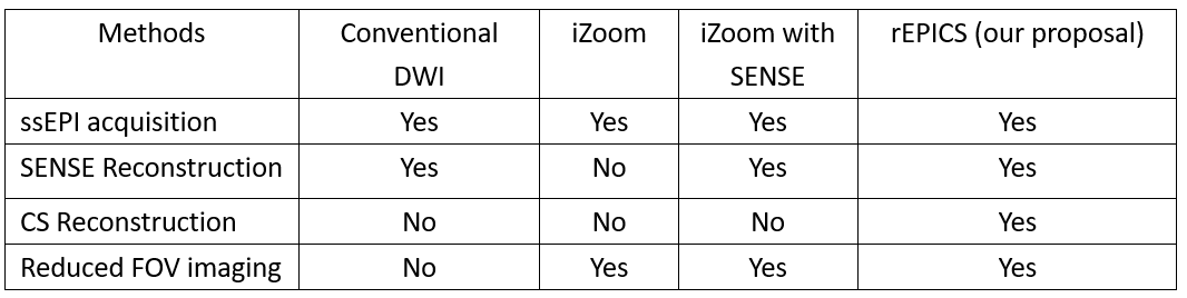
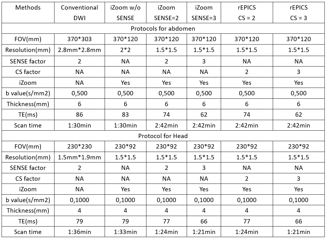
EPICS is based on single shot EPI acquisition utilizing the Compressed SENSE framework, it could dramatically reduce noise-like artifacts, so it can be used for much higher accelerator than conventional parallel imaging methods. Conventional reduced FOV imaging base on 2D RF pulse without parallel imaging is called iZoom which is implemented as ref 2. iZoom applies a tilted 2D Echo-Planar RF excitation with only tilting the k-space along the phase-encoding direction to realize multi-slice imaging. We combined iZoom with EPICS simultaneously, we call it as rEPICS, the sequence diagram and reconstruction workflow for rEPICS were showed as Fig. 1.To evaluate the performance of rEPICS, a pilot study was done for head and abdomen, conventional DWI based on ssEPI, iZoom without SENSE, iZoom with SENSE and our proposed rEPICS were acquired on a Philips 3.0T Elition system (Philips Healthcare, Suzhou, China) , a 16-ch head & spine coil for head and 32-ch torso & spine coil for abdomen. The study was approved by the local IRB. The characteristics of these DWI acquisition schemes were summarized on Table 1. For conventional DWI using ssEPI, SENSE was used, the SENSE factor is 2. For reduced FOV imaging, three different acceleration schemes were used, it means iZoom without SENSE, iZoom with SENSE and rEPICS (iZoom with EPICS). Detailed scan parameters were summarized in Table 2.
Results
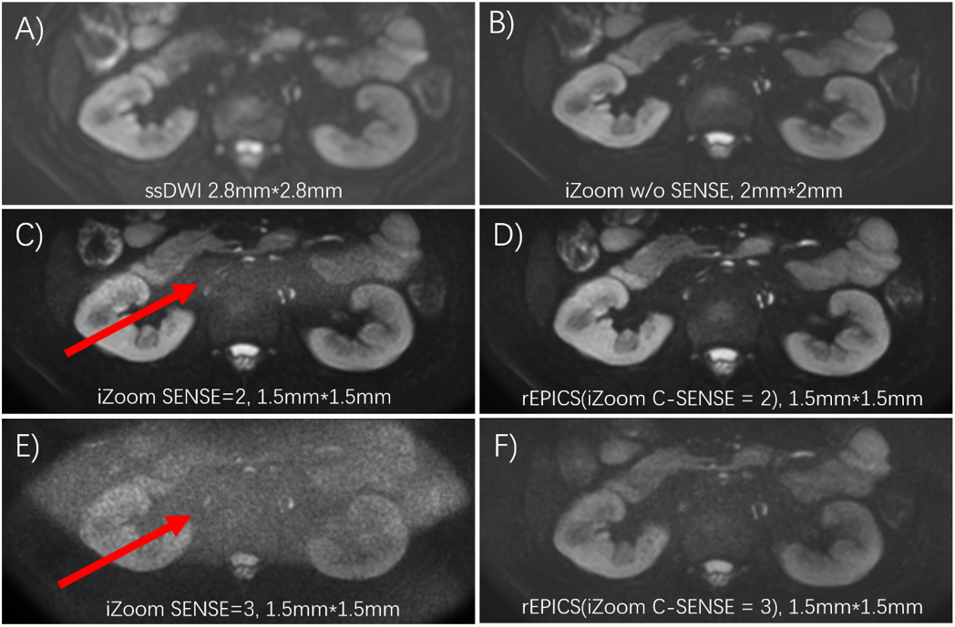
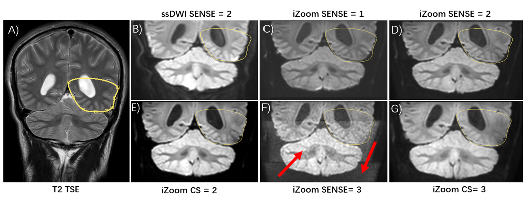
Fig. 2 and Fig. 3 shows the comparison between conventional DWI based on single shot EPI, iZoom without SENSE, iZoom with SENSE and our proposed rEPICS for head and abdomen separately. Compared with other schemes, our proposal rEPICS has the higher resolution, lower distortion and better SNR than other techniques. The image quality was dramatically improved with rEPICS, as the red arrow showed in Fig. 2 and Fig.3. Both iZoom with SENSE 2 and SENSE 3 show noise breakthrough issue. rEPICS with acceleration factor 2 and 3 shows good SNR and better resolution than other acquisition schemes. rEPICS also shows less distortion than other acquisition techniques in Fig3. The results shows that our proposed rEPICS has better performance on distortion, SNR and resolution than other methods. Although further clinical investigation is needed, rEPICS might be clinically useful and promising for DWI in high resolution, low distortion and without noise breakthrough issue.Discussion and conclusions
The proposed method rEPICS combines the reduced FOV imaging with Compressed SENSE framework for EPI (EPICS). rEPICS shows good performance with high resolution, lower distortion and also higher SNR than other schemes, such as conventional diffusion based on single shot EPI, and reduced FOV without or with parallel imaging. A pilot study was done for both head and abdomen. This strategy could enhance the applicability and offer a new solution of DWI in applications that is expected for high resolution and low distortion, such as brain, pancreas, prostate, kidney etc. Because this method doesn’t increase the scan time, it should also be promising for high resolution and low distortion fMRI.Acknowledgements
No.References
1. Bihan DL, et al. Artifacts and pitfalls in diffusion MRI, J. Magn. Reason., 2006; 24: 478-488.
2. Wu ZG, et al. B1 insensitive zoomed FOV imaging, Proc. Intl. Soc. Mag. Reson. Med. 23(2015); 0953.
3. Banerjee S, et al. Reduced field-of-view DWI with robust fat suppression and unrestricted slice coverage using tilted 2D RF excitation. Magn Reson Med. 2016;76:1668-1676.
4. Wilm BJ, et al. Reduced field-of-view MRI using outer volume suppression for spinal cord diffusion imaging. Magn Reson Med 2007;57:625–630.
5. Pruessmann KP, et al. SENSE: sensitivity encoding for fast MRI, Magn. Reason. Med., 1999;42(5):952-62.
6. Geerts-Ossevoort L, et al. Compressed SENSE. Speed done right. Every time. Philips Field Strength Magazine 2018: 6619.
7. Masami Y, et al. Noise Reduction in Prostate Single-Shot DW-EPI utilizing Compressed SENSE Framework. Proc. Intl. Soc. Mag. Reson. Med. 27 (2019) ,1634.
8. Hazuki Y, et al. Reduction of susceptibility artifact using echo-planar imaging with compressed SENSE (EPICS) in the upper abdomen, Proc. Intl. Soc. Mag. Reson. Med. 29 (2021), 1900.
9. Kaga T, et al. Diffusion-weighted imaging of the abdomen using echo planar imaging with compressed SENSE: Feasibility, image quality, and ADC value evaluation. Eur J Radiol. 2021 Sep; 142: 109889.
10. Tamada T, et al. Clinical application of single-shot echo-planar diffusion-weighted imaging with compressed SENSE in prostate MRI at 3T: preliminary experience. MAGMA. 2022 Aug; 35(4):549-556.
Figures