3792
3D Isotropic and high resolution whole brain DWI with single slab and single shot EPI utilizing motion compensated diffusion gradients1Philips Healthcare, Shenzhen, Ltd., Shenzhen, China, 2Philips Health Technology, Suzhou, China, 3MR Clinical Science, Philips Healthcare, Mississauga, ON, Canada, 4BU MR Application, Philips Health Technology, Suzhou, China, 5BU MR R&D, Philips Health Technology, Suzhou, China, 6Philips Healthcare, Beijing, China
Synopsis
Keywords: Pulse Sequence Design, Diffusion/other diffusion imaging techniques, Pulse Sequence Design, Diffusion/other diffusion imaging techniques, Diffusion, 3D
3D Diffusion imaging (3D DWI) has showed great potential in probing tissue microstructure and brain structural connectivity. However, motion-induced phase errors introduced by diffusion gradients will cause severe artifacts in 3D DWI. Multi-slab can be used to overcome this limitation, but it will introduce slab boundary artifacts. We propose a method which utilizes motion-compensated diffusion gradients for 3D DWI to mitigate the phase error between shots. Results from in vivo data demonstrate the proposed method can improve image quality and realize an isotropic high resolution and whole brain 3D DWI in a single slab.Introduction
3D Diffusion imaging (3D DWI) has showed great potential in probing tissue microstructure and brain structural connectivity1. However, it suffers from the motion-induced phase errors, which can severe degrade image quality in multi-shot acquisitions. 3D multi-slab acquisitions with thin slabs have been proposed to minimize phase errors; the whole volume is divided into multiple smaller slabs, each of which is excited and encoded separately along the slice direction and then combined as the whole volume. Remaining challenges for 3D multi-slab acquisitions are the slab boundary artifacts and multi-acquisitions for different slabs2. Several methods were introduced to minimize slab boundary artifacts by inversion of the slab profile3,4,5. The residual boundary artifacts and multi-slab acquisitions limit the application of 3D DWI.Motion-compensated techniques were initially introduced to reduce the bulk motion introduced phase errors and signal loss for diffusion, such as for cardiac applications6. Recently, the technique has been used for multi-shot diffusion MRI of the human brain with motion-compensated oscillating gradients in 2D7. It should be also useful for 3D DWI. In this work, we leverage motion-compensated gradients to reduce phase reconstruction errors for 3D DWI, and investigate the feasibility of realizing an isotropic, high resolution and whole brain 3D DWI in a single slab.
Methods
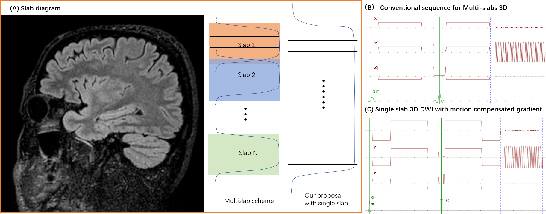
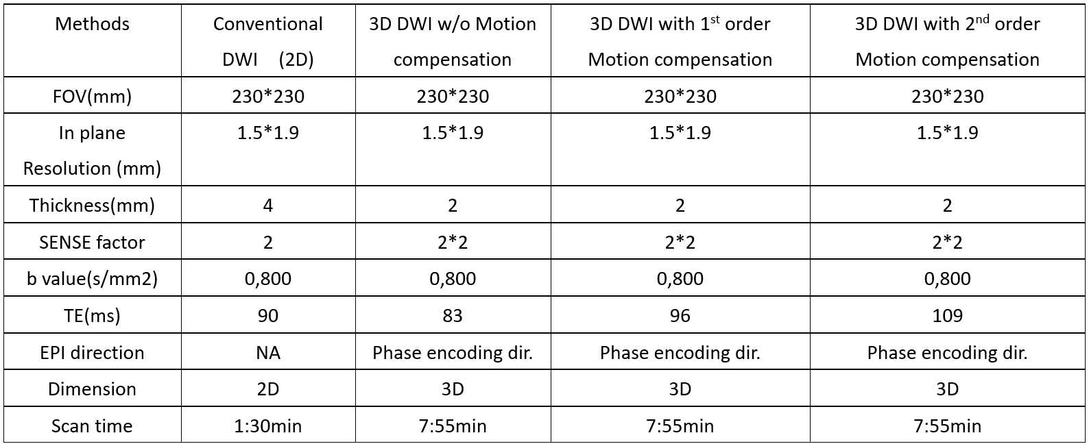
Pulse sequence:Figure 1 shows the conventional multi-slab 3D DWI scheme which acquires the data by multiple slabs with normal pulsed gradients without motion compensation. Figure 1 also shows our proposed scheme; it introduces 2nd-order motion-compensated diffusion gradients to reduce phase errors and to acquires the whole brain volume in one slab. To evaluate the performance of 2nd-order motion-compensated diffusion gradients for 3D DWI, conventional DWI, based on a single-shot 2D EPI acquisition (ssDWI), was used as the reference (ssDWI). Several single-slab 3D diffusion schemes were compared with ssDWI: single-slab 3D DWI with conventional pulsed gradients without motion-compensation, single-slab 3D DWI with 1st-order motion-compensated diffusion gradients and single-slab 3D DWI with 2nd-order motion-compensated diffusion gradients.All scanning was done on a Philips 3.0T Elition system (Philips Healthcare, Suzhou, China) , 16-ch head & spine coil was used. The study was approved by the local IRB. All acquisition schemes used b = 800s/mm2. Detailed scan parameters are listed in Table 1.
Results
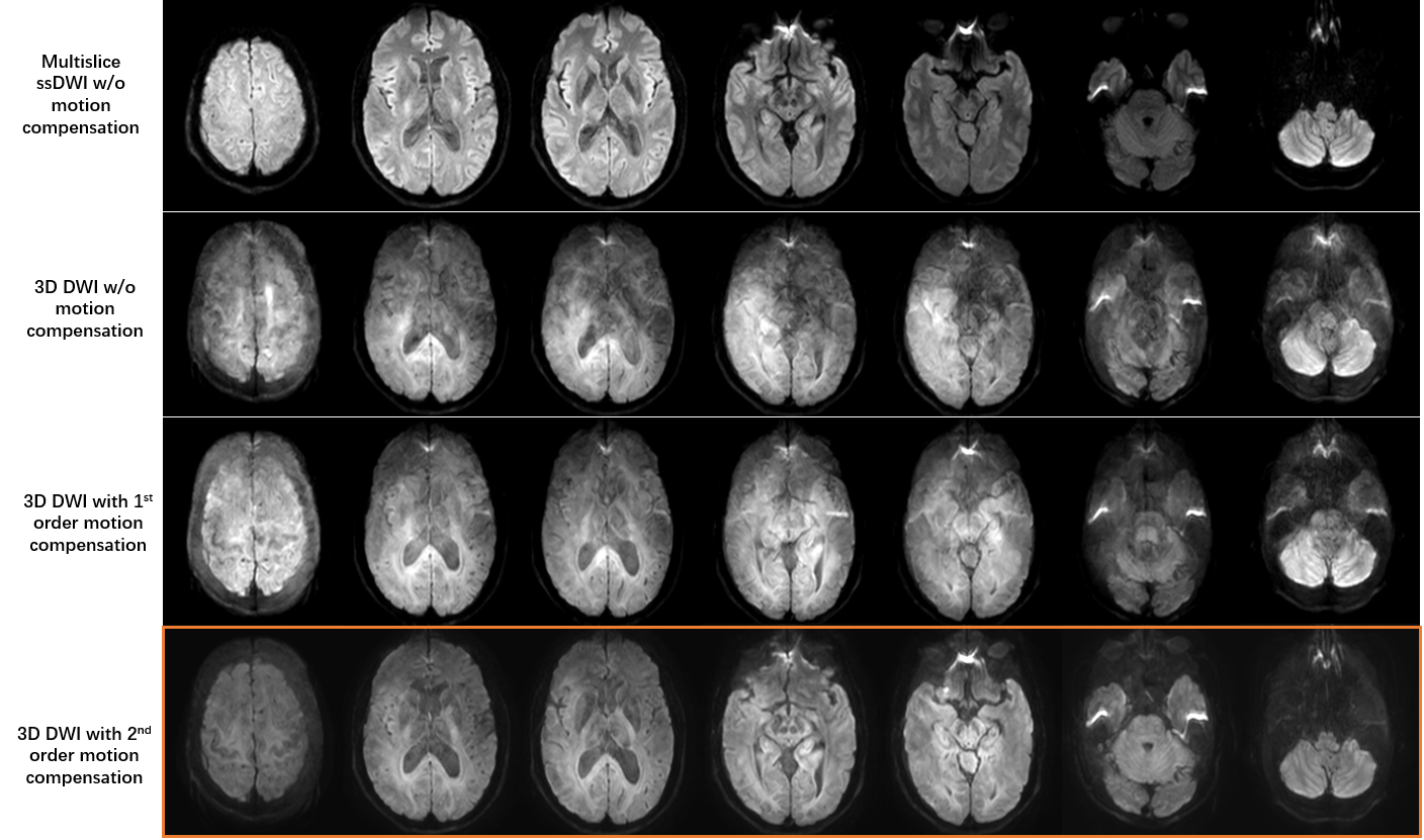
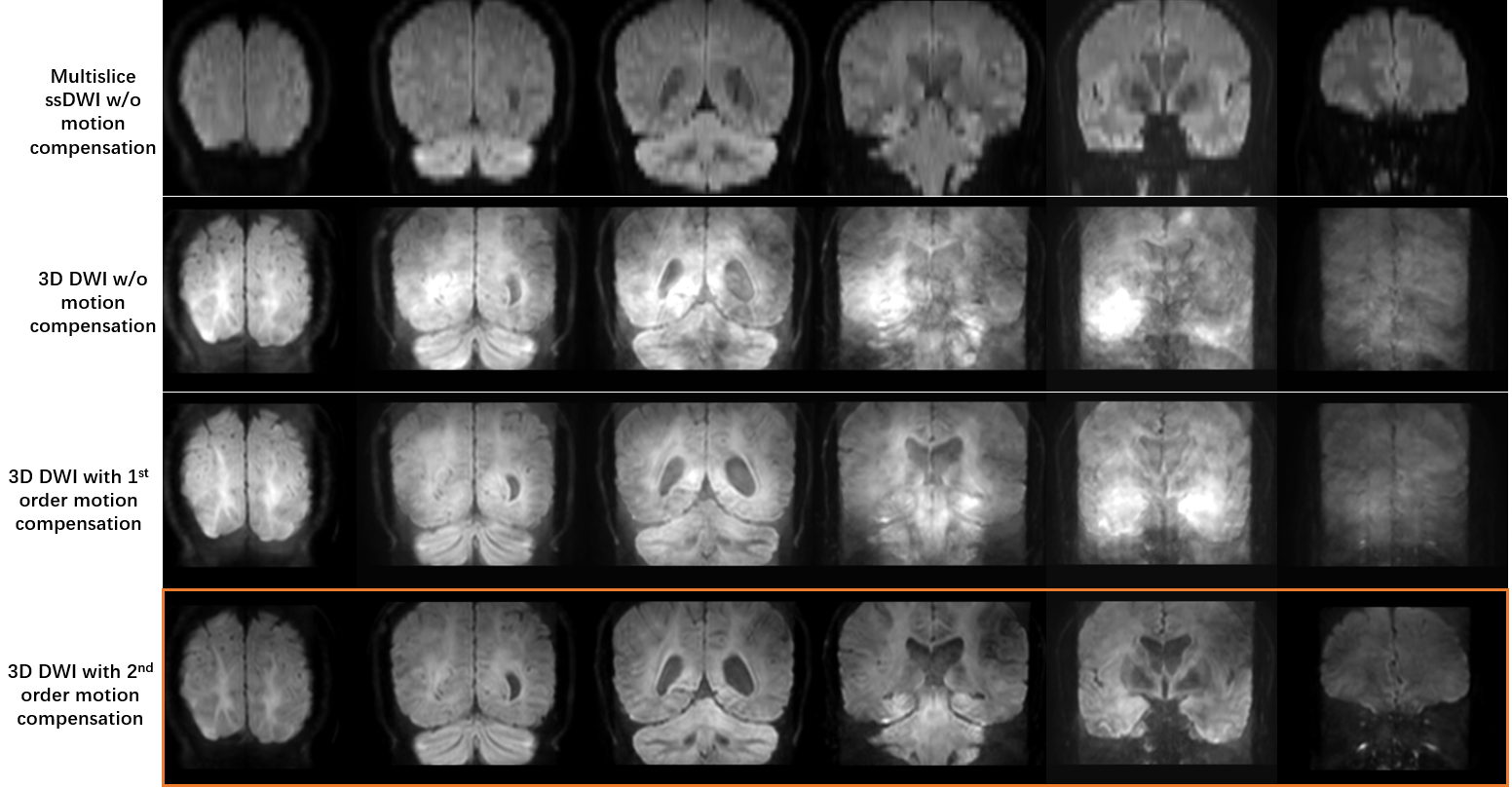
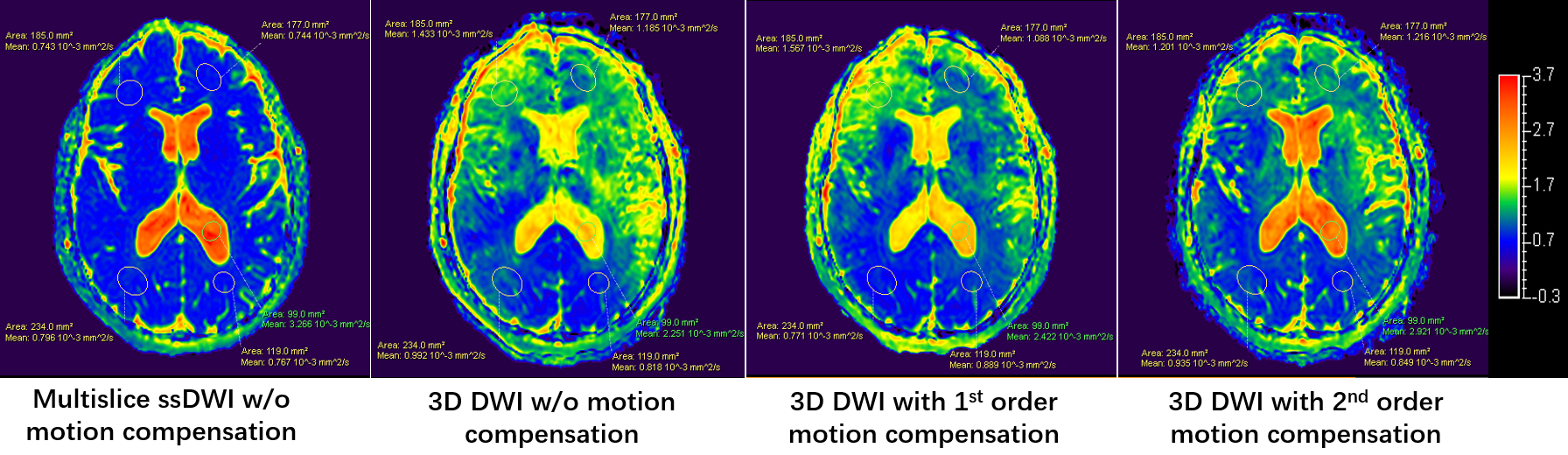
Compared with ssDWI, Fig. 2 shows that 3D DWI with conventional pulsed gradients will suffer from severe artifacts which are introduced by phase errors between shots in a large volume. 3D DWI with 1st-order motion- DWI will reduce the artifacts, but will still have severe residual artifacts. 3D DWI with 2nd-order motion-compensated diffusion gradients will improve the image quality dramatically with little residual artifacts.To evaluate the performance further, Fig. 3 shows coronal images from the different schemes using multiplanar reconstruction, it illustrates that 3D DWI with 2nd-order motion-compensated diffusion gradients shows better image quality than the other two 3D schemes. Fig. 4 also compares ADC values from these schemes; it shows that ADC values have better consistency between ssDWI and 3D DWI with 2nd-order motion-compensated diffusion gradients than when using other 3D schemes.
Discussion
The superior performance of 3D DWI with 2nd-order motion-compensated diffusion gradients shows the capability to realize 3D DWI in a single slab for the whole brain. It could be used to avoid slab boundary artifacts which is common in 3D multi-slab schemes. In this pilot study, 3D DWI with 2nd-order motion-compensated diffusion gradients still shows residual artifacts, which are mainly caused by residual phase errors between shots. It could be resolved by phase correction in the future. As 3D single-slab with motion-compensated gradients would have higher SNR than 3D multi-slab schemes, it could use shorter TR to balance acquisition time. 3D single-slab with motion-compensated gradients could also be combined with advanced k space trajectory and reconstruction methods to reduce scan time and improve image quality, such as compressed-sensing, low-rank methods, etc.Conclusions
In this study, motion-compensated diffusion gradients were used for 3D DWI for the firsttime. They were used to reduce phase errors between shots and could improve the image quality dramatically. Results showed the proposed method has notable advantages to improve image quality and could be used to realize a 3D isotropic high resolution and whole brain DWI in a single slab. This strategy could enhance the applicability and offer a new solution of 3D DWI in a single slab without slab boundary artifacts.Acknowledgements
No.References
1.Engström M, et al. Diffusion‐weighted 3D multislab echo planar imaging for high signal‐to‐noise ratio efficiency and isotropic image resolution. Magnetic resonance in medicine 2013;70(6):1507-1514.
2. Engström M, et al. On the signal-to-noise ratio efficiency and slab-banding artifacts in three-dimensional multislab diffusion-weighted echo-planar imaging. Magn Reson Med. 2015 Feb;73(2):718-25. doi: 10.1002/mrm.25182.
3.Anh TV, et al. Slab profile encoding (PEN) for minimizing slab boundary artifact in three-dimensional diffusion-weighted multislab acquisition. Magn Reson Med. 2015;73(2):605-13.
4.Wu W, et al. Reducing slab boundary artifacts in three-dimensional multislab diffusion MRI using nonlinear inversion for slab profile encoding (NPEN). Magn Reson Med 2016;76(4):1183-1195.
5.Jieying Z, et al. Slab boundary artifact correction in multislab imaging using convolutional-neural-network–enabled inversion for slab profile encoding. Magn Reson Med. 2022;87(3):1546-1560.
6.Stoeck CT, et al. Second-order motion-compensated spin echo diffusion tensor imaging of the human heart. Magn Reson Med. 2016;75(4):1669-1676.
7.Eric SM, et al. Multi-shot diffusion MRI of the human brain with motion-compensated oscillating gradients. Proc. Intl. Soc. Mag. Reson. Med. 29 (2021) 1324.
Figures