3774
An Adaptive Weighted Active Contour Segmentation Model for 3T/5T MRI from the Same Person1Shenzhen Institute of Advanced Technology, Chinese Academy of Sciences, Shenzhen, China, 2Shenzhen United Imaging Research Institute of Innovative Medical Equipment, Shenzhen, China, 3Central Reasearch Institute, United Imaging Healthcare, Shanghai, China
Synopsis
Keywords: Data Analysis, Segmentation, 3T/5T MRI
Image segmentation is a complex and core technique in the medical image domain. However, low-quality images, such as images with weak edges, may bring considerable challenges for radiologists. In this paper, we propose an adaptive weighted curvature-based active contour model by coupling heat kernel convolution and adaptively weighted high-order total variation to improve diagnosis effectiveness. The numerical experimental results on 3T/5T MRI datasets demonstrate that the proposed model is quite efficient and robust compared with several traditional segmentation methods, which would exert great value in quantitative image evaluation of MRI diagnosis for the same person.Introduction
In the actual application field of computer-aided diagnosis (CAD) and image-guided surgery systems, the segmentation of organs or tumors from a medical scan helps clinicians make an accurate diagnosis, plan the surgical procedure, and propose treatment strategies [1]. In the past few decades, many researchers have made great progress in developing many segmentations. For different application scenarios, different segmentation models, such as data-driven models [2, 3, 4, 5] and model-driven models [6, 7], have been proposed. Among the model-driven models, some models need to employ the total variation term to describe the length of the segmentation curves. However, we notice that the weights used in the total variation are not suitable since the weight is more robust in high-order total variation problems. Motivated by the above observations, this paper proposes a novel adaptive weighted curvature-based active contour for the image segmentation problem and then proposes an efficient numerical algorithm to solve it.Materials and methods
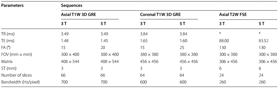
Patient studies: Data from 10 patients (range of 26-45 years old) were acquired to validate the performance of the proposed method. For each subject, MRI examination was performed with a 3.0-Tesla MRI scanner (uMR 790, United Imaging Healthcare, Shanghai, China) and a 5.0-Tesla MRI scanner (uMR Jupiter, United Imaging Healthcare, Shanghai, China). A custom-built 24-channel body coil was used for all studies at 5 T using local B1+shimming for B1+optimization. The following MR sequences were acquired: a. transverse breath-hold T1-weighted volume interpolated gradient-echo sequence (QUICK 3D) with fat suppression; b. coronal breath-hold T1-weighted QUICK 3D with fat suppression; c. transverse T2-weighted fatsaturated FSE sequence with respiratory trigger. The detailed MR protocols for anatomical imaging are listed in Table 1.Method Implements: To reduce the computational complexity, the heat kernel convolution operation is applied to approximate the perimeter of a segmentation curve. In addition, the weighted parameter included in the high-order total variation term can be automatically evaluated based on an adaptive input image to emphasize local structures and increase segmentation accuracy. Since the proposed method is a smoothing optimization model, the alternating direction method of multipliers is introduced to translate the original problems into several easily solvable subproblems. In summary, the proposed method is detailed in Algorithm 1.
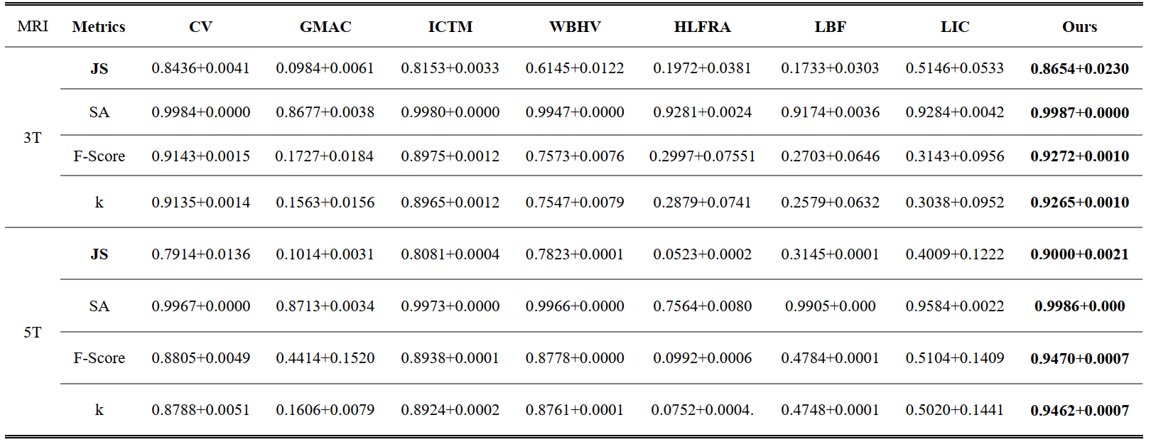
Data analysis: To compare the segmentation quality, we choose several indexes, such as the Jaccard similarity (JS), segmentation accuracy (SA), F1-score and Kappa coefficient (κ), to quantify the segmentation effectiveness. We use the proposed method to segment 3T/5T MR images to show its reasonability and robustness compared with several state-of-the-art model-based methods, such as the CV [8], GMAC [9], ICTM [10], WBHV [11], HLFRA [12], LBF [13] and LIC [14] methods.
Results
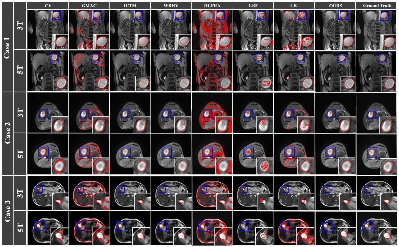
Figure 1 presents the visual comparison results for different methods on 3T/5T MRI from the same person. For patient case 1, the segmentation results on 3T/5T MRI illustrate that our method could gain better edges than other methods on the stomach. Then, our method provides better anatomical information for bones of the knee joint, as for patient case 2. Finally, we consider the segmentation performance of abdominal tissues and prove the effectiveness of the proposed methods. Moreover, we also calculate the evaluation metrics (including JS, SA, F1-Score and K) for the test datasets, as shown in Table 2. Our method shows superior performance compared with the other methods.Conclusion
In conclusion, this work represents a novel adaptive weighted curvature-based active contour for medical image segmentation. To describe local structures and establish an efficient numerical algorithm, we employed weights to adaptively penalize the high-order total variation and used the heat kernel convolution operation to approximate the total variation to improve the numerical method. Since the improved model is nonsmooth, the alternating direction method of multipliers can be used to solve the proposed model. The experimental results on 3T/5T MR images from the same persons are compared with those of other algorithms to show the robustness of our proposed method. In the future, we will conduct accurate quantitative analysis for the clinical comparison of the same person under 3T/5T MRI clinical metrics standards.Acknowledgements
This work was supported by the National Natural Science Foundation of China (32022042, 81871441, and 62101540), the Shenzhen Excellent Technological Innovation Talent Training Project of China (RCJC20200714114436080), and the Shenzhen Science and Technology Program (RCBS20210706092218043), the China Postdoctoral Science Foundation (2022M713290), and the Guangdong Innovation Platform of Translational Research for Cerebrovascular Diseases of China.References
[1] D. Shen, G. Wu, and H. Suk. Deep learning in medical image analysis. Annual review of biomedical engineering, 19:221-248, 2017.
[2] M. Gou, Y. Rao, M. Zhang, J. Sun, and K. Cheng. Automatic image annotation and deep learning for tooth CT image segmentation. International Conference on Image and Graphics, 519-528, 2019.
[3] M. Khan, M. Gajendran, Y. Lee, and M. Khan. Deep neural architectures for medical image semantic segmentation: review. IEEE Access, 9:83002-83024, 2021.
[4] S. Minaee, Y. Boykov, F. Porikli, A. Plaza, N. Kehtarnavaz, and D. Terzopoulos. Image Segmentation Using Deep Learning: A Survey. IEEE Transactions on Pattern Analysis and Machine Intelligence, 2021.
[5] F. Sultana, A. Sufian, and P. Dutta. Evolution of Image Segmentation using Deep Convolutional Neural Network: A Survey. KnowledgeBased Systems, 201:106062, 2020.
[6] M. Falcone, G. Paolucci, and S. Tozza. A high-order scheme for image segmentation via a modified level-set method. SIAM Journal on Imaging Sciences, 13(1):497-534, 2020.
[7] M. Unger, T. Pock, W. Trobin, D. Cremers, and H. Bischof. TVSeg-Interactive total variation based image segmentation. Proceedings of the British Machine Vision Conference, 2008.
[8] T. Chan and L. Vese. Active contours without edges. IEEE Transactions on Image Processing, 10(2):266-277, 2001
[9] X. Bresson, S. Esedoglu, P. Vandergheynst, J. Thiran, and S. Osher. Fast global minimization of the active contour/snake model. Journal of Mathematical Imaging and Vision, 28:151-167, 2007.
[10] D. Wang and X. Wang. The iterative convolution-thresholding method (ICTM) for image segmentation. Pattern Recognition, 2022.
[11] Y. Yang, Q. Zhong, Y. Duan, and T. Zeng. A weighted bounded Hessian variational model for image labeling and segmentation. Signal Processing, 2020.
[12] J. Fang, H. Liu, L. Zhang, and H. Liu. Region-edge-based active contours driven by hybrid and local fuzzy region-based energy for image segmentation. Information Sciences, 546:397-419, 2021.
[13] C. Li, C. Kao, J. Gore, and Z. Ding. Minimization of region-scalable fitting energy for image segmentation. IEEE Transactions on Image Processing, 17:1940-1949, 2008.
[14] C. Li, R. Huang, Z. Ding, C. Gatenby, D. Metaxas, and J. Gore. A level set method for image segmentation in the presence of intensity inhomogeneities with application to MRI. IEEE Transactions on Image Processing, 20(7):2007-2016, 2011.
Figures