3733
15-Channel Head Cap Array using Twisted-Pair Elements for MRI
Julian Adolfo Maravilla1, Colin Taylor Knizek2, Nazem Khairallah1, Ana Claudia Arias1, and Michael Lustig1
1EECS, University of California, Berkeley, Berkeley, CA, United States, 2University of California, Berkeley, Berkeley, CA, United States
1EECS, University of California, Berkeley, Berkeley, CA, United States, 2University of California, Berkeley, Berkeley, CA, United States
Synopsis
Keywords: RF Arrays & Systems, RF Arrays & Systems, Flexible RF Array, Conformal RF Array, fMRI Sleep Studies, TMS/EEG/fMRI
A tight fitting and wearable 15-Channel Head Cap Array using Twisted-Pair Elements was designed and characterized for potential use in a multimodal imaging system. The RF Array was characterized in terms of safety and SNR. The elements in the array (Twisted-Pair coil) show >40dB isolation for active detuning with no significant component heating under harsh RF conditions (ΔT<4°C). SNR evaluations revealed a 3x increase in peripheral SNR and retainment of SNR under parallel imaging conditions when compared to a commercial head array. An array of this caliber can be used for fMRI sleep studies and simultaneous TMS/EEG/fMRI.Purpose
Bulky commercial receive arrays prevent multimodal imaging techniques, such as TMS/EEG/fMRI, from being studied and utilized in research and clinical settings1. Therefore, receive arrays that are thin, flexible, and body conformal are necessary in order to provide adequate image quality without limiting the ability to capture EEG signals and excite the brain with TMS2.Although Coaxial coils3,4,5 have been proposed as receiving elements for such arrays, Twisted-Pair coils could provide lower losses in a thinner, more flexible form factor resulting in improved SNR6. This work aims to evaluate Twisted-Pair receiver elements in a conformal 15-Channel Head Cap Array (Fig. 1). The design of the array is compatible with fMRI sleep studies and creates a new platform for simultaneous TMS/EEG/fMRI. The tight fitting array provides increased comfort, and more options for head placement when compared to traditional head arrays.
Methods
Utilizing techniques inspired by coaxial and twinax coils by GE7,8, thin and flexible receiver elements made from Twisted-Pair (TP) wire were designed. To manufacture a TP Coil, 24-gauge polytetrafluoroethylene (PTFE) insulated TP wire was formed into a loop (Fig. 2a,b). One wire was split to act as a feed port to interface with the scanner, while the complementary wire was split on the opposing side to make the loop resonant at 127.7MHz (Fig. 2c). The two-wire PTFE structure provides a low-loss inherent distributed capacitance, allowing the coil to remain flexible even after tuning and matching. Coil dimensions are reported in Fig. 2. The TP coil was compared to a coaxial coil and a control coil in terms of quality factor, and single coil image SNR when heavily loaded.A 15-Channel TP Head Cap Array was fabricated using 9.5cm diameter coils in three rows sewn to a NomexTM ski mask for a 3T GE MR750W system (Waukesha, WI) (Fig. 1). A double probe measurement was used to evaluate active detuning performance. Component heating under excessive RF conditions was characterized using a heat sequence (TR/TE/FA=18ms/7ms/90°, M:256×256, FOV:400×400mm2, SL:10mm, BW=122.1Hz/Pixel, 15 min. acquisition). The SNR (optimal combine9,10), noise correlation matrix, and retained SNR maps11 of this array were compared to that of a commercial 22-Channel GE Head Coil under similar imaging conditions (Fig. 4). 2D-imaging was performed axially on a spherical loading phantom (18 cm diameter) using GRE PD-weighted images (TR/TE/FA=30ms/6ms/30°, M:192×192, FOV:260×260 mm2, SL:5mm, BW=200Hz/Pixel) (Fig. 4a). In-vivo brain images were obtained using a 3D T1-Bravo sequence with 2x2 acceleration in the AP/SI direction (1mm isotropic resolution, 2:43min. acquisition) (Fig. 5).
Results
The TP and coaxial coils were able to achieve body-noise dominance12 when heavily loaded (Qul and Ql reported in Fig. 3a). A TP coil had similar SNR performance when compared to a control coil, while the coaxial coil had slightly reduced sensitivity. For the Head Cap, safety testing revealed >40dB of isolation for active detuning with no significant heating after a heat sequence (ΔT<4°C). Close proximity of the Twisted-Pair elements to the phantom led to increased SNR along the periphery when compared to the commercial head coil (142 vs. 53.6) and maintained 76.3% percent of SNR at the center while having less average noise correlations between elements (7.6% vs. 18.2%) (Fig. 4a,b). Retained SNR maps reveal higher retained SNR under different acceleration factors for parallel imaging with the Head Cap Array (Fig. 4c). In-vivo scanning showed high quality brain images under accelerated conditions and with different head orientations using the proposed array (Fig. 1b,c, 5). The head orientation in Fig. 1c is not possible with the commercial array.Discussion
Improved sensitivity of the TP coil is emphasized by lower dielectric and conductive losses when compared to a coaxial coil. Consequently, arrays made from TP elements retain SNR while providing flexibility in a thin structure. The sensitivity of the Head Cap at the center could be improved by adding more channels to increase coverage13. Cabling must be modified depending on the application of the array. The current configuration is suitable for fMRI sleep studies as the cables do not perturb the head placement of a subject. However, for TMS/EEG/fMRI the cables must be routed down to prevent collisions with the TMS coil. Hygiene concerns using a ski-mask can be solved by removing the mouth cover, making the elements removable to wash the cap, or by using a head cap that can be easily disinfected. Additionally, a guard must be added to prevent the elements in front from making contact with the subject.Conclusion
A 15-Channel Head Cap Array using Twisted-Pair elements was designed and characterized. The tight fitting array resulted in a 3x increase in peripheral SNR with better performance under accelerated conditions when compared to a 22-channel commercial head coil and has the potential to be used for fMRI sleep studies, as a platform for TMS/EEG/fMRI, and for general brain imaging by offering increased comfort and more options for head placement.Acknowledgements
We would like to acknowledge support from R01MH127104, U01EB029427, and U01EB023829. Julian Maravilla acknowledges the NSF for funding under DGE 2146752 and mentorship from Dr. Jason Stockmann (Martinos Center).References
- Peters, Judith C., et al. "On the feasibility of concurrent human TMS-EEG-fMRI measurements." Journal of Neurophysiology 109.4 (2013): 1214-1227.
- Navarro de Lara, Lucia I., et al. "A novel coil array for combined TMS/fMRI experiments at 3 T." Magnetic resonance in medicine 74.5 (2015): 1492-1501.
- Navarro de Lara, Lucia I., et al. "A wearable “RF-EEG Cap” for full head coverage concurrent TMS/EEG/fMRI experiments at 3T: a feasibility study." Proc. ISMRM (2021).
- Zhang, Bei, Daniel K. Sodickson, and Martijn A. Cloos. "A high-impedance detector-array glove for magnetic resonance imaging of the hand." Nature biomedical engineering 2.8 (2018): 570-577.
- Ruytenberg, Thomas, Andrew Webb, and Irena Zivkovic. "Shielded‐coaxial‐cable coils as receive and transceive array elements for 7T human MRI." Magnetic resonance in medicine 83.3 (2020): 1135-1146.
- Maravilla, Julian Adolfo, et al. "Transmission Line Receiver Coils (TLCs) for MRI." Proc. ISMRM (2022).
- Cogswell, Petrice M., et al. "Application of adaptive image receive coil technology for whole-brain imaging." AJR. American journal of roentgenology 216.2 (2021): 552.
- General Electric Co. US20190353722A1
- Roemer, Peter B., et al. "The NMR phased array." Magnetic resonance in medicine 16.2 (1990): 192-225.
- Kellman, Peter, and Elliot R. McVeigh. "Image reconstruction in SNR units: a general method for SNR measurement." Magnetic resonance in medicine 54.6 (2005): 1439-1447.
- Pruessmann, Klaas P., et al. "SENSE: sensitivity encoding for fast MRI." Magnetic resonance in medicine 42.5 (1999): 952-962.
- Beer, R. de, and D. van Ormondt. "Analysis of NMR data using time domain fitting procedures." In-Vivo Magnetic Resonance Spectroscopy I: Probeheads and Radiofrequency Pulses Spectrum Analysis (1992): 201-248.
- Vaidya, Manushka V., Daniel K. Sodickson, and Riccardo Lattanzi. "Approaching ultimate intrinsic SNR in a uniform spherical sample with finite arrays of loop coils." Concepts in Magnetic Resonance Part B: Magnetic Resonance Engineering 44.3 (2014): 53-65.
- Reykowski, Arne, Steven M. Wright, and Jay R. Porter. "Design of matching networks for low noise preamplifiers." Magnetic resonance in medicine 33.6 (1995): 848-852.
Figures
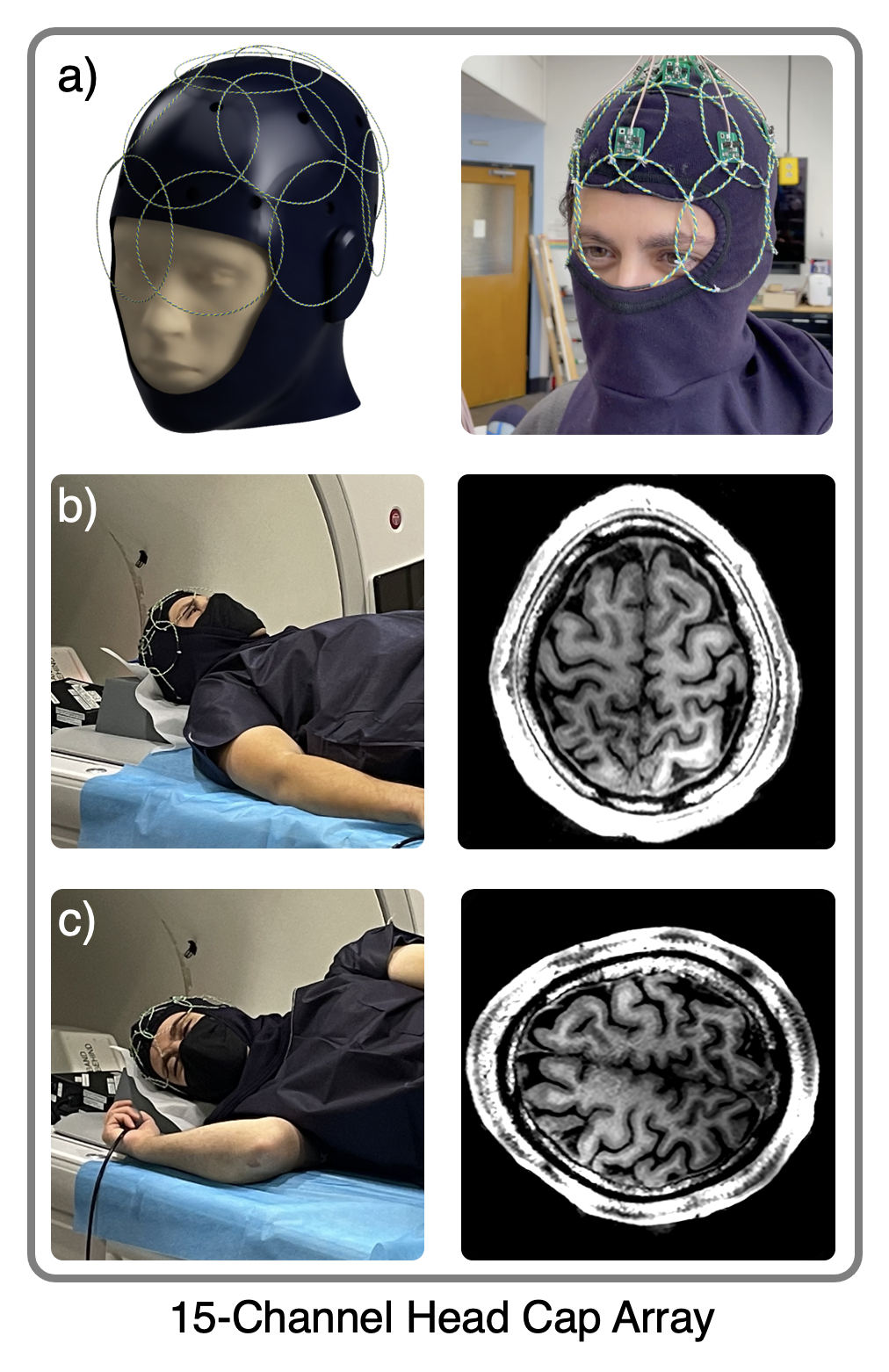
Figure 1: 15-Channel Head Cap Array using Twisted-Pair Elements. a) The Twisted-Pair elements are sewn onto a NomexTM ski mask in three rows (7x7x1). Individual coils are sewn independently to ensure that different sized heads do not strain the elements. The conformal and low profile design consumes less than 1cm of space around the head. b,c) With this array, subjects can be placed in different head orientations and are not constrained by a single supine position.
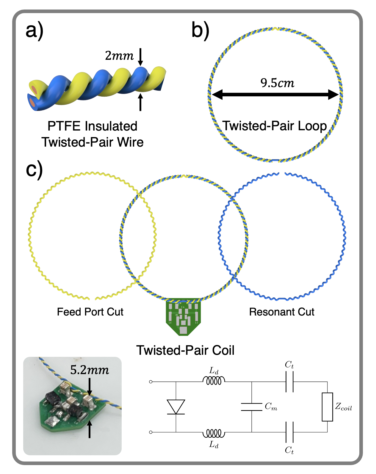
Figure 2: Twisted-Pair Element Assembly. a) A Twisted-Pair coil is constructed by starting with 24-gauge PTFE insulated Twisted-Pair wire. This provides a low-loss and flexible distributed capacitance. b) The thin wire is then formed into a loop of desired diameter. For the Head Cap Array, a diameter of 9.5cm was used. c) Two cuts are then made along the loop. The feed port cut allows the coil to interface with the detuning/matching network, and the resonant cut allows the coil to couple to induced EMF signals. The interface electronics only consume 5.2mm of vertical space14.
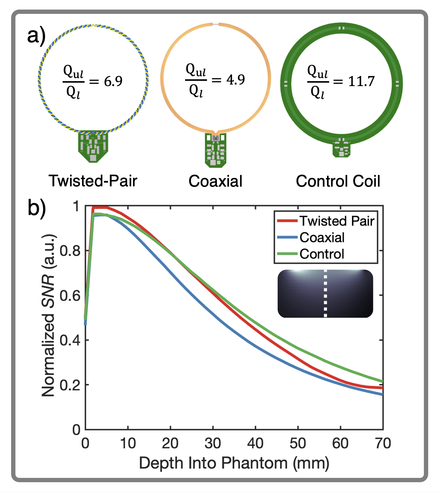
Figure 3: Single Coil Performance a) Twisted-Pair, Coaxial, and a Control Coil were tuned and matched to 127.7MHz using the same matching network shown in Fig. 2c and evaluated in terms of quality factor (unloaded and loaded). b) Single Coil SNR performance was evaluated under extreme loading conditions. The Twisted-Pair and Control coils slightly outperform the Coaxial coil in terms of SNR.
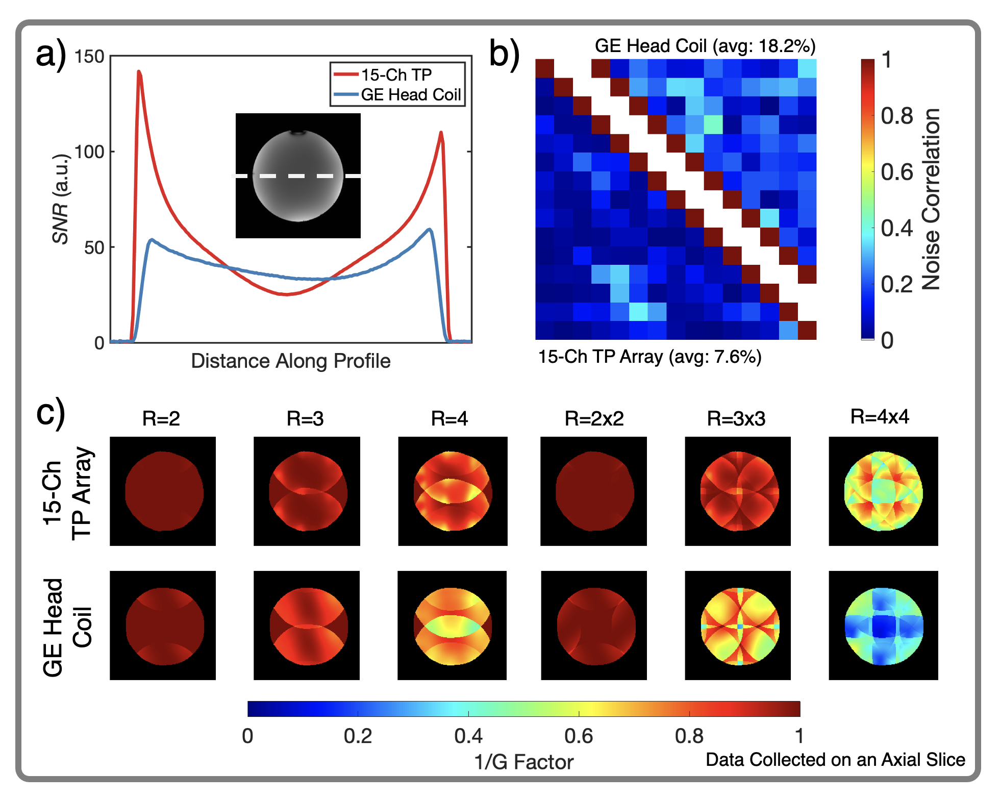
Figure 4: 15-Channel Head Cap Array Performance a) SNR Comparison between the Head Cap Array and commercial 22-Channel GE Head Coil using an axial image of a spherical loading phantom. Up to 3x increase in peripheral SNR with the Head Cap Array and 76.3% retainment in the center. b) Noise Correlation Matrix comparison. Average off-diagonal values are 7.6% for the Head Cap, and 18.2% for the commercial array. c) Retained SNR maps reveal better geometric conditions for parallel imaging with the Head Cap Array leading to more retained SNR for various accelerated conditions.
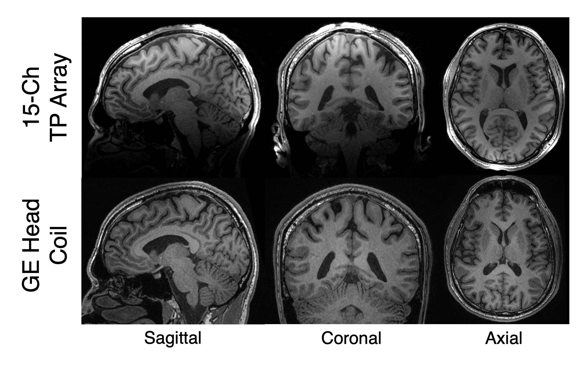
Figure 5: In-Vivo 3D T1-Bravo brain scan with the Head Cap Array and a 22-Channel GE Head Coil. R=2x2 acceleration in the AP/SI direction (1mm isotropic resolution, 2:43min. acquisition). Sagittal, Coronal, and Axial slices shown. The Head Cap Array provides increased peripheral SNR. More elements on the array will extend coverage in the lower part of the head, and improve SNR in the center.
DOI: https://doi.org/10.58530/2023/3733