3731
Development of 1H/23Na dual-tuned RF probe using inductive coupling for 9.4T vertical wide-bore superconducting MRI for rat’s kidney1Graduate School of Science and Technology, University of Tsukuba, Tsukuba, Japan, 2Department of Radiological Sciences, International University of Health and Welfare, Narita, Japan
Synopsis
Keywords: Non-Array RF Coils, Antennas & Waveguides, Non-Proton
A superconducting magnet for vertical NMR, commonly used as a high-field magnet, is effective for the MRI of X nuclides with low detection sensitivity. Even in a narrow magnetic field space, an inductively coupled RF coil with a simple structure can be used to image small animals one size larger. In this study, to visualize 23Na distribution in the kidney region of anesthetized rats, we developed 1H/23Na dual-tuned RF coils using inductive coupling with a 9.4T wide bore magnet (room temperature bore diameter 89 mm) and performed in vivo imaging.Introduction
MRI studies of X-nuclides such as 23Na and 13C are underway because of the advantage of obtaining metabolic information in addition to anatomical structures. Although in vivo visualization of X-nuclides in mice and rats has been performed as preclinical research, the number of MRI systems for small animals is limited, and maintenance costs will rise because of the recent depletion of liquid helium (LHe). In this context, 1H imaging of rats in vivo using a 17.6T wide-bore vertical NMR magnet (room temperature bore diameter: 89 mm) has been reported1. The advantage of using a general-purpose vertical NMR magnet is that it is easy to take measures to prevent LHe evaporation and has a higher cost advantage over horizontal animal superconducting magnets (φ120 mm or larger) in terms of both installation and maintenance costs. Meanwhile, live rats are slightly large with the wide bore 89 mm magnet, so the optimal design and manufacturing of the RF coil and gradient field coil are key points. Therefore, we focused on the dual-tuned (DT-) RF coil2, which utilizes inductive coupling.By using mutual induction, the trap circuit3 and other components necessary for decoupling can be omitted, allowing the subject space to be secured inside the bore of a wide-bore magnet, which is too narrow for rats. To optimize the size of the gradient coil, we utilized a thin gradient coil4 using a printed circuit board. In this study, we developed 1H/23Na DT RF coils using inductive coupling, added a thin gradient coil, and successfully visualized 23Na distribution in the kidney region of a live rat (male, four weeks old) using a 9.4T vertical wide-bore magnet.
Method
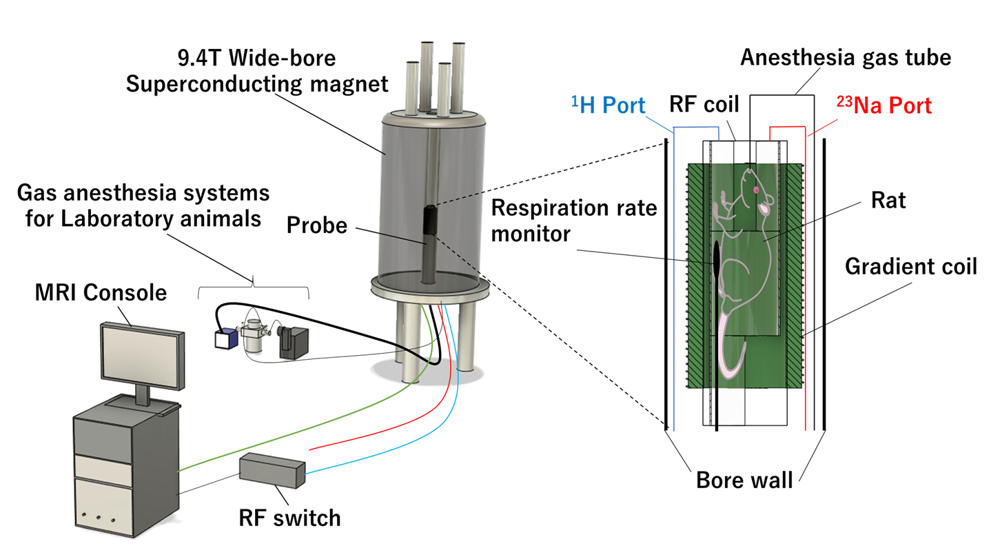
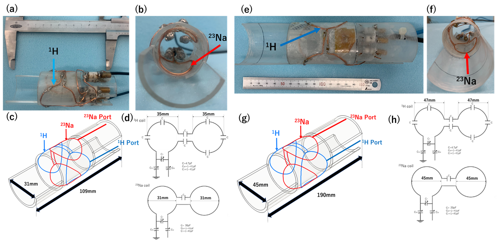
Fig. 1 shows a vertical 9.4T wide-bore superconducting magnet (room temperature bore diameter 89 mm), a gradient coil fabricated using a printed circuit board, an MRI console, and an anesthetic system for laboratory animals5. In vivo experiments were performed by continuously administering isoflurane anesthetic gas (approximately 1%) and monitoring the respiration of the small animals.We fabricated dual-tuned RF coils with resonance frequencies at 9.4 T (1H: 400.4 MHz, 23Na: 105.9 MHz) for live mice and rats, as shown in Fig. 2. We made the mouse coil because it was smaller than the rat coil, easier to construct, and suitable for prototyping tests. Each RF coil was of Helmholtz type. The coil diameter was 31/35 mm (23Na/1H) for the mouse coil and 45/47 mm (23Na/1H) for the rat coil. Each coil was fixed to the inside and outside of the acrylic pipe.
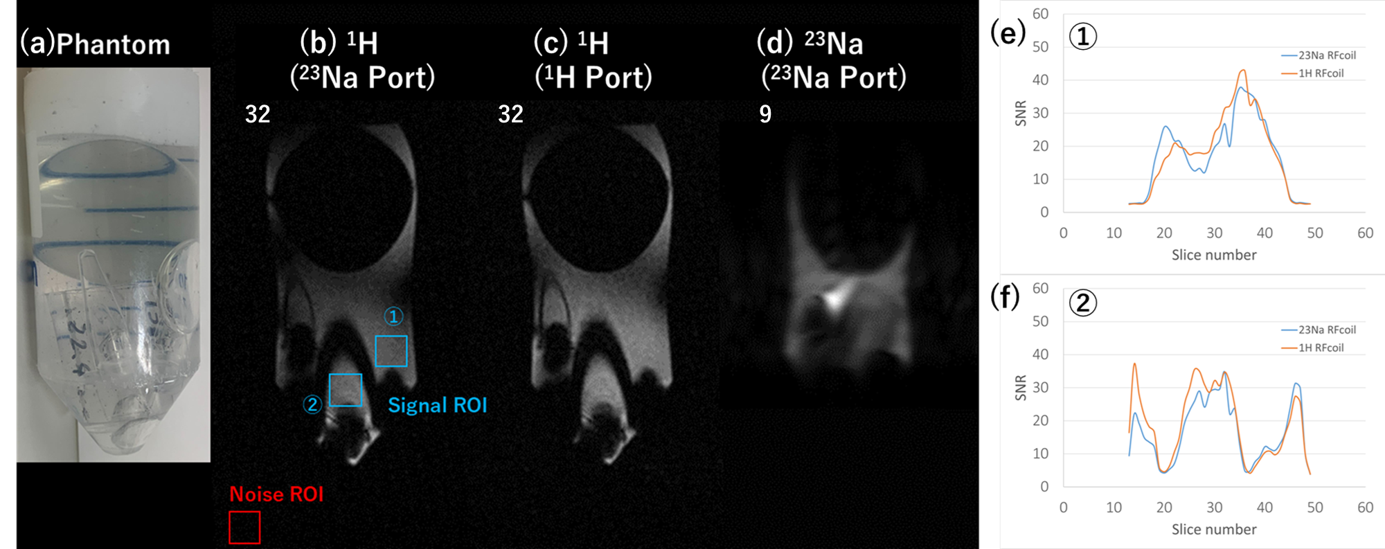
Saline phantoms (Fig. 3(a)) containing a 20mm-diameter acrylic sphere in a test tube and 12-week-old live mice (~20 g) were imaged using the mouse coil. Four-week-old live rats (male, 116g) were imaged using the rat coil. The imaging was performed using a gradient echo sequence under the following conditions.
(1) 1H image with a coaxial cable connected to the 23Na port.
(2) 1H image with a coaxial cable connected to the 1H Port
(3) 23Na image with a coaxial cable connected to the 23Na Port
The compressed sensing was used with an acceleration factor (AF) of 5.
We measured signal-to-noise (SNR) in the phantom, mouse, and rat images acquired under (1) and (2) conditions. The 23Na distribution in the kidney was visualized by creating a fusion image on the 1H image.
Results
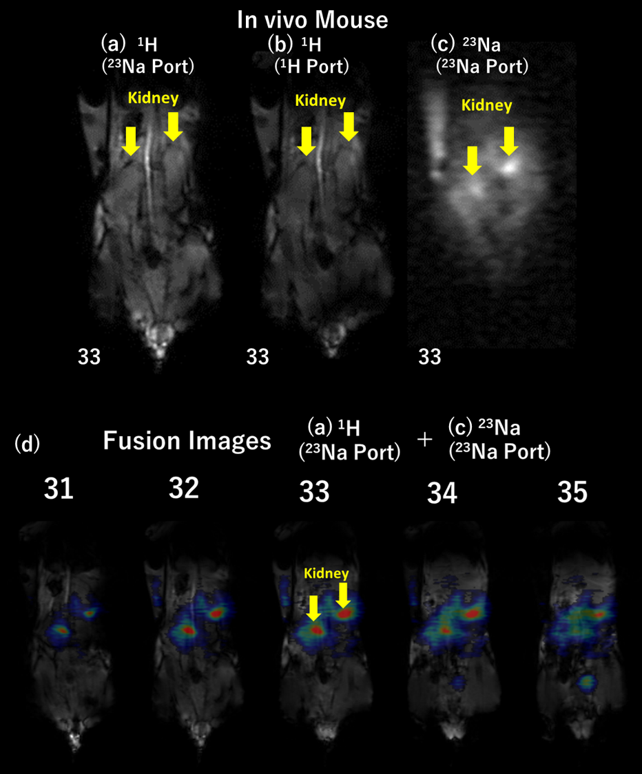
As shown in Figs. 3(e) and (f), the SNR of the saline phantom did not change significantly between ROIs 1 and 2. For 1H images, the SNR was almost the same for both port connections (conditions (1) and (2)), except that the SNR was slightly higher for the 23Na port connection, depending on the slice plane.As shown in Fig. 4, the mouse kidneys were visible under conditions (1) and (2). The SNR of the mouse kidney was 51.83 dB under condition (1) and 51.31 dB under condition (2).
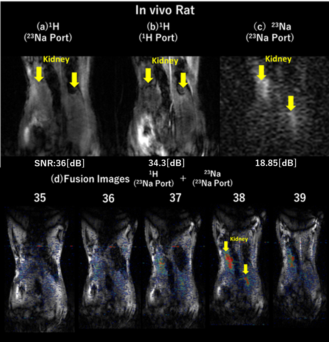
As shown in Fig. 5, the rat kidneys were also visible under conditions (1) and (2). The SNR of the rat kidney was 36 dB under condition (1) and 34.3 dB under condition (2).
In the 23Na and its fusion images of the mouse (Fig. 4(d)) and rat (Fig. 5(d)), regions of high 23Na signal intensity (red regions) were observed in the kidney area.
Discussion
We developed 1H/23Na RF coils using inductive coupling and compared 1H images of the kidney region of the live rat. The 1H images obtained by inductively coupled imaging did not show any deterioration in imaging performance compared to those obtained by conventional imaging with decoupled RF coils. In some cases, the signal intensity was higher with inductive coupling. The fusion images also showed a strong 23Na signal in the kidney region. These results indicate that we were able to visualize the distribution of 1H and 23Na using only the 23Na Port.Conclusion
Here we developed 1H/23Na DT RF coils using inductive coupling. We confirmed that simultaneous acquisition of 23Na distribution and 1H anatomical images of the kidney region of live rats is possible using a 9.4T vertical wide-bore magnet.Acknowledgements
No acknowledgement found.References
[1] V. C. Behr, T. Weber, T. Neuberger. et al. High-resolution MR imaging of the rat spinal cord in vivo in a wide-bore magnet at 17.6 Tesla. MAGMA 17, 353–358 (2004).
[2] M. V. Gulyaev, O. S. Pavlova, D. V. Volkov. et al. The Use of Strong Inductively Coupled Wireless Surface Coil and Transmit/Receive Volume Coil for 1H/19F MRI. Appl Magn Reson 50, 403–413 (2019).
[3] M. Wilcox, S. M. Wright, M. P. McDougall. Multi-Tuned Cable Traps for Multinuclear MRI and MRS. IEEE Transactions on Biomedical Engineering 67, 1221-1228 (2020).
[4] J. Matsuzaki, T. Haishi, Y. Terada. Low-cost gradients using commercially-available printed circuit boards. ISMRM 27th Joint Annual Meeting Montreal, Canada, 1465 (2019).
[5] T. Haishi, S. Sasaki, R. Kaseda. et al. 9.4 Tesla MR microscope of C57BL/6 live mouse kidney using a standard vertical-bore NMR magnet. JSMRM 45th Annual Meeting Tochigi, Japan, 264 (2017).
Figures