3718
Feasibility of Deep Learning Reconstruction in the Clinical Application of MRI for patients with Bladder Cancer: a preliminary prospective study1National Cancer Center/National Clinical Research Center for Cancer/Cancer Hospital, Chinese Academy of Medical Sciences and Peking Union Medical College, Beijing, 100021, China, Beijing, China, 2GE Healthcare, MR Research China, Beijing, Beijing, China
Synopsis
Keywords: Urogenital, Bladder, Deep Learning Reconstruction
The application of DLR significantly shortened scan times and improved the overall image quality score and image artifacts score and SNR and CNR of FSE-T2WI. DLR fast FSE-T2WI demonstrated significantly higher SNR (256.7±102.9 VS 94.7±40.8, p < 0.05) and CNR (168.0±77.3 VS 59.6±29.8, p < 0.05) and overall image quality scores (median, 4.0 vs. 3.0 for reader1 and 4.0 vs. 3.5 for reader2) than those of conventional FSE-T2WI. DLR may be useful in reducing the acquisition time of bladder MRI without compromising image quality.Background
Accelerated MRI acquisition has been a research focus for many years. The main techniques to achieve fast imaging include parallel imaging, compressed sensing, and the latest deep learning-based reconstruction (DLR) technology. DLR can reduce image noise and shorten scanning time while maintaining image quality. DLR uses deep convolutional neural networks to reconstruct images, specifically designed for performing image denoising and Gibbs ringing artifact removal, and ultimately produces images with high SNR and sharp edges. This technique has recently been applied to a rapid MRI scan protocol for the prostate and skeletal muscles, where acceleration sequences using DLR have improved image quality and artifacts compared with acceleration sequences without DLR and have shown diagnostic accuracy similar to standard sequences. However, in bladder MRI, no study has evaluated the feasibility of using DLR to improve image quality on short acquisition time MRI.Purpose
To investigate the performance of deep learning-based reconstruction in improving fast MRI image quality of bladder cancer.Methods
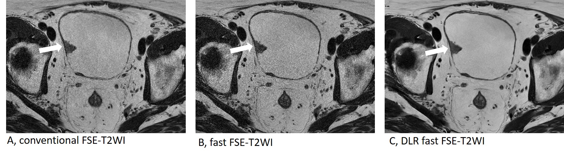
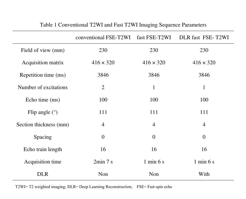
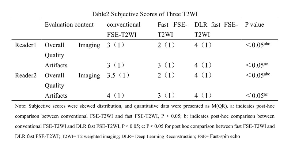
This is a prospective clinical cohort study approved by the institutional ethic board (NCC 3685). Patients suspected with clinically suspected bladder cancer were continuously enrolled into the scanning cohort. All MRI examinations were performed on GE 3.0T SIGNA Architect. Conventional Fast spin echo (FSE) T2-weighted imaging (T2WI), and DLR fast FSE-T2WI scanning were performed, respectively. The original fast FSE-T2WI without DLR was saved. Detailed parameters are shown in Table 1. One radiologist measured the signal-noise ratio (SNR) and contrast-to-noise ratio (CNR). The regions of interest (ROIs) were placed on the image background, iliopsoas muscle, and lesion. The SNR of the lesion and the CNR between the lesion and iliopsoas muscle were calculated according to the following formula: SNR=SI lesion /SD background, CNR = (SI lesion - SI iliopsoas)/SD background, SD background is the standard deviation of signal intensity in the background ROI, which is considered as noise; SI lesions and SI iliopsoas represent the mean signal intensity of lesion and iliopsoas ROI, respectively. The overall image quality and artifacts of three T2WI (conventional FSE-T2WI, fast FSE-T2WI, and DLR fast FSE-T2WI) were assessed subjectively by two radiologists using LIKERT 5-point scales. The radiologist's subjective assessment of artifacts requires consideration of artifacts in the image caused by bladder urine, intestinal cavity contents, and abdominal breathing (1 score is a large number of artifacts; 2 scores are visible significant artifacts; 3 scores are moderate artifacts, 4 scores are rare artifacts and 5 is no artifact). The overall image quality evaluation should comprehensively consider the image clarity, anatomical structure display, and artifacts (1 is non-diagnostic, 2 is poor, 3 is acceptable, 4 is good, and 5 is excellent). One-way ANOVA and Friedman test were performed on normally and non-normally distributed data, respectively, to compare and analyze the differences in SNR, CNR, overall image quality score, and artifacts score of three T2WI. The Weighted-Kappa test was used to validate the consistency of subjective scores between groups.Results
A total of 32 patients (mean age, 65 years±11 [SD]; age range, 39–93 years; 27 men) with bladder cancer were enrolled in this study. The application of DLR significantly improved the image quality of fast FSE-T2WI. DLR fast FSE-T2WI demonstrated significantly higher SNR (256.7 ± 102.9 VS 94.7 ± 40.8, p < 0.05) and CNR (168.0 ± 77.3 VS 59.6 ± 29.8, p < 0.05) and overall image quality scores (median, 4.0 vs. 3.0 for reader1 and 4.0 vs. 3.5 for reader2) than those of conventional FSE-T2WI. Detailed results are shown in Table 2 and Table 3.Conclusion
DLR can significantly improve the image quality of MRI sequences with shortened scan times, which would be beneficial to improve the accuracy of evaluating muscle invasion for bladder cancer and promote the clinical application of fast MRI sequences for bladder cancer.Acknowledgements
nonReferences
[1] Lee D, Yoo J, Tak S, et al. Deep Residual Learning for Accelerated MRI Using Magnitude and Phase Networks. IEEE Trans Biomed Eng 2018;65(9):1985-1995. doi: 10.1109/TBME.2018.2821699
[2] Hammernik K, Klatzer T, Kobler E, et al. Learning a variational network for reconstruction of accelerated MRI data. Magn Reson Med 2018;79(6):3055-3071. doi: 10.1002/mrm.26977
[3] Aggarwal HK, Mani MP, Jacob M. MoDL: Model-Based Deep Learning Architecture for Inverse Problems. IEEE transactions on medical imaging 2019;38(2):394-405. doi: 10.1109/tmi.2018.2865356
[4] Lebel RM. Performance characterization of a novel deep learning-based MR image reconstruction pipeline. 2020; arXiv: 2008.06559. doi:10.48550/arXiv.2008.06559
[5] Park JC, Park KJ, Park MY, et al. Fast T2-Weighted Imaging With Deep Learning-Based Reconstruction: Evaluation of Image Quality and Diagnostic Performance in Patients Undergoing Radical Prostatectomy. J Magn Reson Imaging 2022;55(6):1735-1744. doi: 10.1002/jmri.27992
[6] Hahn S, Yi J, Lee H-J, et al. Image Quality and Diagnostic Performance of Accelerated Shoulder MRI With Deep Learning-Based Reconstruction. AJR Am J Roentgenol 2022;218(3):506-516. doi: 10.2214/AJR.21.26577
Figures