3716
Relative noise variation with Unrolled Neural Networks for Accelerated Cardiac Cine Reconstruction
Suryanarayanan Sivaram Kaushik1, Xucheng Zhu2, Robert Marc Lebel3, Kavitha Manickam1, Ke Li1, Kailash Saravanan1, Florian Wiesinger4, and Martin Janich4
1GE Healthcare, Waukesha, WI, United States, 2GE Healthcare, Menlo Park, CA, United States, 3GE Healthcare, Calgary, AB, Canada, 4GE Healthcare, Munich, Germany
1GE Healthcare, Waukesha, WI, United States, 2GE Healthcare, Menlo Park, CA, United States, 3GE Healthcare, Calgary, AB, Canada, 4GE Healthcare, Munich, Germany
Synopsis
Keywords: Machine Learning/Artificial Intelligence, Cardiovascular
Deep Learning (DL) based reconstructions help alleviate the longer scan times seen in bSSFP Cine acquisitions by offering higher acceleration factors. This work analyzes the spatial and temporal variations in the noise as a function of acceleration factors in DL reconstructions of highly accelerated bSSFP Cine data.Introduction
bSSFP Cine acquisitions remain a staple in all cardiac exams and are used to image both cardiac structure, anatomy and function1. Especially in cardiac views that require multiple slices, these acquisitions still have relatively long scan times despite using conventional parallel imaging approaches. Recently, we have developed a Deep Learning (DL) reconstruction using a physics-based unrolled architecture which can overcome typical limitations seen with conventional parallel imaging techniques to offer higher acceleration factors (12x) and consequently, lower scan times and single heart-beat free-breathing scans1. This work seeks to extend the work by Lauer et al.2 in understanding the spatial and temporal variations in noise that may be seen in DL based reconstruction of Cardiac Cine acquisitions.Methods
A supervised learning approach was used to train a physics-based unrolled neural network (DLCine) using fully sampled retrospectively cardiac gated 2D bSSFP Cine acquisitions. The network consisted of 12 unrolls, each with a data consistency and CNN based regularizer term. The model was trained using an L1 loss function, an Adam optimizer, and had a total of 6.6M trainable parameters.The multiple replica (N=1000) approach by Robson et al. was used to estimate the relative noise (RN) retained by the neural network3. The relative noise was defined as: RN = SDaccelerated / SDfully sampled
This RN metric is the g-factor3 including the √R SNR penalty. This was tested using a single slice of a fully sampled bSSFP Cine acquisition (matrix: 200x160, 16 phases, 8 channels), which was retrospectively undersampled to study the relative noise changes spatially and temporally.
Results
Figure 1 shows a typical DL based reconstruction of a highly undersampled (prospective) bSSFP Cine scan acquired within a single heart-beat (1RR), 3 heart-beats (3RR) and 6 heart-beats (6RR). The image on the top left used a SENSE based parallel imaging reconstruction and the 2x acceleration yielded a scan time of 44s. The 6RR acquisition used a higher matrix size, and an acceleration factor of 9x to yield a scan time of 37s. The 3RR used a 9x acceleration factor to lower the scan time to 20s, and the 1RR used a 12x factor to lower the scan time to 10s.Figure 2 shows a montage of the original fully sampled data set used for the experiment, the relative noise maps for 4x acceleration and 12x acceleration. Overall, both 4x and 12x RN maps show variations in the noise based on local tissue structure. RN values are higher around the boundaries of the heart, and lower in regions of static and/or uniform tissue. The 4x RN maps show lower values compared to the 12x version, and the higher values in the 12x maps are more heavily concentrated on structural edges. In the temporal dimension, as the cardiac phases go from systole to diastole, both RN maps show a loss in structural dependence. As the diastolic phases have reduced cardiac motion, relative noise maps show a more diffuse presentation. The structural dependence is regained as the cardiac phase returns to systole. Relatively, the 12x RN maps retains more structural dependence during both end-diastole and end-systole.
The variability in the RN metric is shown in Figure 3. Figure 3A shows the global average and coefficient of variation (CV) for RN. The mean stays constant, while the CV increases with the acceleration factors. Figure 3B shows the coefficient of variation in RN with each cardiac phase. As noticed in Figure 2, the systolic phases show a higher mean RN value while the diastolic phases show lower RN values.
Discussion
The results indicate that there are both spatial and temporal variations in the RN metric for DLCine reconstruction. Regions with greater motion show a higher RN value, indicating lower denoising, and a greater emphasis by the network to retain structural information. Static and/or uniform tissues show a lower RN value which generates higher SNR images in regions with stable signal.Conclusions
DL based reconstruction of highly undersampled bSSFP Cine data show noise variations that depends on the structural boundaries of the images, and further shows variations temporally. Higher RN values around cardiac boundaries indicate a greater emphasis in reconstructing dynamic tissues with lesser denoising.Acknowledgements
No acknowledgement found.References
1. Sandino et al., "Accelerating cardiac cine MRI using a deep learning‐based ESPIRiT reconstruction." Magnetic Resonance in Medicine 2021
2. Lauer et al., “Assessment of resolution and noise in MR images reconstructed by data driven approaches”, Proceedings of the ISMRM 2022.
3. Robson et al., “Comprehensive Quantification of Signal-to-Noise Ratio and g-Factor for Image-Based and k-Space-Based Parallel Imaging Reconstructions”, Magnetic Resonance in Medicine 2008
Figures
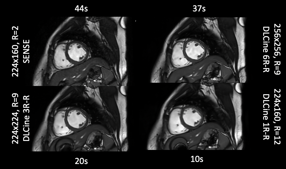
Single phase of a bSSFP cardiac cine acquisition. Conventional SENSE based reconstruction on the top left with a modest acceleration factor. The 3 heart beat (3R-R) offered about a 50% reduction in the scan time, and 6 heart beat 6R-R offered a 15% reduction in scan time, both with higher matrix sizes. The single heart-beat (1R-R) DLCine reconstruction was acquired with an acceleration factor of 12 and lowered the scan time significantly.
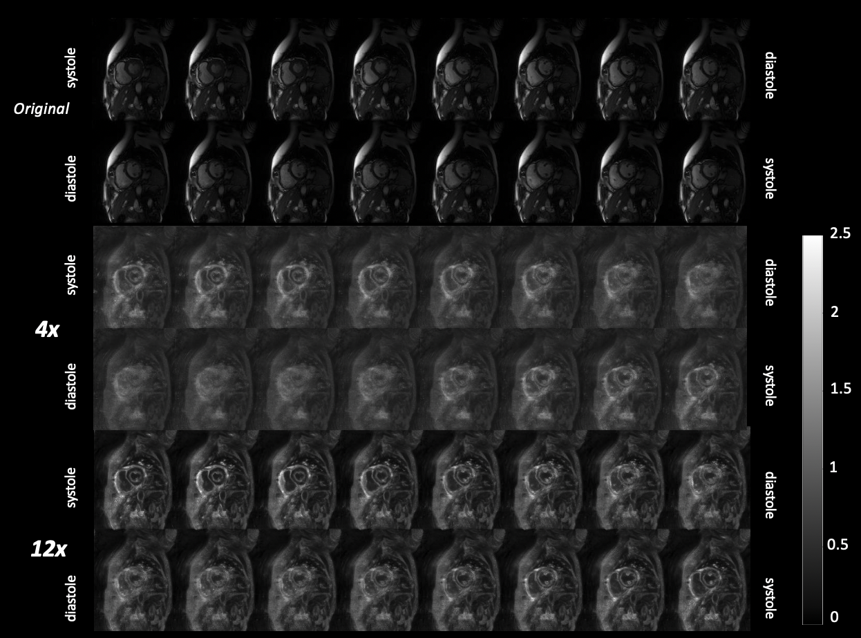
Top: Original fully sampled images used for the relative noise (RN) simulation. Middle: RN maps for 4x undersampled DLCine data. Bottom: RN maps for 12x undersampled data.
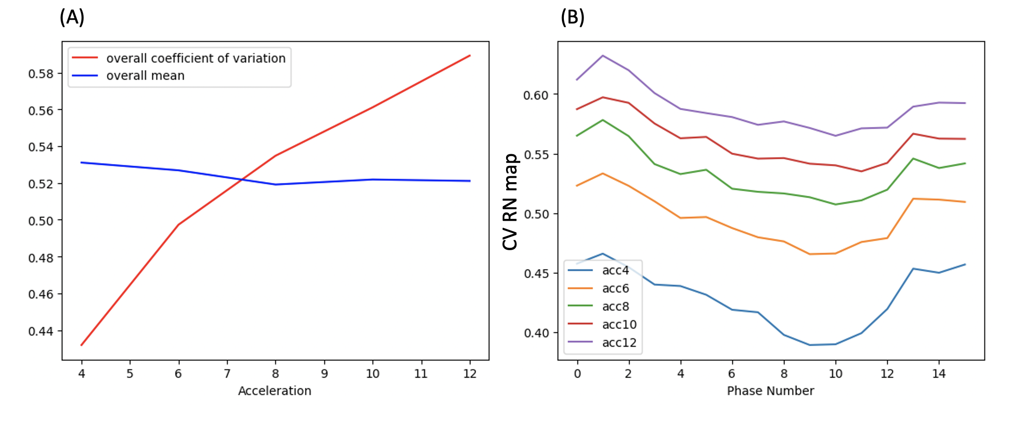
(A) Global variations in the RN metric as a function of acceleration. (B) Coefficient of variation (CV) of the RN values as a function of cardiac phase.
DOI: https://doi.org/10.58530/2023/3716