3707
Deep learning reconstruction algorithm for 100-second rapid Ischemic stroke imaging
Xin Fang1, Ailian Liu1, Qingwei Song1, Guobin Li2, and Shuheng Zhang2
1the First Affiliated Hospital of Dalian Medical University, Dalian, China, 2Shanghai United Imaging Healthcare Co., Ltd, Shanghai, China
1the First Affiliated Hospital of Dalian Medical University, Dalian, China, 2Shanghai United Imaging Healthcare Co., Ltd, Shanghai, China
Synopsis
Keywords: Brain Connectivity, Brain
Head Computed Tomography (HCT) is a common imaging method for the diagnosis of emergency cerebral infarction. However, it is easy to miss the diagnosis due to the poor performance of the ultra-early or early cerebral infarction lesions. Compared with CT, head Magnetic Resonance Imaging (MRI) has the characteristics of high tissue contrast, no ionizing radiation damage and can make functional imaging sequences, so it has significant advantages in the diagnosis of posterior fossa lesions and ultra-early cerebral infarctionSynopsis
To explore the feasibility of deep learning reconstruction algorithm in reducing the scanning time of emergency patients with ischemic stroke.Materials and methods
21 patients (10 males, 11 females, mean age 69.86 years) with suspected acute cerebral infarction underwent head MR imaging using a 3.0T MR (uMR Omega, United Imaging Healthcare, Shanghai, China). Protocol comprised T2WI-FSE, T1WI-FLAIR, T2WI-FLAIR, and DWI sequences. Each patient completed both the regular imaging and "100-second" rapid imaging. The conventional filtering reconstruction algorithm and deep learning reconstruction algorithm (AIFI index level 2; Pretreatment grade 2; Ripple suppression level 0; Brightness coefficient level 2) were used in different imaging methods, respectively. ROI were located in the pons, cerebellum, corpus callosum, white matter and infarct lesions, and were drawn by two radiologists with more than 5 years of experience in image reading. SI and SD values were recorded, and SNR and CNR values were calculated with white matter as the background. Images were scored on a 4-point scale based on the presence of artifacts, noise level and anatomical details. Kappa test was performed to investigate the consistency of scores from two observers. Wilcoxon was used to analyze the differences in SNR, CNR and subjective score for images of each sequence.Results
Among 21 patients, 8 patients had motion artifacts in conventional imaging, but not in the rapid imaging. SNR of T2WI-FSE, CNR of T1WI-FLAIR were significantly better than conventional imaging in 100-second imaging (P < 0.05). CNR of T2WI-FSE, SNR of T1WI-FLAIR , SNR and CNR of T2WI-FLAIR and DWI did not show significant differences between the two imaging approaches (P >0.05). There was good agreement between the subjective scores of two observers (Kappa: 0.709 ~ 0.818), and there was no statistical difference in scores .Discussion and Conclusions
Deep learning reconstruction technology can significantly reduce image noise in rapid head imaging, can improve image quality and increase lesion contrast, and therefore have certain clinical applications for rapid acquisition of routine head images of emergency patients and reduce motion artifacts.Acknowledgements
No acknowledgement found.References
[1] Ramgopal S, Karim S A, Subramanian S, et al. Rapid brain MRI protocols reduce head computerized tomography use in the pediatric emergency department[J]. BMC Pediatr,2020,20(1):14.
[2] Avey G. Technical Improvements in Head and Neck MR Imaging: At the Cutting Edge[J]. Neuroimaging Clin N Am,2020,30(3):295-309.
[3] Hahn S, Yi J, Lee H J, et al. Image Quality and Diagnostic Performance of Accelerated Shoulder MRI With Deep Learning-Based Reconstruction[J]. AJR Am J Roentgenol,2022,218(3):506-516.
Figures
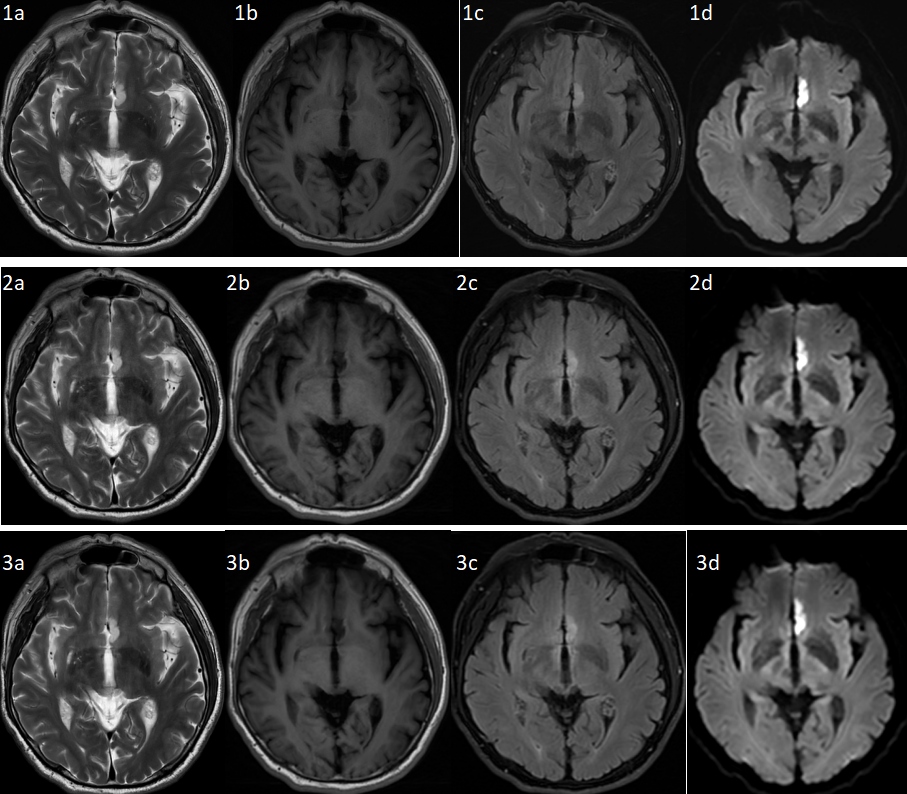
A 64-year-old male patient with infarction in the left frontal gyrus. Figure a-d shows the sequences of T2-WI-FSE, T1WI-FLAIR, T2-Wi-FLAIR and DWI respectively. Where, 1a-d is the conventional acquisition of traditional reconstructed image; 2a-d is the "hundred-second" image with traditional reconstruction; 3a-d is the "hundred-second image" reconstructed by deep learning.
DOI: https://doi.org/10.58530/2023/3707